. 2016 Jun 1;126(6):2167-80.
doi: 10.1172/JCI83967. Epub 2016 May 9.

Proteolytic activation defines distinct lymphangiogenic mechanisms for VEGFC and VEGFD

Proteolytic activation defines distinct lymphangiogenic mechanisms for VEGFC and VEGFD

Hung M Bui et al. J Clin Invest. .

Abstract

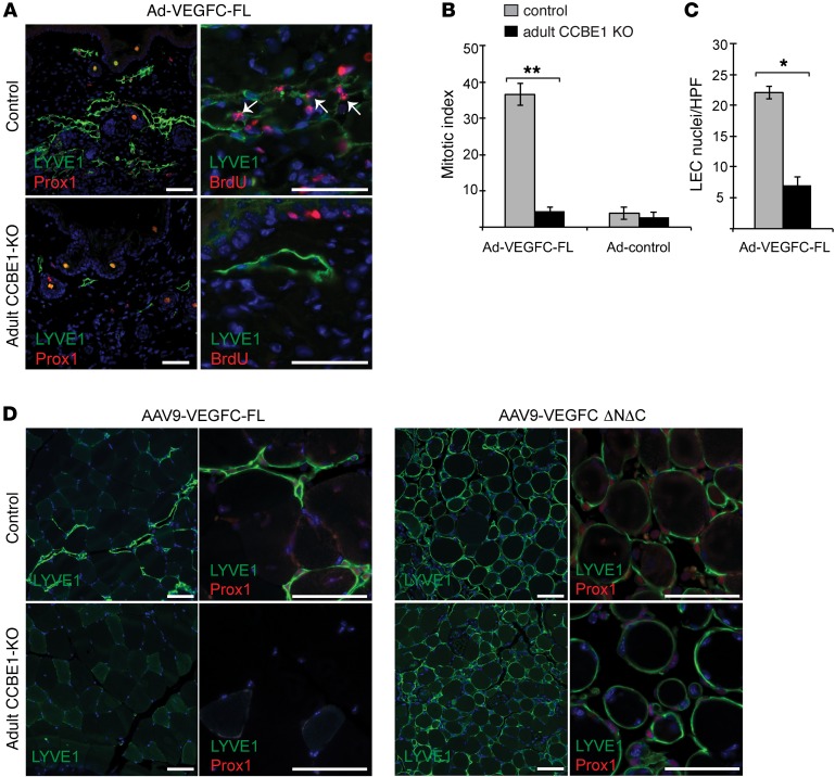
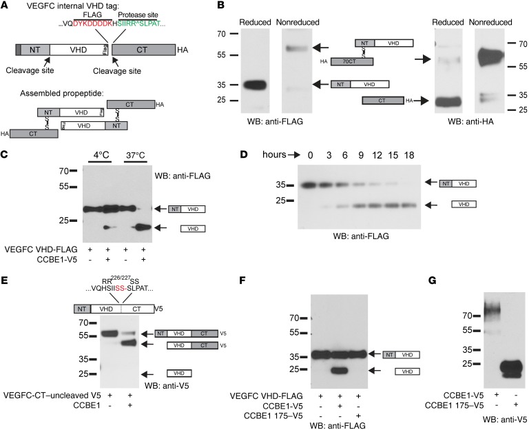
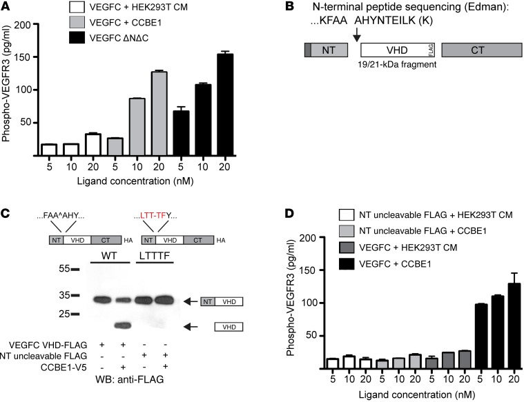
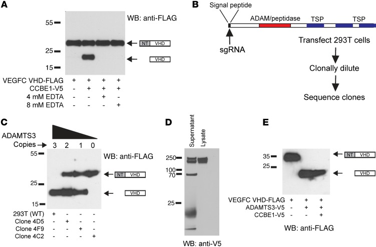
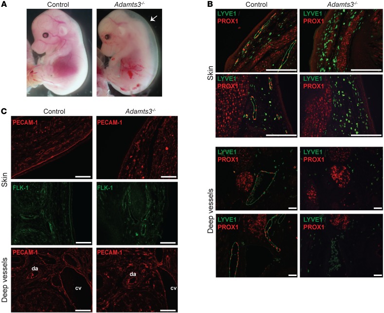
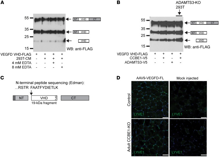
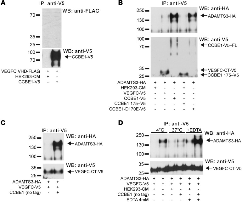
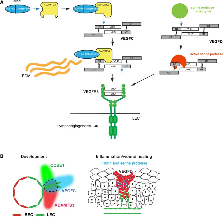
Lymphangiogenesis is supported by 2 homologous VEGFR3 ligands, VEGFC and VEGFD. VEGFC is required for lymphatic development, while VEGFD is not. VEGFC and VEGFD are proteolytically cleaved after cell secretion in vitro, and recent studies have implicated the protease a disintegrin and metalloproteinase with thrombospondin motifs 3 (ADAMTS3) and the secreted factor collagen and calcium binding EGF domains 1 (CCBE1) in this process. It is not well understood how ligand proteolysis is controlled at the molecular level or how this process regulates lymphangiogenesis, because these complex molecular interactions have been difficult to follow ex vivo and test in vivo. Here, we have developed and used biochemical and cellular tools to demonstrate that an ADAMTS3-CCBE1 complex can form independently of VEGFR3 and is required to convert VEGFC, but not VEGFD, into an active ligand. Consistent with these ex vivo findings, mouse genetic studies revealed that ADAMTS3 is required for lymphatic development in a manner that is identical to the requirement of VEGFC and CCBE1 for lymphatic development. Moreover, CCBE1 was required for in vivo lymphangiogenesis stimulated by VEGFC but not VEGFD. Together, these studies reveal that lymphangiogenesis is regulated by two distinct proteolytic mechanisms of ligand activation: one in which VEGFC activation by ADAMTS3 and CCBE1 spatially and temporally patterns developing lymphatics, and one in which VEGFD activation by a distinct proteolytic mechanism may be stimulated during inflammatory lymphatic growth.


Figures

Figure 1
Figure 1. CCBE1 is required for the activity of full-length but not processed VEGFC in vivo.
(A) VEGFC-FL was not lymphangiogenic in CCBE1-deficient animals. Adenovirus expressing VEGFC-FL (Ad-VEGFC-FL) was injected into the skin of Ub-CreERT2 Ccbe1fl/– animals in which Ccbe1 had been deleted by administration of tamoxifen (adult CCBE1-KO) or of control animals. Lymphatic vessels were analyzed 72 hours after adenovirus administration and 12 hours after BrdU administration by immunostaining with the indicated Abs. Arrows indicate BrdU+ LEC nuclei. (B) Percentage of LEC nuclei that were BrdU+ (mitotic index) in the skin of adult CCBE1-KO and control animals following exposure to adenovirus expressing VEGFC-FL (Ad-VEGFC-FL). n = 5 in each group; **P < 0.001, by unpaired, 2-tailed Student’s t test. (C) LEC proliferation in the skin of adult CCBE1-KO and control animals following exposure to adenovirus expressing VEGFC-FL was calculated by counting LEC nuclei per high power field. n = 5 in each group; *P < 0.01. HPF, high-power field. (D) Proteolytically processed VEGFC ΔNΔC was lymphangiogenic in CCBE1-deficient mice. AAV expressing either VEGFC-FL (AAV9-VEGFC-FL) or the core domain of VEGFC released by proteolysis of the N and C termini (AAV9-VEGFC-ΔNΔC) was injected into the tibialis muscle of adult CCBE1-KO or control animals. Lymphatic growth was detected using LYVE1 and PROX1 immunostaining. Note that lymphatic growth stimulated by VEGFC-ΔNΔC tightly encircles muscle fibers (right) and lacks the typical branched vascular appearance associated with VEGFC-FL (left). Data shown are representative of 3 separate experiments. Scale bars: 50 μm.
Figure 2
Figure 2. VEGFC VHD-FLAG demonstrates increased VEGFC proteolysis with full-length but not truncated CCBE1.
(A) Schematic of the VEGFC VHD-FLAG protein, in which a FLAG epitope (shown in red) is inserted in frame at the C-terminal end of the VHD. The CT cleavage site is shown in green; an HA tag is placed in frame at the end of the protein CT. Disulfide bonds are predicted to link 2 full-length VEGFC VHD-FLAG molecules as shown in the bottom panel. (B) Detection of VEGFC VHD-FLAG with a C-terminal HA tag under reduced and nonreduced conditions using anti-FLAG and anti-HA Abs. The protein domains within the detected bands are illustrated schematically. (C and D) CCBE1-FL drives VEGFC proteolysis in vitro. HEK293T conditioned media containing VEGFC VHD-FLAG alone or VEGFC VHD-FLAG plus CCBE1-V5 were incubated for 24 hours at 4°C or 37°C prior to detection of FLAG by immunoblotting (C). Temporal analysis revealed slow proteolysis of VEGFC in the presence of CCBE1 (D). (E) N-terminal cleavage of VEGFC was independent of C-terminal cleavage. A VEGFC VHD-FLAG protein containing a mutation that prevents C-terminal cleavage (RR226-227SS) was incubated with CCBE1-V5 or control conditioned medium for 24 hours. (F and G) A truncated form of CCBE1 lacking its C-terminal collagen-like domain failed to drive VEGFC cleavage. VEGF VHD-FLAG was incubated with control conditioned medium, conditioned medium containing full-length CCBE1-V5, and conditioned medium containing CCBE1 175-V5 for 24 hours prior to anti-FLAG immunoblot analysis (F). Full-length CCBE1-V5 was detected as a 70- to 100-kDa smear that was expressed at lower levels than the 25-kDa CCBE1 175-V5 (G). Data shown are representative of 3 separate experiments. WB, Western blot.
Figure 3
Figure 3. CCBE1-dependent proteolysis is required for VEGFC to activate VEGFR3 signaling.
(A) Phospho-VEGFR3 was measured by ELISA following LEC exposure to 5 to 20 nM VEGFC that was incubated with conditioned medium from control HEK293T cells (HEK293T CM) or CCBE1-V5 (CCBE1) or following exposure to VEGFC ΔNΔC-Fc. (B) Schematic representation of the VEGFC cleavage site generated following exposure to CCBE1 in HEK293T conditioned medium. (C) The VEGFC FAAAH109-113LTTTF mutant was not N-terminally cleaved in the presence of CCBE1. (D) N-terminally uncleavable VEGFC (NT uncleavable) was unable to activate VEGFR3. Phospho-VEGFR3 ELISA was performed as described in A using the indicated concentrations of WT and NT uncleavable VEGFC, with and without CCBE1. n = 3 for each concentration. Biochemical data shown are representative of 3 separate experiments. CM, conditioned medium.
Figure 4
Figure 4. ADAMTS3 is required for N-terminal VEGFC proteolysis in HEK293T cell supernatant.
(A) Incubation with EDTA revealed that CCBE1-dependent VEGFC proteolysis was cation dependent. (B) The ADAMTS3 gene was disrupted in HEK293T cells using CRISPR-Cas9 to generate disabling mutations in the sequence encoding the ADAMTS3 signal peptide. TSP, thrombospondin 1 motif; sgRNA, single-guide RNA. (C) Incubation of VEGFC VHD-FLAG and CCBE1-V5 generated by expression in HEK293T cell clones with 3 (WT), 2 (4D5), 1 (4F9), or 0 (4C2) WT ADAMTS3 alleles revealed a dose-dependent loss of VEGFC proteolysis, with loss of ADAMTS3. (D) Expression of ADAMTS3-V5 in HEK293T cells revealed a 220- to 240-kDa doublet in cell lysate and a number of smaller bands in conditioned supernatant, consistent with extracellular proteolysis. (E) Expression of ADAMTS3-V5 rescued proteolytic processing of VEGFC VHD-FLAG in ADAMTS3–/– HEK293T cells. Data shown are representative of 3 separate experiments.
Figure 5
Figure 5. ADAMTS3 is required for lymphatic development in mouse embryos.
(A) Loss of ADAMTS3 resulted in severe cutaneous edema at E14.5. Arrow indicates translucent space between the skin and body, indicative of edema. (B) Loss of ADAMTS3 resulted in a complete lack of PROX1+LYVE1+ LECs in the E14.5 embryo. Note the presence of smaller lymphatic vessels in the skin and larger lymphatic vessels adjacent to the dorsal aorta and cardinal vein in the WT littermate control embryo. (C) ADAMTS3 was not required for blood vessel development. Blood vessels from an ADAMTS3-deficient and control embryo were identified using anti-PECAM and anti-FLK1 immunostaining. cv, cardinal vein; da, dorsal aorta. Data shown are representative of 3 separate experiments. Scale bars: 50 μm.
Figure 6
Figure 6. VEGFD proteolysis and activity are independent of ADAMTS3 and CCBE1.
(A) Incubation with EDTA revealed that VEGFD VHD-FLAG proteolysis in HEK293T conditioned medium was cation independent. (B) VEGFD VHD-FLAG proteolysis was not increased by incubation with conditioned medium containing CCBE1 (lane 2) or ADAMTS3 (lane 3). VEGFD VHD-FLAG proteolysis was also not reduced when expressed by ADAMTS3-deficient HEK293T cells (lane 4, right). (C) N-terminal sequencing of the 21-kDa band observed in VEGFD VHD-FLAG–expressing medium revealed cleavage after R88, a site that is 3 aa N-terminal of that detected in VEGFC VHD-FLAG. frag, fragment. (D) VEGFD was lymphangiogenic in CCBE1-deficient animals. AAV expressing VEGFD-FL was injected into the tibialis muscle of adult CCBE1-KO and control animals. Lymphatic growth was detected using LYVE1 immunostaining and compared with that in mock-injected animals. Data shown are representative of 3 separate experiments. Scale bars: 50 μm.
Figure 7
Figure 7. VEGFC, ADAMTS3, and CCBE1 form a molecular complex prior to VEGFC cleavage.
(A) VEGFC did not directly bind CCBE1. VEGFC VHD-FLAG and CCBE1-V5 conditioned media were mixed prior to immunoprecipitation with anti-V5 Abs and to immunoblot analysis with anti-FLAG Abs. Precipitated CCBE1-V5 is shown in the bottom panel. (B) ADAMTS3 coprecipitated with full-length, but not N-terminal, CCBE1. The indicated conditioned media were mixed, and CCBE1-V5 was immunoprecipitated. Coprecipitated ADAMTS3-HA is shown at the top, and precipitated CCBE1-V5 proteins are shown at the bottom. (C) VEGFC coprecipitated with ADAMTS3 in a CCBE1-dependent manner. Conditioned media containing ADAMTS3-HA and VEGFC-V5 were mixed alone and with media containing untagged CCBE1 prior to anti-V5 immunoprecipitation. (D) ADAMTS3-CCBE1 release of VEGFC required ADAMTS3 enzymatic activity. VEGFC coprecipitation with ADAMTS3 was performed in the presence of untagged CCBE1, as described in C, at 4°C and 37°C and in the presence of EDTA to inhibit metalloprotease activity. Note that VEGFC was retained at 4°C and in the presence of EDTA, conditions that block ADAMTS3 enzymatic activity. Data shown are representative of 3 separate experiments.
Figure 8
Figure 8. Distinct mechanisms of proteolytic activation support distinct biological roles for VEGFC and VEGFD.
(A) Molecular mechanisms of VEGFC and VEGFD activation. VEGFC is activated by the formation of a VEGFC-ADAMTS3-CCBE1 complex (left). CCBE1 binding to ADAMTS3 via the CCBE1 CT (left) is predicted to confer a conformational change that permits the enzyme to associate with and cleave VEGFC (middle). N-terminal cleavage of VEGFC releases the VHD that is able to activate VEGFR3 on the LEC. The CCBE1 NT may bind extracellular matrix (ECM) to localize the complex spatially during lymphatic growth. In contrast, VEGFD is activated independently of ADAMTS3 and CCBE1, most likely through a serine protease generated at sites of inflammation (right). (B) Proposed lymphangiogenic roles of VEGFC and VEGFD in vivo. VEGFC activation by ADAMTS3 and CCBE1 provides a mechanism for spatial patterning of the developing lymphatic vasculature (left). Schematic shows the mid-gestation cardinal vein with newly specified LECs that are in the process of sprouting to form the lymphatic network. The area encircled by the dotted line represents a zone of active VEGFC. VEGFD activation by inflammatory proteases such as those generated during wound healing in the skin provides a mechanism for lymphangiogenesis in mature animals with preexisting lymphatic networks (right).

References

    1. Tammela T, Alitalo K. Lymphangiogenesis: Molecular mechanisms and future promise. Cell. 2010;140(4):460–476. doi: 10.1016/j.cell.2010.01.045. - DOI - PubMed
    1. Randolph GJ, Angeli V, Swartz MA. Dendritic-cell trafficking to lymph nodes through lymphatic vessels. Nat Rev Immunol. 2005;5(8):617–628. doi: 10.1038/nri1670. - DOI - PubMed
    1. Alitalo K. The lymphatic vasculature in disease. Nat Med. 2011;17(11):1371–1380. doi: 10.1038/nm.2545. - DOI - PubMed
    1. Joukov V, et al. A novel vascular endothelial growth factor, VEGF-C, is a ligand for the Flt4 (VEGFR-3) and KDR (VEGFR-2) receptor tyrosine kinases. EMBO J. 1996;15(2):290–298. - PMC - PubMed
    1. Kaipainen A, et al. Expression of the fms-like tyrosine kinase 4 gene becomes restricted to lymphatic endothelium during development. Proc Natl Acad Sci U S A. 1995;92(8):3566–3570. doi: 10.1073/pnas.92.8.3566. - DOI - PMC - PubMed

Publication types

MeSH terms