. 2016 Jul;12(7):497-503.
doi: 10.1038/nchembio.2079. Epub 2016 May 9.

Global survey of cell death mechanisms reveals metabolic regulation of ferroptosis

Affiliations

Global survey of cell death mechanisms reveals metabolic regulation of ferroptosis

Kenichi Shimada et al. Nat Chem Biol. 2016 Jul.

Abstract

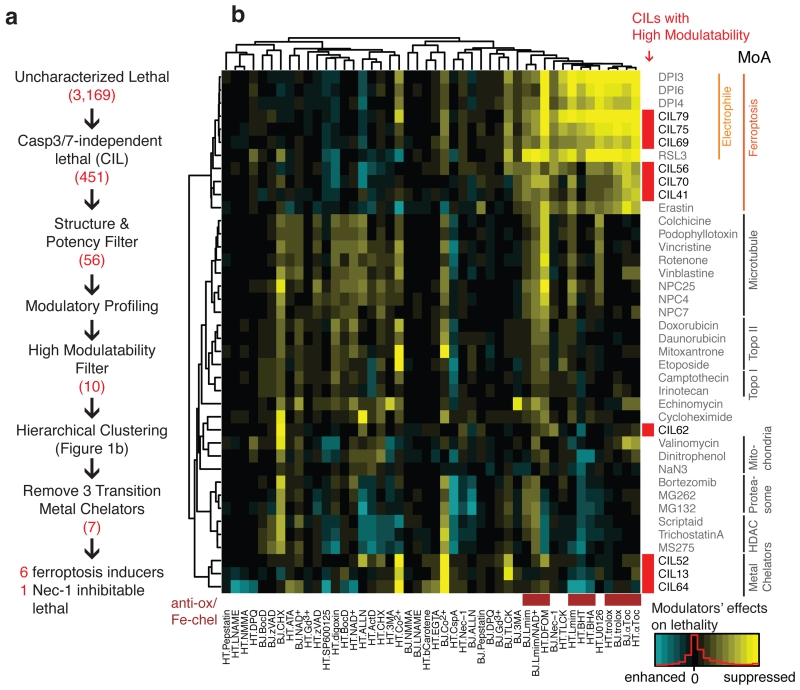
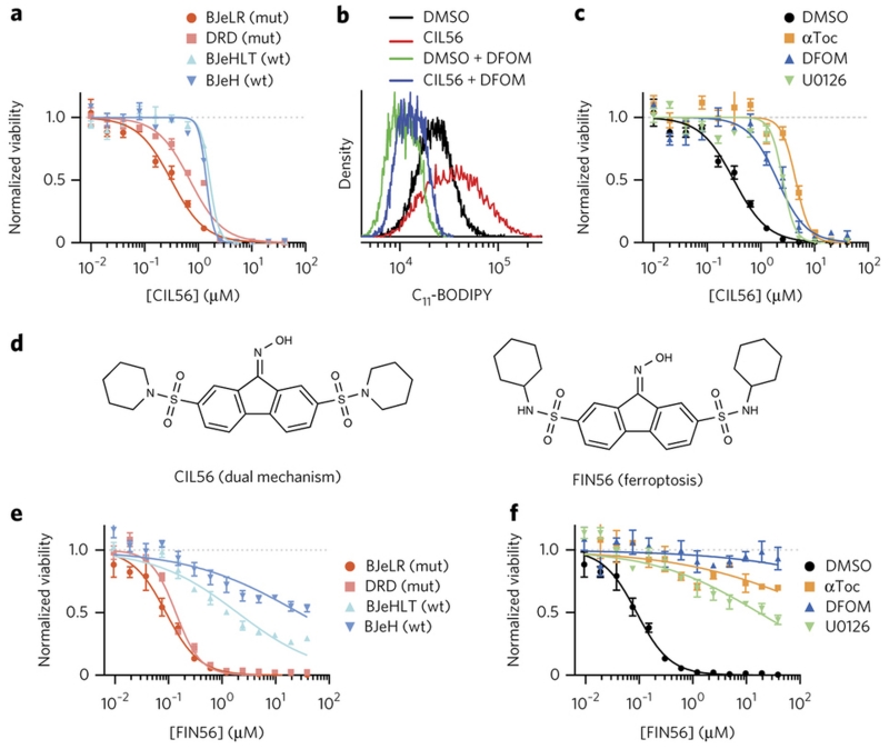
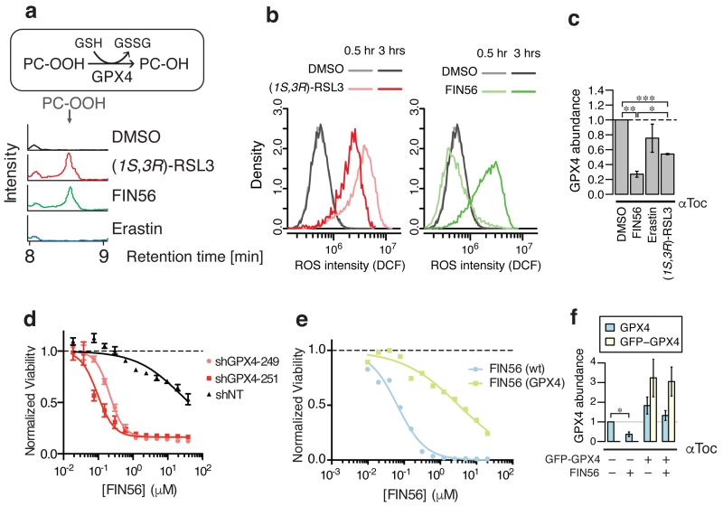
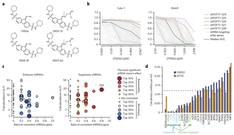
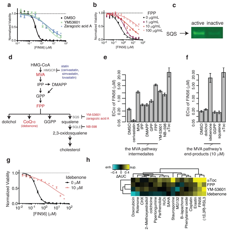
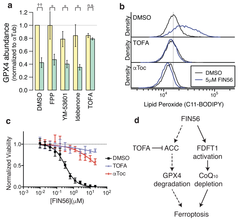
Apoptosis is one type of programmed cell death. Increasingly, non-apoptotic cell death is recognized as being genetically controlled, or 'regulated'. However, the full extent and diversity of alternative cell death mechanisms remain uncharted. Here we surveyed the landscape of pharmacologically accessible cell death mechanisms. In an examination of 56 caspase-independent lethal compounds, modulatory profiling showed that 10 compounds induced three different types of regulated non-apoptotic cell death. Optimization of one of those ten resulted in the discovery of FIN56, a specific inducer of ferroptosis. Ferroptosis has been found to occur when the lipid-repair enzyme GPX4 is inhibited. FIN56 promoted degradation of GPX4. FIN56 also bound to and activated squalene synthase, an enzyme involved in isoprenoid biosynthesis, independent of GPX4 degradation. These discoveries show that dysregulation of lipid metabolism is associated with ferroptosis. This systematic approach is a means to discover and characterize novel cell death phenotypes.


Figures

Figure 1
Figure 1. Modulatory profiling revealed three types of regulated non-apoptotic cell death
a. Experimental scheme to identify regulated non-apoptotic cell death inducers with high modulatability. The numbers in red are the number of compounds satisfying each criterion. b. Hierarchical clustering of modulatory profiles of 10 CILs with high modulatability and 30 characterized lethal compounds from several classes of lethal mechanisms. Lethal compounds are shown on the right. 10 CILs are indicated in red. 46 modulators are shown on the bottom (28 death modulators in 2 cell lines, HT-1080 or BJeLR). Antioxidants and iron-chelators are indicated in brown. A detailed list of modulators is shown in the Supplementary Table 2. Supplementary Fig. 1–4 show additional data on the CIL screening scheme, modulatory profiling scheme, and structures and characterization of ten regulated non-apoptotic cell death inducers.
Figure 2
Figure 2. Optimization of CIL56 revealed a potent and selective ferroptosis inducer
a, e. HRASG12V selectivity. Viability of four engineered BJ cell lines treated with (a) CIL56 or (e) FIN56 for 48 hrs. mut: cells tumor-transformed due to HRASG12V overexpression, wt: isogenic cells without HRASG12V. b. Lipid ROS generation. Flow cytometry analysis with BODIPY-581/591 C11 staining in HT-1080 cells incubated with test compounds for six hours. DFOM: 152 μM deferoxamine. c, f. Effects of ferroptosis inhibitors on viability of HT-1080 cells co-treated with (c) CIL56 or (f) FIN56 for 48 hrs. αToc: 100 μM α-tocopherol, U0126: 3.8 μM. d. Chemical structures of CIL56 and FIN56. See Supplementary Fig. 5 for structure activity relationship around the CIL56 scaffold. Experiments in a-f were performed in biological triplicates, and single representative results were shown; error-bars indicate s.e.m. of technical triplicates.
Figure 3
Figure 3. FIN56-induced ferroptosis decreases GPX4 expression
a. GPX4 enzymatic activity in BJeLR cells upon ferroptosis inducer treatment. b. Kinetics of ROS generation upon 0.5 μM (1S, 3R)-RSL3 or 5 μM FIN56 treatment detected with 25 μM H2-DCFDA staining in BJeLR cells. c. GPX4 protein abundance in BJeLR cells upon co-treatment with 100 μM αToc and ferroptosis inducers for ten hours. d. The effect of shGPX4 on FIN56-induced ferroptosis in BJeLR cells. e. The effects of GFP-GPX4 fusion protein overexpression on sensitivity to FIN56 in BJeLR cells. f. The effects of GFP-GPX4 fusion protein overexpression on endogenous and exogenous GPX4 protein abundance upon FIN56 treatment. Supplementary Fig. 6 supports more connection between FIN56 and GPX4 as well as corresponding westerns to c and f. Statistical significance (paired two-tailed t-test) – *: p < 0.05, **: p < 0.005, ***: p < 0.0005, †: p < 0.01, ††: p < 0.001, n.s.: not significant. Experiments in a-f were done in biological triplicates. Single representative results are shown for a,b,d,e; error-bars in d indicate s.e.m. of technical triplicates.
Figure 4
Figure 4. Squalene synthase (SQS) encoded by FDFT1 as FIN56’s target protein
a. Active and inactive FIN56 analogs with PEG linkers. Supplementary Fig. 7 shows their potency in HT-1080 cells. b. Effects of five shRNAs against FDFT1 on FIN56. The results in two of the four cell lines were shown. Five shRNA clones targeting FDFT1 are shown in polychromatic lines. A black line indicates a shRNA which gives no effect (median AUC among tested shRNAs) in each cell line. Grey lines indicate shRNAs targeting other genes. c. Summary of proteomic target identification and shRNA screening targeting 70 identified genes on FIN56. Each dot summarizes the result of multiple shRNAs targeting a gene. Each shRNA is considered ‘consistent’ when it exerts indicated effect (enhancing or suppressing FIN56). X-axis shows the ratio: the number of consistent shRNAs inducing indicated (i.e., enhancing or suppressing FIN56) effects to the total number of shRNAs targeting the gene. Y-axis shows fold-enrichment of protein abundance on active versus inactive probe-beads in pull-down assay. See Supplementary Fig. 8 for more description. d. Effect of siRNAs against ‘loss-of-function’ candidates on BJeLR cell viability. Cells were grown under DMSO or ATOC (α-tocopherol)’s existence. shRNA screens in b,c were performed once in four cell lines. siRNA experiment in d was performed in BJeLR twice and mean of biological replicates.
Figure 5
Figure 5. Validating SQS as the functionally relevant target to FIN56’s lethality
a. Effects of chemical inhibitors of SQS on FIN56’s lethality. b. Effects of Farnesyl-PP on FIN56’s lethality. c. Detection of SQS using pull-down assay from HT-1080 whole cell lysate with active or inactive probes. Note that the probes are the same as what were used for chemoproteomic target identification. d. Schematics of the mevalonate pathway. Large letters are metabolites, small letters are enzymes responsible for the reactions or small molecules. Red and blue letters indicate the molecules (inhibitors or metabolites) suppressed or enhanced FIN56’s lethality. The detailed results are shown in e and f. e. Perturbation of the mevalonate pathway and their effects on FIN56’s lethality. Concentrations: cerivastatin (1 μM), metabolites (100 μM), YM-53601 (5 μM), NB-598 (25 μM), αToc (100 μM). f. Supplementation of 10 μM end-products of the MVA pathway and their effects on FIN56. g. Effect of 10 μM idebenone on FIN56 in HT-1080. h. Modulatory profiling between the modulators of the MVA pathway and various lethal compounds inducing oxidative stress. See Supplementary Fig. 9-–10 for SQS pull-down with competition and effect of statins on FIN56 lethality. a,b,g were performed in biological replicates and error-bars are s.e.m. of technical triplicates; e,f in biological duplicates and error-bars are standard errors of EC50 estimation from sigmoidal curve-fitting; h in singlicate. Full image of c is in Supplementary Figure 14.
Figure 6
Figure 6. ACC inhibitor prevents GPX4 protein degradation
a. Effects of the mevalonate pathway modulators and ACC inhibitor on GPX4 abundance with or without FIN56. The corresponding gel is shown in Supplementary Fig. 12. b. Lipid peroxide levels upon TOFA or αToc treatments. c. Effects of TOFA and αToc on FIN56 lethality. d. Model of FIN56’s mechanism of action. Dotted arrows indicate the mechanistic details are still elusive. Statistical significance (paired two-tailed t-test) – *: p < 0.05, ††: p < 0.001, n.s.: not significant. Supplementary Fig. 11d also supports that the mevalonate pathway modulators did not change GPX4 abundance. a–c were performed in biological triplicates. mean and s.e.m. are shown in a; single representative results were shown in b and c. error-bars are s.e.m. of technical triplicates in c.

Comment in

References

    1. Fuchs Y, Steller H. Programmed Cell Death in Animal Development and Disease. Cell. 2011;147:742–758. - PMC - PubMed
    1. Aravind L, Dixit VM, Koonin EV. The domains of death: evolution of the apoptosis machinery. Trends Biochem. Sci. 1999;24:47–53. - PubMed
    1. Degterev A, et al. Identification of RIP1 kinase as a specific cellular target of necrostatins. Nat. Chem. Biol. 2008;4:313–321. - PMC - PubMed
    1. Berghe TV, Linkermann A, Jouan-Lanhouet S, Walczak H, Vandenabeele P. Regulated necrosis: the expanding network of non-apoptotic cell death pathways. Nat. Rev. Mol. Cell Biol. 2014;15:135–147. - PubMed
    1. Kaczmarek A, Vandenabeele P, Krysko DV. Necroptosis: The Release of Damage-Associated Molecular Patterns and Its Physiological Relevance. Immunity. 2013;38:209–223. - PubMed

Methods-only references

    1. Yagoda N, et al. RAS-RAF-MEK-dependent oxidative cell death involving voltage-dependent anion channels. Nature. 2007;447:865–869. - PMC - PubMed
    1. Cholody WM, et al. Derivatives of fluorene, anthracene, xanthene, dibenzosuberone and acridine and uses thereof. 2008 at < http://www.google.com/patents/WO2008140792A1>.
    1. Backman TWH, Cao Y, Girke T. ChemMine tools: an online service for analyzing and clustering small molecules. Nucleic Acids Res. 2011:gkr320. doi:10.1093/nar/gkr320. - PMC - PubMed
    1. Alegre-Aguarón E, et al. Growth Factor Priming Differentially Modulates Components of the Extracellular Matrix Proteome in Chondrocytes and Synovium-Derived Stem Cells. PLoS ONE. 2014;9:e88053. - PMC - PubMed

Publication types