. 2016 Aug;68(2):127-42.
doi: 10.1097/FJC.0000000000000395.

Microsomal Prostaglandin E Synthase-1 Expression by Aortic Smooth Muscle Cells Attenuates the Differentiated Phenotype

Affiliations

Microsomal Prostaglandin E Synthase-1 Expression by Aortic Smooth Muscle Cells Attenuates the Differentiated Phenotype

Oreoluwa O Adedoyin et al. J Cardiovasc Pharmacol. 2016 Aug.

Abstract

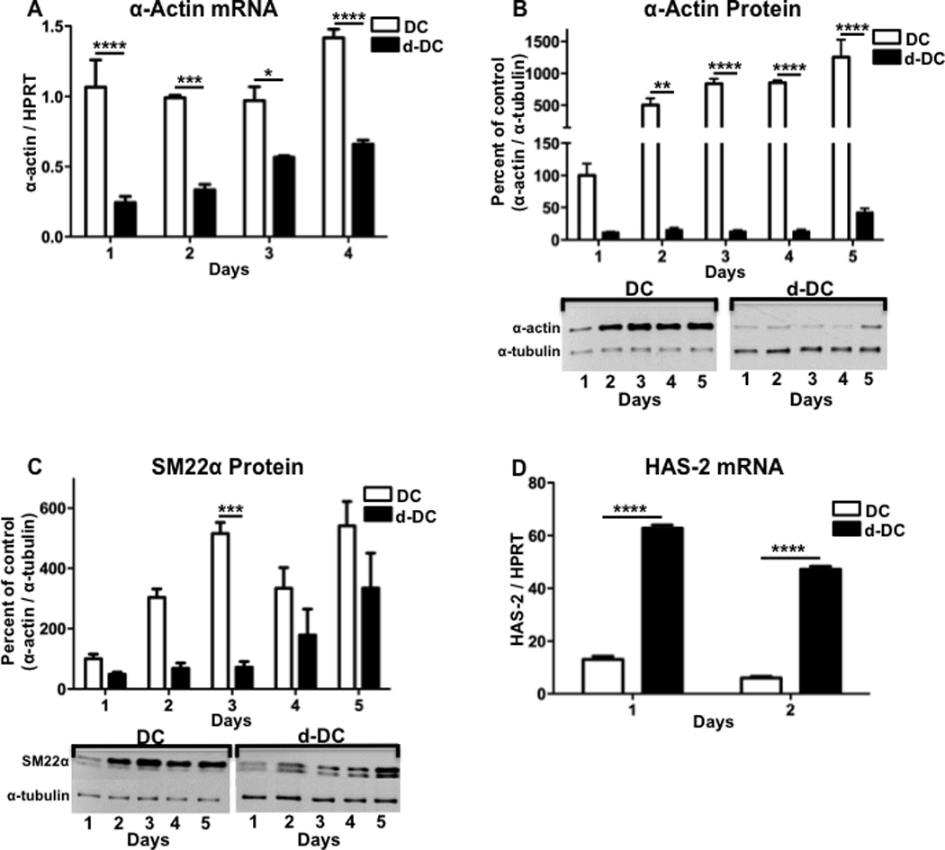
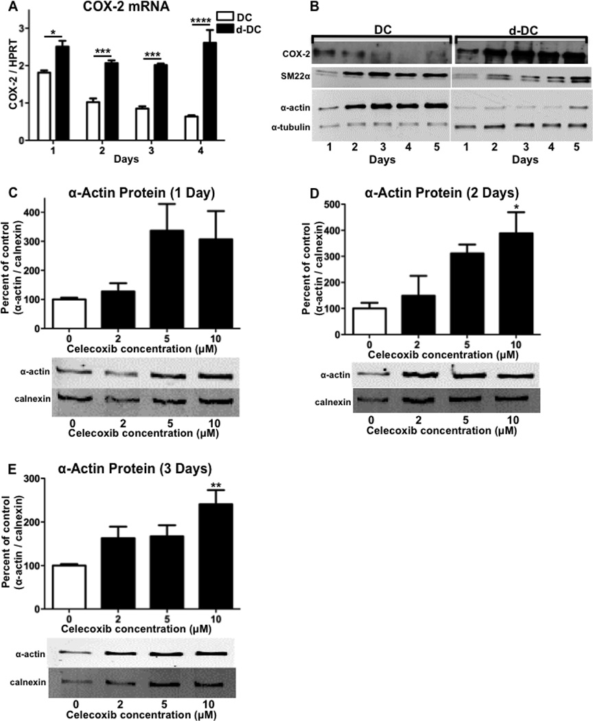
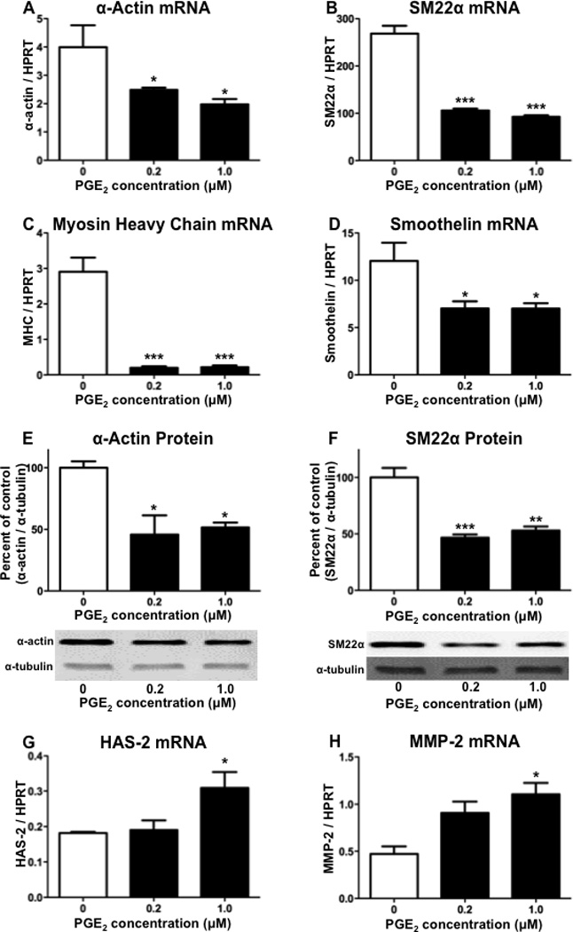
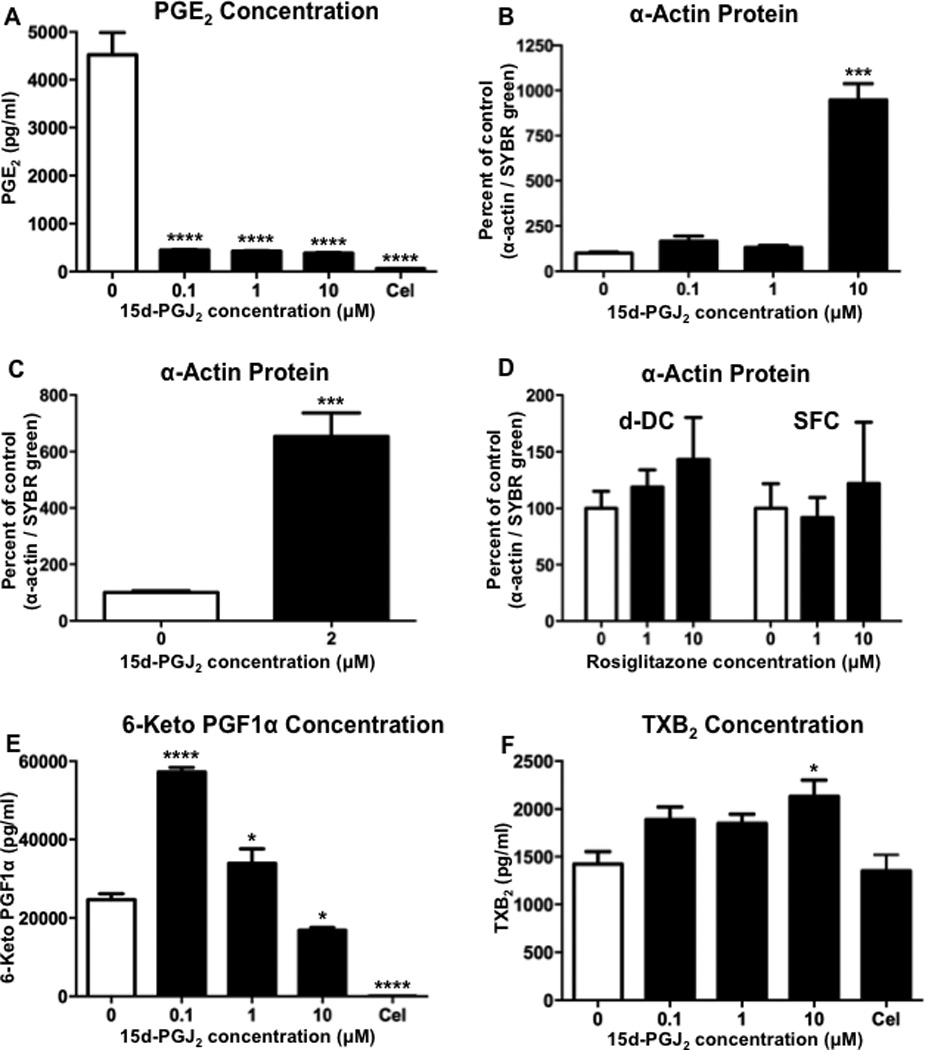
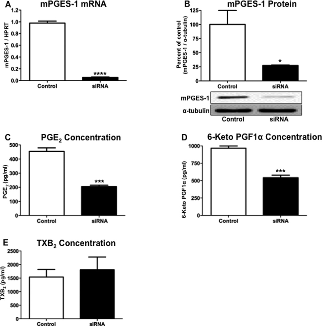
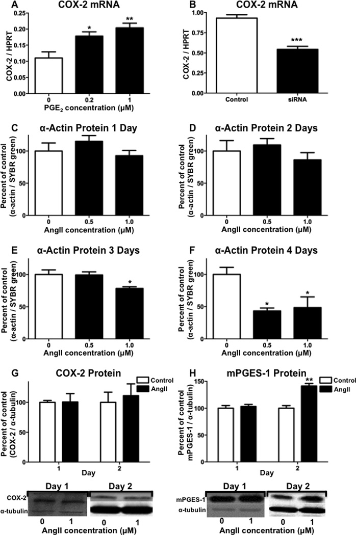
The development of numerous types of cardiovascular disease is associated with alteration of the vascular smooth muscle cell (SMC) phenotype. We have previously shown that abdominal aortic aneurysm progression in a mouse model of the disease is associated with reduced differentiation of SMCs within the lesion and that cyclooxygenase-2 (COX-2) is critical to initiation and progression of the aneurysms. The current studies used human aortic SMC (hASMC) cultures to better characterize mechanisms responsible for COX-2-dependent modulation of the SMC phenotype. Depending on the culture conditions, hASMCs expressed multiple characteristics of a differentiated and contractile phenotype, or a dedifferentiated and secretory phenotype. The pharmacological inhibition of COX-2 promoted the differentiated phenotype, whereas treatment with the COX-2-derived metabolite prostaglandin E2 (PGE2) increased characteristics of the dedifferentiated phenotype. Furthermore, pharmacological inhibition or siRNA-mediated knockdown of microsomal prostaglandin E synthase-1 (mPGES-1), the enzyme that functions downstream of COX-2 during the synthesis of PGE2, significantly increased expression of characteristics of the differentiated SMC phenotype. Therefore, our findings suggest that COX-2 and mPGES-1-dependent synthesis of PGE2 contributes to a dedifferentiated hASMC phenotype and that mPGES-1 may provide a novel pharmacological target for treatment of cardiovascular diseases where altered SMC differentiation has a causative role.


Conflict of interest statement

The authors have no conflicts of interest to disclose.

Figures

Figure 1
Figure 1. Time-course of hASMC phenotypic modulation in response to differentiation or de-differentiation culture conditions
hASMCs were cultured for up to 5 days under conditions promoting either differentiation (DC) or de-differentiation (d-DC) and analyzed for (A) α-actin mRNA, (B) α-actin protein, (C) SM22α protein or (D) HAS-2 mRNA. For quantitation of protein expression, densitometry was performed on bands from Western blots and a representative Western blot is shown for each graph. The data represent mean + SEM (n=4–5), * = p < 0.05, ** = p < 0.01, *** = p < 0.001, and **** = p < 0.0001; two-way ANOVA used to determine differences due to culture condition and time.
Figure 2
Figure 2. COX-2 contributes to reduced differentiation of hASMCs
hASMCs were cultured for up to 5 days under conditions promoting either differentiation (DC) or de-differentiation (d-DC) and analyzed for (A) COX-2 mRNA, (B) COX-2 protein, SM22α protein, α-actin protein, and α-tubulin loading control. hASMCs were treated with varying concentrations of the COX-2 inhibitor celecoxib for (C) 1 day, (D) 2 days, or (E) 3 days and analyzed for expression of the differentiation marker α-actin. For quantitation of α-actin and the loading control calnexin, densitometry was performed on bands from Western blots and a representative Western blot is shown for each graph. The data represent mean + SEM (n=4–5), * = p < 0.05, ** = p < 0.01, *** = p < 0.001, and **** = p < 0.0001; (A) two-way ANOVA used to determine differences due to culture condition and time; (C–D) one-way ANOVA.
Figure 3
Figure 3. PGE2 treatment decreases hASMC differentiation and increases de-differentiation
Following treatment with PGE2 for 1 day, hASMCs were analyzed for expression of (A) α-actin mRNA, (B) SM22α mRNA, (C) myosin heavy chain mRNA, (D) smoothelin mRNA, (E) α-actin protein, (F) SM22α protein, (G) HAS-2 mRNA, or (H) MMP-2 mRNA. For quantitation of protein expression, densitometry was performed on bands from Western blots and a representative Western blot is shown for each graph. The data represent mean + SEM (n=4–5), * = p < 0.05, ** = p < 0.01, *** = p < 0.001; one-way ANOVA.
Figure 4
Figure 4. mPGES-1 inhibition increases hASMC differentiation
Following treatment with the mPGES-1 inhibitor 15d-PGJ2 or the COX-2 inhibitor celecoxib (Cel, 2 µM) for 1 day in de-differentiation conditions, hASMCs were analyzed for (A) PGE2 in the culture media, or (B) α-actin protein expression by immunohistochemistry. (C) hASMCs were also treated with 15d-PGJ2 in serum-free conditions and analyzed for α-actin protein expression by immunohistochemistry. (D) hASMCs were treated with rosiglitazone under de-differentiation (d-DC) and serum-free conditions (SFC) and analyzed for α-actin protein expression by immunohistochemistry. (E) hASMCs were treated for 1 day with 15d-PGJ2 or celecoxib (Cel, 2 µM) under de-differentiation conditions and analyzed for the stable PGI2 product 6-keto PG F1α in the culture media. (F) hASMCs were treated with 15d-PGJ2 or celecoxib (Cel, 2 µM) under de-differentiation conditions and analyzed for the stable TXA2 product TXB2. The data represent mean + SEM (n=4), * = p < 0.05, *** = p < 0.001, and **** = p < 0.0001; one-way ANOVA.
Figure 5
Figure 5. mPGES-1 knock-down decreases PGE2 and PGI2 synthesis
Following incubation with mPGES-1 siRNA for 3 days, hASMCs were analyzed for expression of (A) mPGES-1 mRNA, (B) mPGES-1 protein, (C) PGE2 in the culture media, (D) 6-keto PG F1α, or (E) TXB2. The data represent mean + SEM (n=3–5), * = p < 0.05, *** = p < 0.001, **** = p < 0.0001; unpaired two-tailed t-test.
Figure 6
Figure 6. mPGES-1 knock-down increases differentiation markers and decreases de-differentiation markers
Following incubation with mPGES-1 siRNA for 3 days, hASMCs were analyzed for expression of (A) α-actin mRNA, (B) smoothelin mRNA, (C) SM22α mRNA, (D) α-actin protein, (E) SM22α protein, (F) HAS-2 mRNA, or (G) MMP-2 mRNA. The data represent mean + SEM (n=3–5), ** = p < 0.01, *** = p < 0.001; unpaired two-tailed t-test.
Figure 7
Figure 7. Regulation of COX-2 and mPGES-1 expression in hASMCs
(A) Following treatment with PGE2 for 1 day, hASMCs were analyzed for expression of COX-2 mRNA. (B) Following incubation with mPGES-1 siRNA for 3 days, hASMCs were analyzed for expression of COX-2 mRNA. hASMCs were incubated with angiotensin II (AngII), for (C) 1 day, (D) 2 days, or (E) 3 days, or (F) 4 days, hASMCs were analyzed for expression of α-actin protein by immunohistochemistry. Following incubation with AngII, hASMCs were analyzed for (G) COX-2 protein or (H) mPGES-1 protein. For quantitation of protein expression, densitometry was performed on bands from Western blots and a representative Western blot is shown for each graph. The data represent mean + SEM (n=3–5), * = p < 0.05, ** = p < 0.01, *** = p < 0.001; (A, C–F) one-way ANOVA, (B, G–H) unpaired two-tailed t-test, (G–H).

References

    1. Owens GK, Kumar MS, Wamhoff BR. Molecular regulation of vascular smooth muscle cell differentiation in development and disease. Physiol Rev. 2004 Jul;84(3):767–801. - PubMed
    1. Regan CP, Adam PJ, Madsen CS, Owens GK. Molecular mechanisms of decreased smooth muscle differentiation marker expression after vascular injury. J Clin Invest. 2000 Nov;106(9):1139–1147. - PMC - PubMed
    1. Rzucidlo EM, Martin KA, Powell RJ. Regulation of vascular smooth muscle cell differentiation. J Vasc Surg. 2007 Jun;(45 Suppl A):A25–A32. - PubMed
    1. Roan JN, Tsai YC, Chen IW, Chang SW, Huang CC, Lam CF. Inhibition of cyclooxygenase-2 modulates phenotypic switching of vascular smooth muscle cells during increased aortic blood flow. Heart and vessels. 2012 May;27(3):307–315. - PubMed
    1. Zhu L, Vranckx R, Khau Van Kien P, Lalande A, Boisset N, Mathieu F, Wegman M, Glancy L, Gasc JM, Brunotte F, Bruneval P, Wolf JE, Michel JB, Jeunemaitre X. Mutations in myosin heavy chain 11 cause a syndrome associating thoracic aortic aneurysm/aortic dissection and patent ductus arteriosus. Nat Genet. 2006 Mar;38(3):343–349. - PubMed

MeSH terms