. 2016 Jul;68(1):114-22.
doi: 10.1161/HYPERTENSIONAHA.116.07084. Epub 2016 May 9.

Increasing Regulatory T Cells With Interleukin-2 and Interleukin-2 Antibody Complexes Attenuates Lung Inflammation and Heart Failure Progression

Affiliations

Increasing Regulatory T Cells With Interleukin-2 and Interleukin-2 Antibody Complexes Attenuates Lung Inflammation and Heart Failure Progression

Huan Wang et al. Hypertension. 2016 Jul.

Abstract

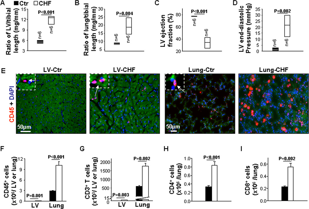
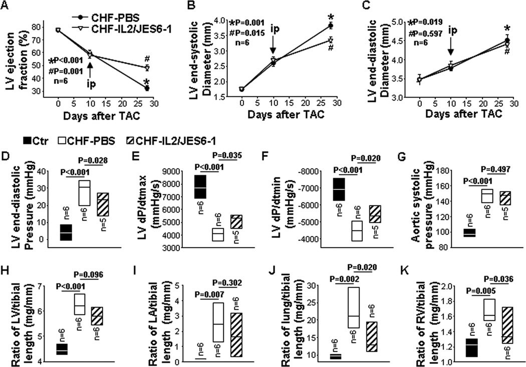
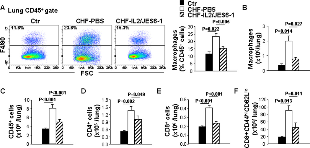
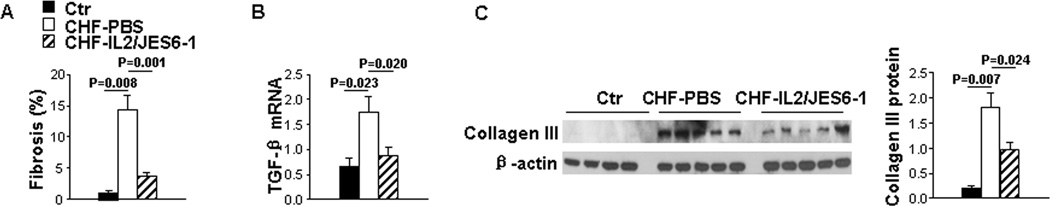
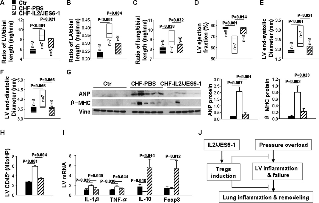
Congestive heart failure (CHF) is associated with an increase of leukocyte infiltration, proinflammatory cytokines, and fibrosis in the heart and lung. Regulatory T cells (Tregs, CD4(+)CD25(+)FoxP3(+)) suppress inflammatory responses in various clinical conditions. We postulated that expansion of Tregs attenuates CHF progression by reducing cardiac and lung inflammation. We investigated the effects of interleukin-2 (IL-2) plus IL-2 monoclonal antibody clone JES6-1 complexes (IL2/JES6-1) on induction of Tregs, transverse aortic constriction-induced cardiac and lung inflammation, and CHF progression in mice. We demonstrated that end-stage CHF caused a massive increase of lung macrophages and T cells, as well as relatively mild left ventricular (LV) leukocyte infiltration. Administration of IL2/JES6-1 caused an ≈6-fold increase of Tregs within CD4(+) T cells in the spleen, lung, and heart of mice. IL2/JES6-1 treatment of mice with existing transverse aortic constriction-induced LV failure markedly reduced lung and right ventricular weight and improved LV ejection fraction and LV end-diastolic pressure. Mechanistically, IL2/JES6-1 treatment significantly increased Tregs; suppressed CD4(+) T-cell accumulation; dramatically attenuated leukocyte infiltration, including decreasing CD45(+) cells, macrophages, CD8(+) T cells, and effector memory CD8(+); and reduced proinflammatory cytokine expressions and fibrosis in the lung of mice. Furthermore, IL2/JES6-1 administered before transverse aortic constriction attenuated the development of LV hypertrophy and dysfunction in mice. Our data indicate that increasing Tregs through administration of IL2/JES6-1 effectively attenuates pulmonary inflammation, right ventricular hypertrophy, and further LV dysfunction in mice with existing LV failure, suggesting that strategies to properly expand Tregs may be useful in reducing CHF progression.

Keywords: fibrosis; heart; heart failure; inflammation; lung; regulatory T cell.


Figures

Figure 1
Figure 1. End-stage congestive heart failure (CHF) exhibits robust leukocyte infiltration in lungs and moderate leukocyte infiltration in hearts
Data were collected from mice under basal (Ctr) and CHF conditions. A and B, The ratio of left ventricle (LV) and lung weight to tibial length of mice. C, Echocardiographic measurements of LV ejection fraction from hearts. D, Hemodynamics of LV end-diastolic pressure from hearts. E and F, Representative images of immunostaining and flow cytometry quantitative data of CD45+ cells in the LV and lung. G, Flow cytometry quantitative data represent the number of CD3+ T cells in the LV and lung. H and I, Flow cytometry quantitative data represent the number of CD4+ and CD8+ T cells in the lung. n=5 per group.
Figure 2
Figure 2. IL2/JES6-1 treatment attenuates progression of congestive heart failure (CHF) in mice with existing left ventricle (LV) failure
The treatment was commenced when LV ejection fraction (EF) reached around 55% due to TAC. Data were collected from mice under basal conditions (Ctr), or treated with IL2/JES6-1 or PBS under TAC conditions (CHF-IL2/JES6-1 or CHF-PBS). A to C, Echocardiographic measurements of LV EF, LV end-systolic diameter (LVESD) and LV end-diastolic diameter (LVEDD) from hearts. D to G, Hemodynamics of LV end-diastolic pressure, LV maximum rate of rise of pressure (dP/dtmax), LV maximum rate of decline of pressure (dP/dtmin) and aortic systolic pressure from hearts. H to K, The ratio of LV, LA, lung and right ventricle weight to tibial length of mice. *p as compared with control before TAC. #p as compared with CHF-PBS.
Figure 3
Figure 3. IL2/JES6-1 treatment significantly reduces lung inflammatory cytokines and mildly affects LV inflammatory cytokines in mice with existing left ventricle (LV) failure
Data were collected from mice under basal conditions (Ctr), or treated with IL2/JES6-1 or PBS under TAC conditions (CHF-IL2/JES6-1 or CHF-PBS). A, Flow cytometry plots and quantitative data represent the percentage of Tregs (CD25+Foxp3+) within the CD4+ T-cell population of lungs. B, Quantitative data of flow cytometry represent the total number of Tregs in the lung. C, Quantitative RT-PCR results of IL-1β, TNF-α, MCP-1 and IL-10 mRNA levels in lung lysates. D, Quantitative RT-PCR results of IL-1β, TNF-α, IL-10 and Foxp3 mRNA levels in LV lysates. n=4–6 per group.
Figure 4
Figure 4. IL2/JES6-1 treatment suppresses immune-cell infiltration in the lung of mice with existing left ventricle (LV) failure
Data were collected from mice under basal conditions (Ctr), or treated with IL2/JES6-1 or PBS under TAC conditions (CHF-IL2/JES6-1 or CHF-PBS). A, Flow cytometry plots and quantitative data represent the percentage of macrophages (F4/80+) within the CD45+ cell population of lungs. B, Quantitative data of flow cytometry represent the total number of macrophages in the lung. C to F, Quantitative data of flow cytometry represent the total number of CD45+ cells, CD4+ T, CD8+ T cells and effector memory (CD44highCD62Llow) CD8+ T cells in the lung. n=4–6 per group.
Figure 5
Figure 5. IL2/JES6-1 treatment attenuates lung fibrosis in mice with existing left ventricle (LV) failure
Data were collected from mice under basal conditions (Ctr), or treated with IL2/JES6-1 or PBS under TAC conditions (CHF-IL2/JES6-1 or CHF-PBS). A, Quantitative data of Sirius red/Fast green staining for detection of fibrosis in lungs. B, Quantitative RT-PCR results of TGF-β mRNA levels in lung lysates. C, Western blot of collagen III and β-actin (loading control) in lung lysates. n=5 per group.
Figure 6
Figure 6. IL2/JES6-1 treatment attenuates the development of transverse aortic constriction (TAC)-induced left ventricular (LV) hypertrophy and cardiac dysfunction
The treatment was commenced 3 days before TAC. Data were collected from mice under basal conditions (Ctr), or treated with IL2/JES6-1 or PBS under TAC conditions (CHF-IL2/JES6-1 or CHF-PBS). A to C, The ratio of LV, left atria (LA) and lung weight to tibial length of mice. D to F, Echocardiographic measurements of LV ejection fraction (EF), LV end-systolic diameter (LVESD) and LV end-diastolic diameter (LVEDD) from hearts. G, Western blot of ANP, β-MHC and vinculin (loading control) in LV lysates. H, Quantitative data of CD45 immunostaining of LVs. I, Quantitative RT-PCR results of IL-1β, TNF-α, IL-10 and Foxp3 mRNA levels in LV lysates. n=5 per group. J, Diagram of the proposed underlying mechanism.

Comment in

References

    1. Bursi F, McNallan SM, Redfield MM, Nkomo VT, Lam CS, Weston SA, Jiang R, Roger VL. Pulmonary pressures and death in heart failure: a community study. J Am Coll Cardiol. 2012;59:222–231. - PMC - PubMed
    1. Vachiery JL, Adir Y, Barbera JA, Champion H, Coghlan JG, Cottin V, De Marco T, Galie N, Ghio S, Gibbs JS, Martinez F, Semigran M, Simonneau G, Wells A, Seeger W. Pulmonary hypertension due to left heart diseases. J Am Coll Cardiol. 2013;62:D100–D108. - PubMed
    1. Epelman S, Liu PP, Mann DL. Role of innate and adaptive immune mechanisms in cardiac injury and repair. Nat Rev Immunol. 2015;15:117–129. - PMC - PubMed
    1. El-Menyar AA. Cytokines and myocardial dysfunction: state of the art. J Card Fail. 2008;14:61–74. - PubMed
    1. Nevers T, Salvador AM, Grodecki-Pena A, Knapp A, Velazquez F, Aronovitz M, Kapur NK, Karas RH, Blanton RM, Alcaide P. Left Ventricular T-Cell Recruitment Contributes to the Pathogenesis of Heart Failure. Circ Heart Fail. 2015;8:776–787. - PMC - PubMed

Publication types

MeSH terms