

Sustained expression of MCP-1 by low wall shear stress loading concomitant with turbulent flow on endothelial cells of intracranial aneurysm
- PMID: 27160403
- PMCID: PMC4862234
- DOI: 10.1186/s40478-016-0318-3
Sustained expression of MCP-1 by low wall shear stress loading concomitant with turbulent flow on endothelial cells of intracranial aneurysm
Abstract
Introduction: Enlargement of a pre-existing intracranial aneurysm is a well-established risk factor of rupture. Excessive low wall shear stress concomitant with turbulent flow in the dome of an aneurysm may contribute to progression and rupture. However, how stress conditions regulate enlargement of a pre-existing aneurysm remains to be elucidated.
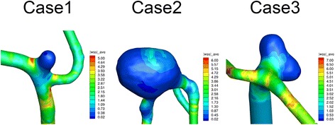
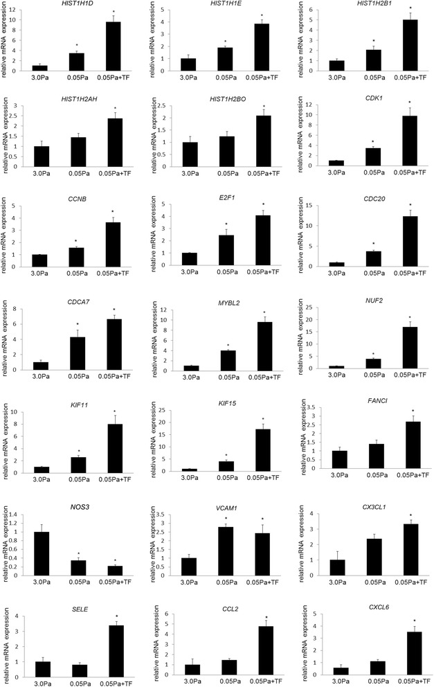
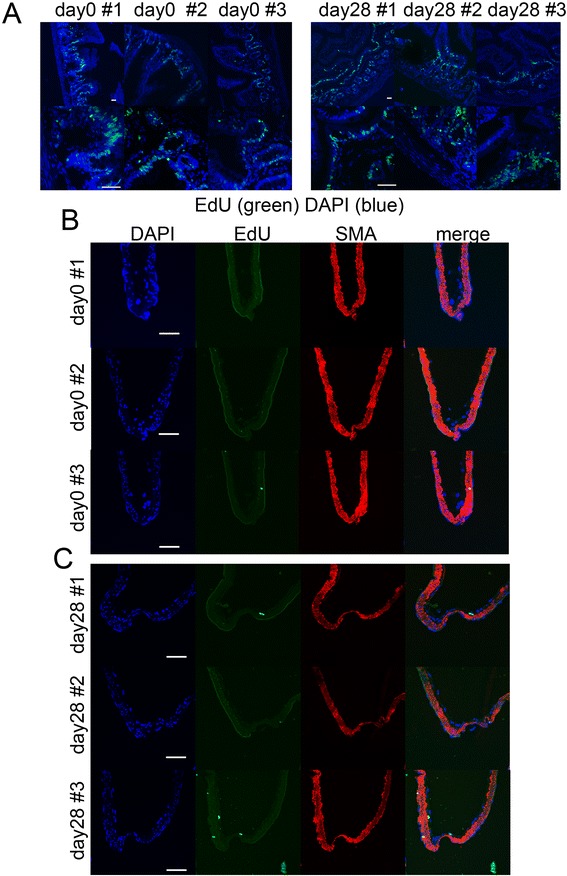
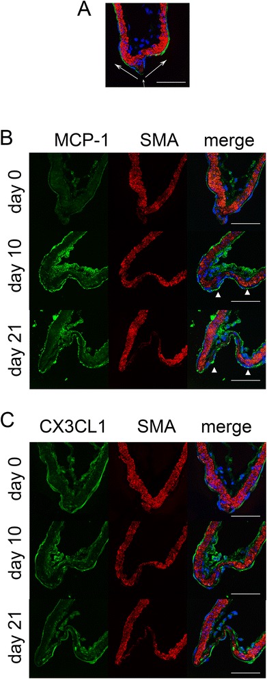
Results: Wall shear stress was calculated with 3D-computational fluid dynamics simulation using three cases of unruptured intracranial aneurysm. The resulting value, 0.017 Pa at the dome, was much lower than that in the parent artery. We loaded wall shear stress corresponding to the value and also turbulent flow to the primary culture of endothelial cells. We then obtained gene expression profiles by RNA sequence analysis. RNA sequence analysis detected hundreds of differentially expressed genes among groups. Gene ontology and pathway analysis identified signaling related with cell division/proliferation as overrepresented in the low wall shear stress-loaded group, which was further augmented by the addition of turbulent flow. Moreover, expression of some chemoattractants for inflammatory cells, including MCP-1, was upregulated under low wall shear stress with concomitant turbulent flow. We further examined the temporal sequence of expressions of factors identified in an in vitro study using a rat model. No proliferative cells were detected, but MCP-1 expression was induced and sustained in the endothelial cell layer.
Conclusions: Low wall shear stress concomitant with turbulent flow contributes to sustained expression of MCP-1 in endothelial cells and presumably plays a role in facilitating macrophage infiltration and exacerbating inflammation, which leads to enlargement or rupture.
Figures
References
-
- Unruptured intracranial aneurysms--risk of rupture and risks of surgical intervention. International Study of Unruptured Intracranial Aneurysms Investigators (1998). N Engl J Med. 339 (24):1725-1733. doi:10.1056/NEJM199812103392401. - PubMed
-
- Aoki T, Nishimura M, Matsuoka T, Yamamoto K, Furuyashiki T, Kataoka H, Kitaoka S, Ishibashi R, Ishibazawa A, Miyamoto S, Morishita R, Ando J, Hashimoto N, Nozaki K, Narumiya S. PGE2–EP2 signalling in endothelium is activated by haemodynamic stress and induces cerebral aneurysm through an amplifying loop via NF-kappaB. Br J Pharmacol. 2011;163(6):1237–1249. doi: 10.1111/j.1476-5381.2011.01358.x. - DOI - PMC - PubMed
Publication types
MeSH terms
Substances
LinkOut - more resources
Full Text Sources
Other Literature Sources
Medical
Miscellaneous
