. 2016 Jun;5(6):754-63.
doi: 10.5966/sctm.2015-0197. Epub 2016 May 9.

Mesenchymal Stromal Cell-Derived Extracellular Vesicles Protect the Fetal Brain After Hypoxia-Ischemia

Affiliations

Mesenchymal Stromal Cell-Derived Extracellular Vesicles Protect the Fetal Brain After Hypoxia-Ischemia

Daan R M G Ophelders et al. Stem Cells Transl Med. 2016 Jun.

Abstract

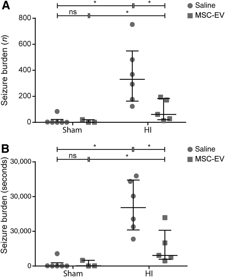
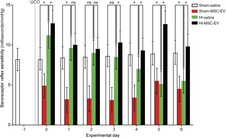
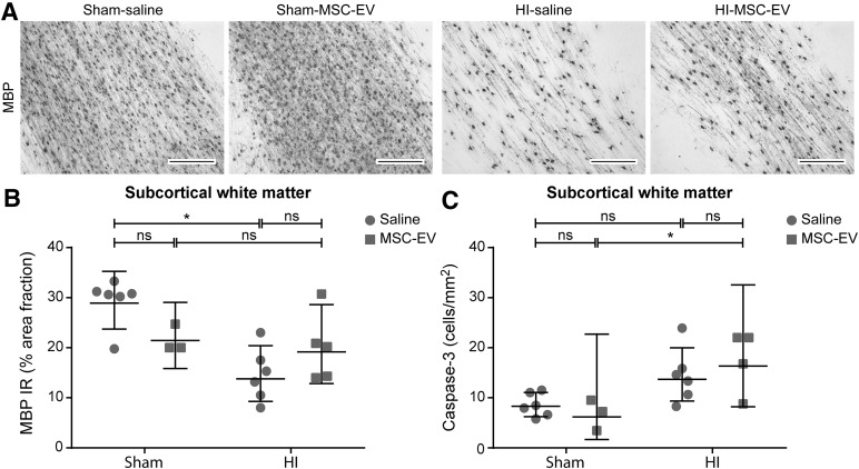
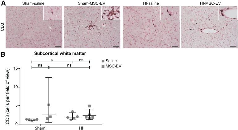
Preterm neonates are susceptible to perinatal hypoxic-ischemic brain injury, for which no treatment is available. In a preclinical animal model of hypoxic-ischemic brain injury in ovine fetuses, we have demonstrated the neuroprotective potential of systemically administered mesenchymal stromal cells (MSCs). The mechanism of MSC treatment is unclear but suggested to be paracrine, through secretion of extracellular vesicles (EVs). Therefore, we investigated in this study the protective effects of mesenchymal stromal cell-derived extracellular vesicles (MSC-EVs) in a preclinical model of preterm hypoxic-ischemic brain injury. Ovine fetuses were subjected to global hypoxia-ischemia by transient umbilical cord occlusion, followed by in utero intravenous administration of MSC-EVs. The therapeutic effects of MSC-EV administration were assessed by analysis of electrophysiological parameters and histology of the brain. Systemic administration of MSC-EVs improved brain function by reducing the total number and duration of seizures, and by preserving baroreceptor reflex sensitivity. These functional protections were accompanied by a tendency to prevent hypomyelination. Cerebral inflammation remained unaffected by the MSC-EV treatment. Our data demonstrate that MSC-EV treatment might provide a novel strategy to reduce the neurological sequelae following hypoxic-ischemic injury of the preterm brain. Our study results suggest that a cell-free preparation comprising neuroprotective MSC-EVs could substitute MSCs in the treatment of preterm neonates with hypoxic-ischemic brain injury, thereby circumventing the potential risks of systemic administration of living cells.

Significance: Bone marrow-derived mesenchymal stromal cells (MSCs) show promise in treating hypoxic-ischemic injury of the preterm brain. Study results suggest administration of extracellular vesicles, rather than intact MSCs, is sufficient to exert therapeutic effects and avoids potential concerns associated with administration of living cells. The therapeutic efficacy of systemically administered mesenchymal stromal cell-derived extracellular vesicles (MSC-EVs) on hypoxia-ischemia-induced injury was assessed in the preterm ovine brain. Impaired function and structural injury of the fetal brain was improved following global hypoxia-ischemia. A cell-free preparation of MSC-EVs could substitute for the cellular counterpart in the treatment of preterm neonates with hypoxic-ischemic brain injury. This may open new clinical applications for "off-the-shelf" interventions with MSC-EVs.

Keywords: Brain injury; Exosomes; Extracellular vesicles; Hypoxia-ischemia; Mesenchymal stromal cells; Preterm.


Figures

Figure 1.
Figure 1.
Study design. Fetuses were instrumented at gestational age 102 days. After a recovery period of 4 days, fetuses were subjected to 25 minutes of umbilical cord occlusion or sham occlusion (d0). One hour and 4 days (d4) after umbilical cord occlusion or sham occlusion, fetuses received either intravenous MSC-EVs (2.0 × 10⁷ cell equivalents; closed arrow) or saline 0.9% (open arrow). After a 7-day reperfusion period, brain tissue was collected. Abbreviations: d, day; END, end of experiment; GA, gestational age; HI, hypoxia-ischemia; IN, instrumentation; MSC-EV, mesenchymal stem cell‐derived extracellular vesicle; UCO, umbilical cord occlusion.
Figure 2.
Figure 2.
MSC-EV treatment induced functional neuroprotection after global HI. Global HI caused a significant seizure burden, indicated by an increased total number (A) and duration (B) of seizures compared with sham-occluded animals. Administration of MSC-EVs reduced electrographic seizure number and duration compared with saline-treated animals. Medians ± interquartile ranges and levels of significance, which were calculated by Mann‐Whitney test, are depicted. ∗, p ≤ .05. Abbreviations: HI, hypoxia‐ischemia; MSC-EV, mesenchymal stem cell‐derived extracellular vesicle; ns, not significant.
Figure 3.
Figure 3.
MSC-EVs prevented loss of baroreflex sensitivity. Global HI (green bars) caused a significant gradual decline of baroreflex sensitivity over time, which was prevented by MSC-EV treatment (black bars). Means ± 95% confidence intervals and levels of significance of the treatment effect (HI‐saline vs. HI‐MSC-EV) are depicted and were calculated by the Bayesian multilevel model. MSC-EV treatment significantly compromised baroreflex sensitivity in healthy controls (red bars). ∗, p ≤ .05. Abbreviations: HI, hypoxia‐ischemia; MSC-EV, mesenchymal stem cell‐derived extracellular vesicle; ns, not significant; UCO, umbilical cord occlusion.
Figure 4.
Figure 4.
MSC-EVs reduced white matter injury after global HI but did not prevent HI-induced apoptosis. (A): Immunohistochemical MBP staining in the subcortical white matter of all four experimental groups. Magnification, ×100. Global HI induced marked hypomyelination in the subcortical white matter. MSC-EVs tended to prevent the decrease in MBP reactivity after global HI. The area fraction of MBP was similar in sham conditions. (B): Graphic representation of MBP immunoreactivity in the subcortical white matter. (C): Graphic presentation of caspase-3 cell density in the subcortical white matter. A mild trend in increase of caspase-3 cell density was found following global HI. MSC-EV treatment did not affect apoptosis in the subcortical white matter. Levels of significance are depicted, which were calculated by the random intercept model with all repeated measures (i.e., brain sections) per animal (sham-saline, n = 6; sham-MSC-EV, n = 3; HI-saline, n = 6; HI-MSC-EV, n = 5). Because of positive skewing, data were log-transformed for statistical testing. For graphic presentation and interpretation, averages on the log scale were transformed to the original scale and presented as geometric means with corresponding 95% confidence intervals. Scale bars = 200 µm. ∗, p ≤ .05. Abbreviations: HI, hypoxia‐ischemia; IR, immunoreactivity; MBP, myelin basic protein 1; MSC-EV, mesenchymal stem cell‐derived extracellular vesicle; ns, not significant.
Figure 5.
Figure 5.
MSC-EVs do not reduce cerebral inflammation in the subcortical white matter and hippocampus. (A, B): Immunohistochemical IBA-1 staining. Global HI induced a significant increase of IBA-1 immunoreactivity in (A) the subcortical white matter (magnification, ×100) and (B) the hippocampus (magnification, ×20), which was not attenuated by MSC-EV treatment. (C, D): Graphic presentation of the area fraction of IBA‐1 immunoreactivity in (C) the subcortical white matter and (D) the hippocampus. Levels of significance are depicted, which were calculated by the random intercept model with all repeated measures (i.e., brain sections) per animal (sham-saline, n = 5; sham-MSC-EV, n = 3; HI-saline, n = 6; HI-MSC-EV, n = 5). Because of positive skewing, data were log-transformed for statistical testing. For graphic presentation and interpretation, averages on the log scale were transformed to the original scale and presented as geometric means with corresponding 95% confidence intervals. Scale bars = 1,000 µm. ∗, p ≤ .05. Abbreviations: HI = hypoxia‐ischemia; IBA‐1, ionized calcium binding adaptor molecule 1; IR, immunoreactivity; MSC-EV, mesenchymal stem cell-derived extracellular vesicles; ns, not significant.
Figure 6.
Figure 6.
MSC-EVs increase cerebral influx of T‐effector cells. (A): Immunohistochemical staining for CD3-positive T lymphocytes in the subcortical white matter of all four experimental groups. Magnification, ×200; inset, ×400. (B): Graphic representation of CD3-positive cells per field of view in the subcortical white matter. MSC-EV treatment increased numbers of CD3-positive T lymphocytes in the subcortical white matter compared with saline-treated animals. Levels of significance are depicted, which were calculated by the random intercept model with all repeated measures (i.e., brain sections) per animal (sham-saline, n = 5; sham-MSC-EV, n = 3; HI-saline, n = 5; HI-MSC-EV, n = 5). Because of positive skewing, data were log-transformed for statistical testing. For graphic presentation and interpretation, averages on the log scale were transformed to the original scale and presented as geometric means with corresponding 95% confidence intervals. Scale bars = 100 µm. ∗, p ≤ .05. Abbreviations: HI, hypoxia‐ischemia; MSC-EV = mesenchymal stem cell‐derived extracellular vesicle; ns = not significant.

Similar articles

Cited by

References

    1. Fleiss B, Tann CJ, Degos V, et al. Inflammation-induced sensitization of the brain in term infants. Dev Med Child Neurol. 2015;57(suppl 3):17–28. - PubMed
    1. Beck S, Wojdyla D, Say L, et al. The worldwide incidence of preterm birth: A systematic review of maternal mortality and morbidity. Bull World Health Organ. 2010;88:31–38. - PMC - PubMed
    1. Jacobs SE, Berg M, Hunt R, et al. Cooling for newborns with hypoxic ischaemic encephalopathy. Cochrane Database Syst Rev. 2013;1:CD003311. - PMC - PubMed
    1. Azzopardi D, Robertson NJ, Cowan FM, et al. Pilot study of treatment with whole body hypothermia for neonatal encephalopathy. Pediatrics. 2000;106:684–694. - PubMed
    1. Briatore E, Ferrari F, Pomero G, et al. EEG findings in cooled asphyxiated newborns and correlation with site and severity of brain damage. Brain Dev. 2013;35:420–426. - PubMed

Publication types