. 2016 May 10;18(1):47.
doi: 10.1186/s13058-016-0705-5.

The distribution of ductal carcinoma in situ (DCIS) grade in 4232 women and its impact on overdiagnosis in breast cancer screening

Affiliations

The distribution of ductal carcinoma in situ (DCIS) grade in 4232 women and its impact on overdiagnosis in breast cancer screening

P A van Luijt et al. Breast Cancer Res. .

Abstract

Background: The incidence of ductal carcinoma in situ (DCIS) has rapidly increased over time. The malignant potential of DCIS is dependent on its differentiation grade.

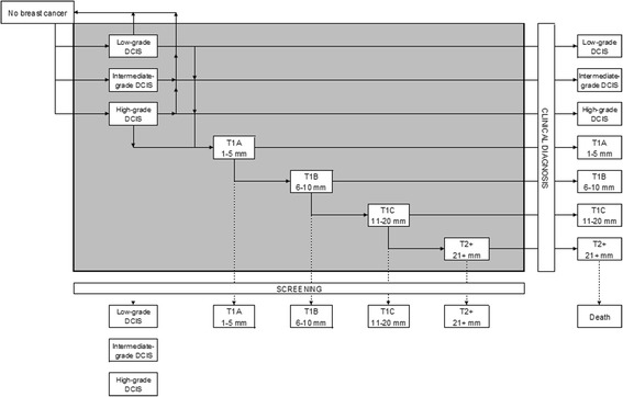
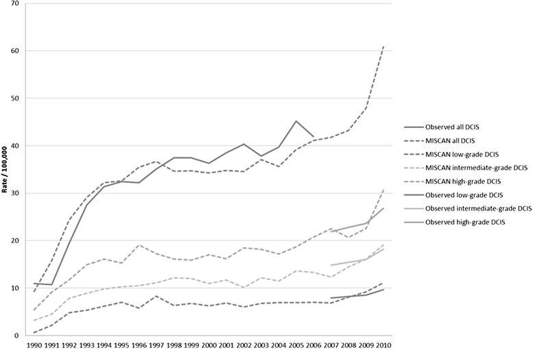
Methods: Our aim is to determine the distribution of different grades of DCIS among women screened in the mass screening programme, and women not screened in the mass screening programme, and to estimate the amount of overdiagnosis by grade of DCIS. We retrospectively included a population-based sample of 4232 women with a diagnosis of DCIS in the years 2007-2009 from the Nationwide network and registry of histopathology and cytopathology in the Netherlands. Excluded were women with concurrent invasive breast cancer, lobular carcinoma in situ and no DCIS, women recently treated for invasive breast cancer, no grade mentioned in the record, inconclusive record on invasion, and prevalent DCIS. The screening status was obtained via the screening organisations. The distribution of grades was incorporated in the well-established and validated microsimulation model MISCAN.

Results: Overall, 17.7 % of DCIS were low grade, 31.4 % intermediate grade, and 50.9 % high grade. This distribution did not differ by screening status, but did vary by age. Older women were more likely to have low-grade DCIS than younger women. Overdiagnosis as a proportion of all cancers in women of the screening age was 61 % for low-grade, 57 % for intermediate-grade, 45 % for high-grade DCIS. For women age 50-60 years with a high-grade DCIS this overdiagnosis rate was 21-29 %, compared to 50-66 % in women age 60-75 years with high-grade DCIS.

Conclusions: Amongst the rapidly increasing numbers of DCIS diagnosed each year is a significant number of overdiagnosed cases. Tailoring treatment to the probability of progression is the next step to preventing overtreatment. The basis of this tailoring could be DCIS grade and age.

Keywords: Breast cancer; Ductal carcinoma in situ; MISCAN; Overdiagnosis; Screening.


Figures

Fig. 1
Fig. 1
Schematic drawing of the extended MISCAN model. Transition possibilities are indicated with arrows. All diseases within the grey area are preclinical disease, after diagnosis they are either clinically detected or detected by mass screening. There is no transition between low-grade DCIS, intermediate-grade DCIS and high-grade DCIS. DCIS ductal carcinoma in situ, MISCAN MIcrosimulation SCreening ANalysis (predicted rates by the model), T1a tumour with a diameter up to 5 mm, T1b tumour with a diameter from 5 mm up to 10 mm, T1c tumour with a diameter from 10 mm up to 20 mm, T2 + any tumour with a diameter larger than 20 mm
Fig. 2
Fig. 2
Screening affecting three women differently. The first box is the life history of a woman who has an onset of breast cancer, is diagnosed clinically, and dies of breast cancer. The second box is the life history of a woman who also has an onset of breast cancer, but who dies of other causes before this would be detected. The third box is the life history of a woman who has an onset of breast cancer, but also a spontaneous regression, this woman would not have been diagnosed without screening. The fourth box indicates the situation for these three women had screening been introduced. The woman in the first box no longer dies from breast cancer; the other two women do not benefit from screening, they have been overdiagnosed
Fig. 3
Fig. 3
Low-grade DCIS, intermediate-grade DCIS and high-grade DCIS per 100,000 women aged 50–60. Observed: the number of DCIS as calculated when applying DCIS grade distribution to the data on total DCIS incidence from the Dutch Cancer Registry. DCIS ductal carcinoma in situ, MISCAN MIcrosimulation SCreening ANalysis (predicted rates by the model)

References

    1. Jones JL. Overdiagnosis and overtreatment of breast cancer: progression of ductal carcinoma in situ: the pathological perspective. Breast Cancer Res. 2006;8(2):204. doi: 10.1186/bcr1397. - DOI - PMC - PubMed
    1. Registry TNC. Cijfersoverkanker. 2013. http://www.cijfersoverkanker.nl. Accessed 17 Sept 2013.
    1. Hofvind S, Lee CI, Elmore JG. Stage-specific breast cancer incidence rates among participants and non-participants of a population-based mammographic screening program. Breast Cancer Res Treat. 2012;135(1):291–299. doi: 10.1007/s10549-012-2162-x. - DOI - PMC - PubMed
    1. de Gelder R, Heijnsdijk EA, van Ravesteyn NT, Fracheboud J, Draisma G, de Koning HJ. Interpreting overdiagnosis estimates in population-based mammography screening. Epidemiol Rev. 2011;33(1):111–121. doi: 10.1093/epirev/mxr009. - DOI - PMC - PubMed
    1. Yen MF, Tabar L, Vitak B, Smith RA, Chen HH, Duffy SW. Quantifying the potential problem of overdiagnosis of ductal carcinoma in situ in breast cancer screening. Eur J Cancer. 2003;39(12):1746–1754. doi: 10.1016/S0959-8049(03)00260-0. - DOI - PubMed

Publication types