. 2016 May 17;15(7):1481-1492.
doi: 10.1016/j.celrep.2016.04.032. Epub 2016 May 5.

The Tumor Suppressor Hace1 Is a Critical Regulator of TNFR1-Mediated Cell Fate

Affiliations

The Tumor Suppressor Hace1 Is a Critical Regulator of TNFR1-Mediated Cell Fate

Luigi Tortola et al. Cell Rep. .

Erratum in

  • The Tumor Suppressor Hace1 Is a Critical Regulator of TNFR1-Mediated Cell Fate.
    Tortola L, Nitsch R, Bertrand MJM, Kogler M, Redouane Y, Kozieradzki I, Uribesalgo I, Fennell LM, Daugaard M, Klug H, Wirnsberger G, Wimmer R, Perlot T, Sarao R, Rao S, Hanada T, Takahashi N, Kernbauer E, Demiröz D, Lang M, Superti-Furga G, Decker T, Pichler A, Ikeda F, Kroemer G, Vandenabeele P, Sorensen PH, Penninger JM. Tortola L, et al. Cell Rep. 2016 Sep 20;16(12):3414. doi: 10.1016/j.celrep.2016.08.072. Epub 2016 Sep 20. Cell Rep. 2016. PMID: 27653700 Free PMC article. No abstract available.

Abstract

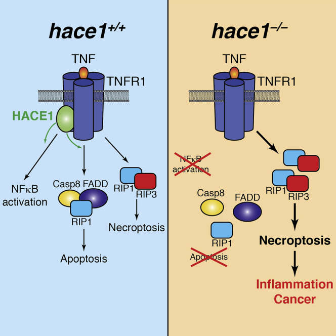
The HECT domain E3 ligase HACE1 has been identified as a tumor suppressor in multiple cancers. Here, we report that HACE1 is a central gatekeeper of TNFR1-induced cell fate. Genetic inactivation of HACE1 inhibits TNF-stimulated NF-κB activation and TNFR1-NF-κB-dependent pathogen clearance in vivo. Moreover, TNF-induced apoptosis was impaired in hace1 mutant cells and knockout mice in vivo. Mechanistically, HACE1 is essential for the ubiquitylation of the adaptor protein TRAF2 and formation of the apoptotic caspase-8 effector complex. Intriguingly, loss of HACE1 does not impair TNFR1-mediated necroptotic cell fate via RIP1 and RIP3 kinases. Loss of HACE1 predisposes animals to colonic inflammation and carcinogenesis in vivo, which is markedly alleviated by genetic inactivation of RIP3 kinase and TNFR1. Thus, HACE1 controls TNF-elicited cell fate decisions and exerts tumor suppressor and anti-inflammatory activities via a TNFR1-RIP3 kinase-necroptosis pathway.


Figures

None
Graphical abstract
Figure 1
Figure 1
HACE1 Is Essential for the TNFR1-NF-κB Response (A) Hace1+/+ and hace1–/– MEFs were stimulated with murine TNF (10 ng/ml) to induce the activation of TNFR1 downstream pathways. β-actin protein levels are shown as controls. Results are representative of three independent experiments. (B) NF-κB activation of TNF (10 ng/ml) stimulated (+) and untreated (−) hace1+/+ MEFs, hace1–/– MEFs, hace1–/– MEFs re-expressing WT HACE1, and hace1–/– MEFs re-expressing the E3 ligase-dead C876S mutant. Triplicate filter plate assays of nuclear extracts are shown as mean values ± SD. HACE1 expression and re-expression were confirmed by western blot. (C) Time course of interleukin-6 (IL-6) induction in hace1+/+ and hace1–/– MEFs following TNF (10 ng/ml) stimulation. IL-6 concentration was determined by ELISA in triplicate (mean values ± SD). (D) In vivo Listeria monocytogenes infections in hace1+/+ and hace1–/– mice. Four days after infection animals were sacrificed, spleen extracts were plated, and bacterial loads in each spleen were determined as colony-forming units (CFUs)/spleen. Data for individual mice are shown. (E) Kaplan-Meier survival curves of hace1+/+ and hace1–/– mice after Listeria infections are shown (p < 0.01). See also Figure S1.
Figure 2
Figure 2
Loss of Hace1 Confers Resistance to TNF-Induced Apoptosis In Vitro (A and B) Hace1–/– MEFs do not undergo apoptosis. Cell viability (A) and representative images (B) are shown for hace1+/+ and hace1–/– MEFs treated for 8 hr with either TNF alone (10 ng/ml) or TNF + ActD (1 μg/ml). Cell viability was determined in quadruplicate cultures (mean values ± SD). For images, cells were stained with crystal violet. Magnifications are ×10. (C) Levels of cleaved caspase-8 (cC8), cleaved caspase-3 (cC3), total RIP1 kinase protein, and cleaved RIP1 kinase (cRIP1) in lysates from hace1+/+ and hace1–/– MEFs treated with TNF + ActD for the indicated time points. GAPDH is shown as a loading control. (D) Cell viability of hace1+/+ and hace1–/– MEFs untreated (control) or exposed for 8 hr to TNF alone or TNF + cycloheximide (CHX; 10 μg/ml). Mean survival (±SD) was determined in quadruplicate cultures. (E and F) Cell viability of hace1–/– MEFs re-expressing WT HACE1 and hace1–/– MEFs re-expressing the E3 ligase-dead C876S mutant left untreated (control) or treated for 8 hr with TNF alone and either TNF + ActD (E) or TNF + CHX (F). Mean survival (±SD) was determined in quadruplicate cultures. Results are representative of more than three independent experiments. See also Figure S1.
Figure 3
Figure 3
Hace1–/– Mice Are Protected from TNFR1-Induced Lethality In Vivo (A–C) Hace1–/– mice are protected from liver damage in response to a high-dose (450 μg/kg) TNF challenge. (A) Kaplan-Meier survival curves are shown for hace1+/+ and hace1–/– mice after TNF injection (n = 6 mice per group, p < 0.05). (B) Serum AST and ALT levels served as markers for liver damage in littermate hace1+/+ and hace1–/– mice following in vivo challenge with high-dose TNF. Data from pooled sera (n = 4 mice per group) are shown. (C) H&E staining of representative histological liver sections 9 hr after the TNF challenge is shown. (D–F) Hace1–/– mice are protected from death in response to LPS/D-Gal (LPS 10 μg/kg and D-Gal 1 g/kg mouse, i.p.). (D) Kaplan-Meier survival curves of hace1+/+ and hace1–/– littermates injected intravenously (i.v.) with a lethal dose of LPS/D-Gal (n = 10 mice per group, p < 0.01). (E) H&E staining and active caspase-3 immunodetection on liver sections 7 hr after LPS/D-Gal injections are shown. (F) Serum AST and ALT levels served as markers for liver damage in littermate hace1+/+ and hace1–/– mice following in vivo challenge with a lethal dose of LPS + D-Gal. Data from pooled sera (n = 5 mice per group) are shown.
Figure 4
Figure 4
Hace1–/– Mutant Cells Undergo Necroptotis (A) Hace1+/+ and hace1–/– MEFs were treated with TNF (10 ng/ml), Z-VAD (5 μg/ml), and Nec-1 (50 nM) plus either ActD (1 μg/ml) or CHX (10 μg/ml) using the indicated combinations. Cell viability was determined in triplicates (mean values ± SD). (B) Hace1+/+, hace1–/–, and hace1–/–ripk3–/– MEFs were treated with TNF (10 ng/ml), Z-VAD (5 μg/ml), Nec-1 (50 nM), and ActD (1 μg/ml). Cell viability was determined in quadruplicates (mean values ± SD). Results are representative of at least three independent experiments. See also Figure S2.
Figure 5
Figure 5
Complex I and Complex II Formation (A) Hace1+/+ and hace1–/– MEFs were stimulated with FLAG-tagged human TNF (FLAG-hTNF, 50 μg/ml) for the indicated time points followed by an anti-FLAG immunoprecipitation to detect proteins associated with the activated TNFR1 (complex I). FLAG-immunoprecipitates (IPs) and input lysates were probed with antibodies to IKKα, RIP1 kinase, TRAF2, and TNFR1. RIP1 kinase ubiquitylation as well as phosphorylation (p-RIP1) are indicated. (B) Serial immunoprecipitation of TRAF2 from the activated TNFR1 complex. Western blots were probed to detect K63-ubiquitylated TRAF2. (C) Interactions of TRAF2 with FADD were determined in anti-TRAF2 IPs from TNF- (10 ng/ml) stimulated (+) and untreated (−) hace1+/+ and hace1–/– MEFs. (D) RIP1/TRAF2 interactions in hace1+/+ and hace1–/– MEFs were determined by western blot on TRAF2 IPs from TNF-stimulated (+) and untreated (−) MEFs. (E) Hace1+/+ and hace1–/– MEFs were treated with TNF (10 ng/ml), ActD (1 μg/ml), Z-VAD (5 μg/ml), and Nec-1 (50 nM) for 90 min followed by immunoprecipitation of FADD to detect FADD-RIP1 kinase and FADD-caspase-8 interactions. Lysates were assayed for protein expression of RIP1, caspase-8 (C8), and FADD. Phosphorylated RIP1 kinase (p-RIP1) is indicated. (F) Hace1+/+ and hace1–/– MEFs were stimulated (+) or untreated (−) with TNF (10 ng/ml), ActD (1 μg/ml), and Z-VAD (5 μg/ml). Lysates were immunoprecipitated with anti-RIP1 antibodies to detect RIP1/RIP3 kinase interactions. (G) Hace1+/+ and hace1–/– MEFs were left untreated (−) or stimulated (+) with TNF (10 ng/ml), ActD (1 μg/ml), and Z-VAD (5 μg/ml). Western blots show levels of total and phosphorylated MLKL (p-MLKL) at the indicated time points. See also Figures S3–S6.
Figure 6
Figure 6
Deletion of RIP3 Kinase or TNFR1 Alleviates the Severe Colitis in Hace1–/– Mice (A) Percentage body weight changes during 7-day administration of DSS (2.5%) in WT, hace1–/–, hace1–/–ripk3–/–, and hace1–/–tnfr1–/– mice are shown. (B) Representative colonic histopathology (H&E staining) from WT, hace1–/–, hace1–/–ripk3–/–, and hace1–/–tnfr1–/– mice after 7 days of treatment with DSS. Lower histology panels depict magnifications of the selected areas. (C and D) Diarrhea and blood scores of WT, hace1–/–, hace1–/–ripk3–/–, and hace1–/–tnfr1–/– mice treated with DSS for the indicated time periods. Data are shown as mean values ± SD (n = 5 mice per group). The p values are relative to DSS-treated hace1–/– versus all other genotypes (WT, hace1–/–ripk3–/–, and hace1–/–tnfr1–/–). (E) Myeloid cell recruitment to the colonic lamina propria of DSS-treated mice. Neutrophils were defined as CD11b+ Gr-1+ Ly6G+, inflammatory monocytes as CD11b+ Gr-1+ Ly6G, and dendritic cells (DCs) as CD11b+/– CD11clo/+ Gr-1. All data in (B)–(E) are from mice at day 7 after DSS administration. Values show average ± SEM. Results are representative of more than three independent experiments. (F) Body weight changes during 7-day administration of DSS (2.5%) in WT, ripk3–/–, and tnfr1–/– mice (n = 5 per group). See also Figure S7.
Figure 7
Figure 7
Genetic Inactivation of ripk3 or tnfr1 Reverts the Sensitivity of hace1–/– Mice to DSS + AOM-Induced Colon Cancer (A) Representative colonoscopies and incidence of colon cancer of hace1+/+, hace1–/–, hace1–/–ripk3–/–, and hace1–/–tnfr1–/– mice (n = 5 mice per cohort) after 7 weeks of treatment with AOM + DSS. Arrow indicates tumor. (B) Colonic histopathology (H&E stainings) on tissues harvested 14 weeks after AOM-DSS challenge. Note the large adenoma in hace1–/– mice, whereas no aberrant hyperplasias or adenomas were observed in hace1+/+, hace1–/–ripk3–/–, and hace1–/–tnfr1–/– mice. Magnifications are ×10; inset magnification are ×40. Experiments were repeated two more times with similar cohort sizes and we always observed analogous results.

Comment in

  • TNF-driven cell fate: till HACE do us part.
    Tortola L, Nitsch R, Penninger JM. Tortola L, et al. Oncotarget. 2016 Jul 19;7(29):44871-44872. doi: 10.18632/oncotarget.10168. Oncotarget. 2016. PMID: 27343549 Free PMC article. No abstract available.

Similar articles

Cited by

  • Non-Canonical Programmed Cell Death in Colon Cancer.
    Pan B, Zheng B, Xing C, Liu J. Pan B, et al. Cancers (Basel). 2022 Jul 7;14(14):3309. doi: 10.3390/cancers14143309. Cancers (Basel). 2022. PMID: 35884370 Free PMC article. Review.
  • Development of a novel necroptosis-associated miRNA risk signature to evaluate the prognosis of colon cancer patients.
    Huang Y, Zou Y, Xiong Q, Zhang C, Sayagués JM, Shelat VG, Wang X. Huang Y, et al. Ann Transl Med. 2021 Dec;9(24):1800. doi: 10.21037/atm-21-6576. Ann Transl Med. 2021. PMID: 35071494 Free PMC article.
  • PARP1 activation increases expression of modified tumor suppressors and pathways underlying development of aggressive hepatoblastoma.
    Valanejad L, Cast A, Wright M, Bissig KD, Karns R, Weirauch MT, Timchenko N. Valanejad L, et al. Commun Biol. 2018 Jun 11;1:67. doi: 10.1038/s42003-018-0077-8. eCollection 2018. Commun Biol. 2018. PMID: 30271949 Free PMC article.
  • Molecular mechanisms of cell death: recommendations of the Nomenclature Committee on Cell Death 2018.
    Galluzzi L, Vitale I, Aaronson SA, Abrams JM, Adam D, Agostinis P, Alnemri ES, Altucci L, Amelio I, Andrews DW, Annicchiarico-Petruzzelli M, Antonov AV, Arama E, Baehrecke EH, Barlev NA, Bazan NG, Bernassola F, Bertrand MJM, Bianchi K, Blagosklonny MV, Blomgren K, Borner C, Boya P, Brenner C, Campanella M, Candi E, Carmona-Gutierrez D, Cecconi F, Chan FK, Chandel NS, Cheng EH, Chipuk JE, Cidlowski JA, Ciechanover A, Cohen GM, Conrad M, Cubillos-Ruiz JR, Czabotar PE, D'Angiolella V, Dawson TM, Dawson VL, De Laurenzi V, De Maria R, Debatin KM, DeBerardinis RJ, Deshmukh M, Di Daniele N, Di Virgilio F, Dixit VM, Dixon SJ, Duckett CS, Dynlacht BD, El-Deiry WS, Elrod JW, Fimia GM, Fulda S, García-Sáez AJ, Garg AD, Garrido C, Gavathiotis E, Golstein P, Gottlieb E, Green DR, Greene LA, Gronemeyer H, Gross A, Hajnoczky G, Hardwick JM, Harris IS, Hengartner MO, Hetz C, Ichijo H, Jäättelä M, Joseph B, Jost PJ, Juin PP, Kaiser WJ, Karin M, Kaufmann T, Kepp O, Kimchi A, Kitsis RN, Klionsky DJ, Knight RA, Kumar S, Lee SW, Lemasters JJ, Levine B, Linkermann A, Lipton SA, Lockshin RA, López-Otín C, Lowe SW, Luedde T, Lugli E, MacFarlane M, Madeo F, Malewicz M, Malorni W, Manic G, Marine JC, Marti… See abstract for full author list ➔ Galluzzi L, et al. Cell Death Differ. 2018 Mar;25(3):486-541. doi: 10.1038/s41418-017-0012-4. Epub 2018 Jan 23. Cell Death Differ. 2018. PMID: 29362479 Free PMC article. Review.
  • The Role of E3 Ubiquitin Ligases and Deubiquitinases in Inflammatory Bowel Disease: Friend or Foe?
    Zou M, Zeng QS, Nie J, Yang JH, Luo ZY, Gan HT. Zou M, et al. Front Immunol. 2021 Dec 8;12:769167. doi: 10.3389/fimmu.2021.769167. eCollection 2021. Front Immunol. 2021. PMID: 34956195 Free PMC article. Review.

References

    1. Anglesio M.S., Evdokimova V., Melnyk N., Zhang L., Fernandez C.V., Grundy P.E., Leach S., Marra M.A., Brooks-Wilson A.R., Penninger J., Sorensen P.H. Differential expression of a novel ankyrin containing E3 ubiquitin-protein ligase, Hace1, in sporadic Wilms’ tumor versus normal kidney. Hum. Mol. Genet. 2004;13:2061–2074. - PubMed
    1. Beg A.A., Baltimore D. An essential role for NF-kappaB in preventing TNF-alpha-induced cell death. Science. 1996;274:782–784. - PubMed
    1. Bergink S., Jentsch S. Principles of ubiquitin and SUMO modifications in DNA repair. Nature. 2009;458:461–467. - PubMed
    1. Bernassola F., Ciechanover A., Melino G. The ubiquitin proteasome system and its involvement in cell death pathways. Cell Death Differ. 2010;17:1–3. - PubMed
    1. Bhoj V.G., Chen Z.J. Ubiquitylation in innate and adaptive immunity. Nature. 2009;458:430–437. - PubMed

Publication types

MeSH terms

LinkOut - more resources