

The role of Onodi cells in sphenoiditis: results of multiplanar reconstruction of computed tomography scanning
- PMID: 27161189
- PMCID: PMC9444771
- DOI: 10.1016/j.bjorl.2016.01.011
The role of Onodi cells in sphenoiditis: results of multiplanar reconstruction of computed tomography scanning
Abstract
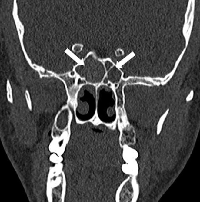
Introduction: Onodi cells are the most posterior ethmoid air cells and extend superolateral to the sphenoid sinus. These cells are also intimately related with the sphenoid sinus, optic nerve, and carotid artery. Radiologic evaluation is mandatory to assess for anatomic variations before any treatment modalities related to the sphenoid sinus.
Objective: To evaluate the effect of Onodi cells on the frequency of sphenoiditis.
Methods: A retrospective analysis was performed in 618 adult patients who underwent high-resolution computed tomography between January 2013 and January 2015. The prevalence of Onodi cells and sphenoiditis was evaluated. Whether the presence of Onodi cells leads to an increase in the prevalence of sphenoiditis was investigated.
Results: Onodi cell positivity was observed in 326 of 618 patients and its prevalence was found to be 52.7%. In the study group, 60.3% (n=73) were ipsilaterally (n=21) or bilaterally (n=52) Onodi-positive, whereas 39.7% (n=48) were Onodi-negative (n=35) or only contralaterally Onodi-positive (n=13). Of the control group, 48.3% (n=240) were Onodi-positive and 51.7% (n=257) were Onodi negative. The co-existence of Onodi cells ipsilaterally was observed to increase the identification of sphenoiditis 1.5-fold, and this finding was statistically significant (p<0.05).
Conclusion: The prevalence of sphenoiditis appears to be higher in patients with Onodi cells. However, it is not possible to state that Onodi cells are the single factor that causes this disease. Further studies are needed to investigate contributing factors related to sphenoiditis.
Introdução: As células de Onodi são as células etmoidais mais posteriores, que se prolongam superolateralmente ao seio esfenoidal. Essas células também se encontram em íntima relação com o seio esfenoidal, o nervo óptico e a artéria carótida. Para análise de variações anatômicas antes da implementação de qualquer modalidade terapêutica relacionada ao seio esfenoidal, a avaliação radiológica é obrigatória,
Objetivo: Nosso objetivo foi avaliar o papel das células de Onodi na frequência de esfenoidite.
Método: Em nosso estudo, foi realizada uma análise retrospectiva em 618 pacientes adultos que se submeteram à tomografia computadorizada de alta resolução entre janeiro de 2013 e janeiro de 2015. Avaliamos a prevalência de células de Onodi e de esfenoidite. Investigamos se a presença de células de Onodi leva a um aumento na prevalência de esfenoidite.
Resultados: A positividade para células de Onodi foi observada em 326 de 618 pacientes, e sua prevalência foi de 52,7%. No grupo de estudo, 60,3% (n = 73) eram CO-positivas: ipsilateral (n = 21) ou bilateralmente (n = 52); e 39,7% (n = 48) eram CO-negativas (n = 35) ou apenas contralateralmente CO-positivas (n = 13). No grupo de controle, 48,3% (n = 240) eram CO-positivas; e 51,7% (n = 257) eram CO-negativas. Observamos que a coexistência de CO ipsilateralmente aumentava em 1,5 vezes a associação com esfenoidite, e esse achado foi estatisticamente significante (p < 0,05).
Conclusão: A prevalência de esfenoidite parece ser maior em pacientes com células de Onodi, mas não é possível afirmar que elas são isoladamente o fator causador desta doença. Novos estudos precisam ser realizados para uma investigação dos fatores contributivos relacionados à esfenoidite.
Keywords: Anatomic variation; Computed tomography; Célula de Onodi; Esfenoidite; Onodi cell; Sphenoiditis; Tomografia computadorizada; Variação anatômica.
Copyright © 2016 Associação Brasileira de Otorrinolaringologia e Cirurgia Cérvico-Facial. Published by Elsevier Editora Ltda. All rights reserved.
Figures


Similar articles
-
Laterally attached superior turbinate is associated with opacification of the sphenoid sinus.Auris Nasus Larynx. 2013 Apr;40(2):194-8. doi: 10.1016/j.anl.2012.07.010. Epub 2012 Jul 31. Auris Nasus Larynx. 2013. PMID: 22854056
-
Is there a relationship between Onodi cell and optic canal?Eur Arch Otorhinolaryngol. 2019 Apr;276(4):1057-1064. doi: 10.1007/s00405-019-05284-0. Epub 2019 Jan 7. Eur Arch Otorhinolaryngol. 2019. PMID: 30617426
-
The prevalence and pattern of pneumatization of Onodi cell in Thai patients.J Med Assoc Thai. 2011 Sep;94(9):1122-6. J Med Assoc Thai. 2011. PMID: 21970203
-
Identification of Onodi cell and new classification of sphenoid sinus for endoscopic sinus surgery.Int Forum Allergy Rhinol. 2015 Nov;5(11):1068-76. doi: 10.1002/alr.21567. Epub 2015 Jun 10. Int Forum Allergy Rhinol. 2015. PMID: 26097234 Review.
-
Chronic Sphenoiditis With Deep Neck Space Extension: Case Report With Review of the Literature and Postulated Mechanisms for Extracranial Extension.Ear Nose Throat J. 2024 Mar;103(3):151-155. doi: 10.1177/0145561321989453. Epub 2021 Jan 20. Ear Nose Throat J. 2024. PMID: 33470832 Review.
Cited by
-
A new CBCT-based classification of posterior extramural ethmoid cells.Surg Radiol Anat. 2025 Jul 16;47(1):173. doi: 10.1007/s00276-025-03686-w. Surg Radiol Anat. 2025. PMID: 40670633
-
A Stepwise Progression of Acute Bilateral Visual Loss Due to Onodi Cell Sinusitis.Cureus. 2023 Oct 20;15(10):e47359. doi: 10.7759/cureus.47359. eCollection 2023 Oct. Cureus. 2023. PMID: 38021640 Free PMC article.
-
Establishment of AI-assisted diagnosis of the infraorbital posterior ethmoid cells based on deep learning.BMC Med Imaging. 2025 Jul 21;25(1):292. doi: 10.1186/s12880-025-01831-w. BMC Med Imaging. 2025. PMID: 40691526 Free PMC article.
-
Investigation of the prevalence and main features of skull-base anomalies and characteristics of the sphenoid sinus using cone-beam computed tomography.J Korean Assoc Oral Maxillofac Surg. 2022 Aug 31;48(4):207-218. doi: 10.5125/jkaoms.2022.48.4.207. J Korean Assoc Oral Maxillofac Surg. 2022. PMID: 36043251 Free PMC article.
-
Type IV optic nerve and Onodi cell: is there a risk of injury during sphenoid sinus surgery?Acta Otorhinolaryngol Ital. 2024 Feb;44(1):36-41. doi: 10.14639/0392-100X-N2462. Epub 2023 Dec 29. Acta Otorhinolaryngol Ital. 2024. PMID: 38165204 Free PMC article.
References
-
- Hwang S.H., Joo Y.H., Seo J.H., Cho J.H., Kang J.M. Analysis of sphenoid sinus in the operative plane of endoscopic transsphenoidal surgery using computed tomography. Eur Arch Otorhinolaryngol. 2014;271:2219–2225. - PubMed
-
- Nomura K., Nakayama T., Asaka D., Okushi T., Hama T., Kobayashi T., et al. Laterally attached superior turbinate is associated with opacification of the sphenoid sinus. Auris Nasus Larynx. 2013;40:194–198. - PubMed
-
- Ozturan O., Yenigun A., Degirmenci N., Aksoy F., Veyseller B. Co-existence of the Onodi cell with the variation of perisphenoidal structures. Eur Arch Otorhinolaryngol. 2013;270:2057–2063. - PubMed
-
- Chee E., Looi A. Onodi sinusitis presenting with orbital apex syndrome. Orbit. 2009;28:422–424. - PubMed
-
- Deshmukh S., DeMonte F. Anterior clinoidal mucocele causing optic neuropathy: resolution with nonsurgical therapy. Case report. J Neurosurg. 2007;106:1091–1093. - PubMed
MeSH terms
LinkOut - more resources
Full Text Sources
Other Literature Sources
