. 2016 May 10:7:11460.
doi: 10.1038/ncomms11460.

Evidence for the involvement of ASIC3 in sensory mechanotransduction in proprioceptors

Affiliations

Evidence for the involvement of ASIC3 in sensory mechanotransduction in proprioceptors

Shing-Hong Lin et al. Nat Commun. .

Abstract

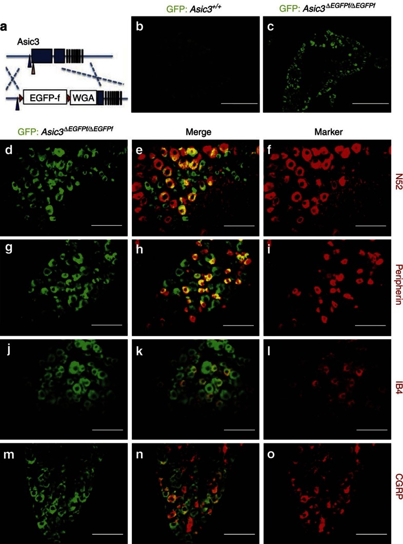
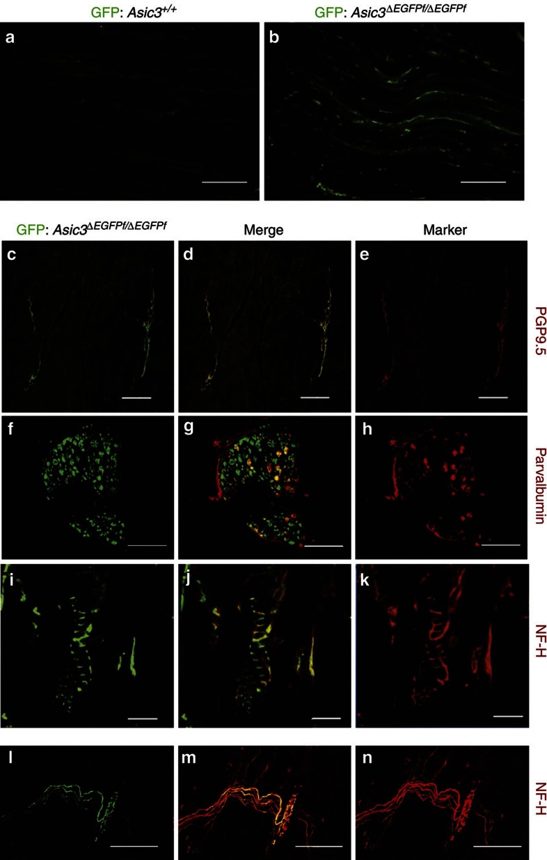
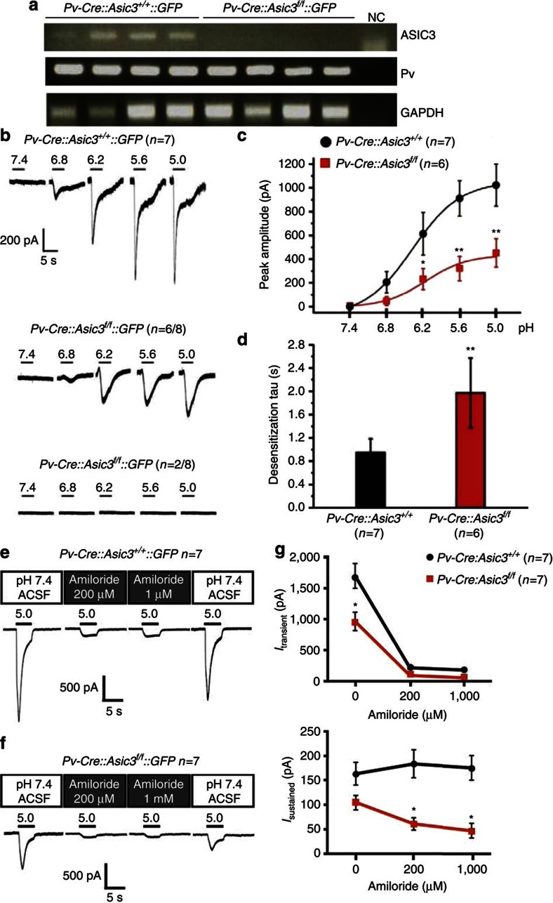
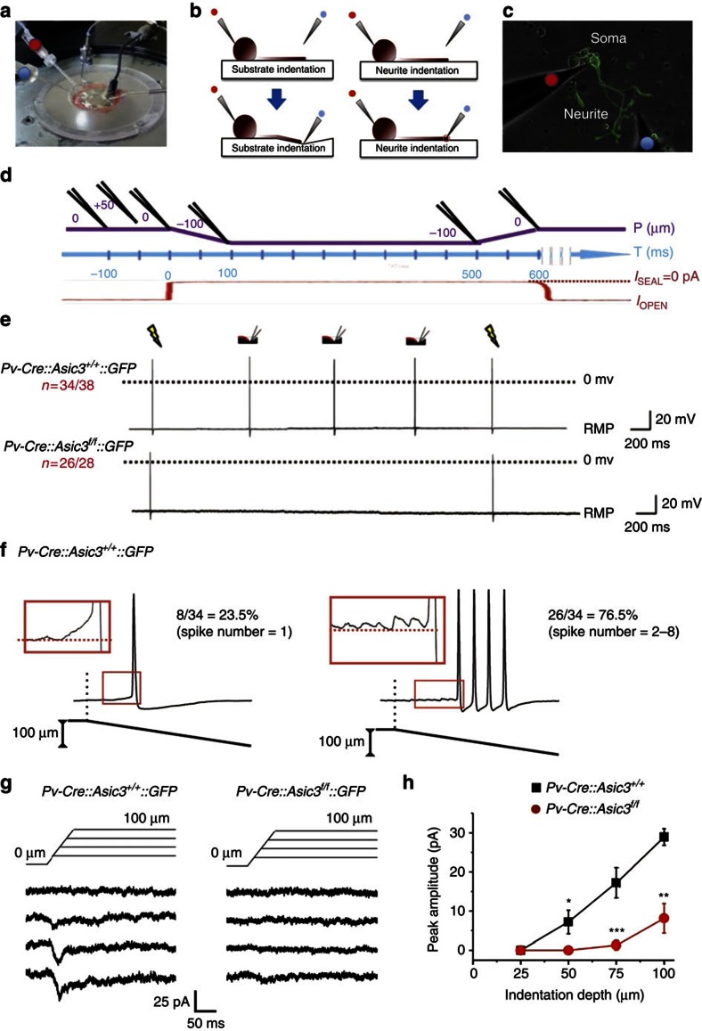
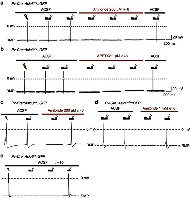
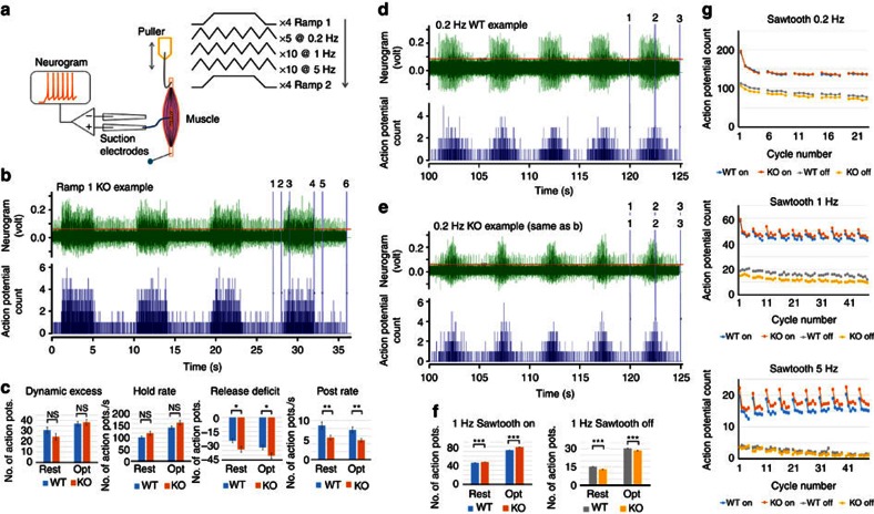
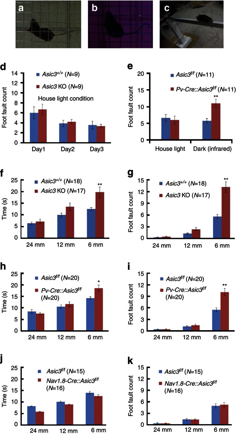
Acid-sensing ion channel 3 (ASIC3) is involved in acid nociception, but its possible role in neurosensory mechanotransduction is disputed. We report here the generation of Asic3-knockout/eGFPf-knockin mice and subsequent characterization of heterogeneous expression of ASIC3 in the dorsal root ganglion (DRG). ASIC3 is expressed in parvalbumin (Pv+) proprioceptor axons innervating muscle spindles. We further generate a floxed allele of Asic3 (Asic3(f/f)) and probe the role of ASIC3 in mechanotransduction in neurite-bearing Pv+ DRG neurons through localized elastic matrix movements and electrophysiology. Targeted knockout of Asic3 disrupts spindle afferent sensitivity to dynamic stimuli and impairs mechanotransduction in Pv+ DRG neurons because of substrate deformation-induced neurite stretching, but not to direct neurite indentation. In behavioural tasks, global knockout (Asic3(-/-)) and Pv-Cre::Asic3(f/f) mice produce similar deficits in grid and balance beam walking tasks. We conclude that, at least in mouse, ASIC3 is a molecular determinant contributing to dynamic mechanosensitivity in proprioceptors.


Figures

Figure 1
Figure 1. Immunofluorescence staining of GFP in ASIC3-expressing DRG neurons in Asic3-KO/eGFP-f-KI (Asic3ΔEGFPf/EGFPf) mice.
(a) Illustration of the EGFPffloxed-WGA construct targeted in Accn3 allele in mice. Blue and pink arrowheads indicate the position of the transcription and translation start sites of mouse Accn3, respectively. Red triangles represent the loxP sites. Blue boxes indicate the exons. (b) GFP signal was very low in wild-type lumbar DRG (scale bar, 50 μm). (c) Expression of GFP was much higher in the Asic3ΔEGFPf/EGFPf lumbar DRG (scale bar, 50 μm). (do) Co-localization of GFP signals with: (df) myelinated neuron marker N52, (gi) small nociceptor marker peripherin, (jl) non-peptidergic nociceptor marker IB4 and (mo) peptidergic nociceptor marker CGRP in the Asic3ΔEGFPf/EGFPf lumbar DRG are illustrated (scale bar, 100 μm).
Figure 2
Figure 2. ASIC3-expressing DRG afferent neurons in muscle and proprioceptors.
(a,b) eGFP immunofluorescence in horizontal sections of gastrocnemius muscle is low in wild-type but high in Asic3ΔEGFPf/EGFPf mice. Scale bar, 200 μm. (ce) eGFP and the nerve marker PGP9.5 co-localize in the nerves of gastrocnemius muscle (transverse section: scale bar, 100 μm). (fh) eGFP is present in most (∼90%) parvalbumin positive, large diameter neurons in the lumbar DRG of the Asic3ΔEGFPf/EGFPf mice, as well as many smaller, parvalbumin-negative somata. Scale bar, 200 μm. (ik) Co-localization of GFP and myelinated-nerve marker neurofilament heavy chain (NF-H) in annulospiral endings characteristic of spindles in the soleus muscle of an Asic3ΔEGFPf/EGFPf mouse (scale bar, 50 μm). (ln) Co-localization of GFP and NF-H in the Golgi tendon organs from an Asic3ΔEGFPf/EGFPf mouse embryo's gluteus muscle (scale bar, 50 μm).
Figure 3
Figure 3. Conditional knockout of Asic3 in DRG Pv+ neurons.
(a) Single-cell RT–PCR indicated that every GFP-positive DRG neuron showed expression of parvalbumin (Pv) and ASIC3 transcripts in Pv-Cre::Asic3+/+::GFP-reporter mice, whereas ASIC3 transcripts were eliminated in the Pv-Cre::Asic3f/f::GFP-reporter samples, showing the Asic3 gene expression was successfully disrupted. (b) Acid-induced inward currents (Iacid) were impaired (n=6/8) or completely undetectable (n=2/8) in Asic3-null Pv+ DRG neurons. (c) Even in responsive neurons, the peak amplitudes of acid-induced currents were significantly smaller (by ∼50%) in Asic3-null Pv+ DRG neurons than in wild-type controls at all pHs below 6.8 (pH 6.8, U=9.0, P=0.053; pH 6.2, U=30.0, P<0.05; pH 5.6, U=35.0, P<0.01; and pH 5.0, U=2.0, P<0.01). (d) In these same neurons, at pH=5.0, the desensitization time constant of Iacid in proprioceptors was also substantially longer in Asic3 knockouts (Mann–Whitney U-test, U=3.0, P<0.01). (e) Only the transient phase of the acid (pH 5.0)-induced current was inhibited by amiloride (200 μM or 1 mM) in wild-type Pv+ DRG neurons. However, (f) both transient and sustained phases of the acid (pH 5.0)-induced current were inhibited by the same doses of amiloride in Asic3-null neurons. These data are summarized in g. For transient current, two-way ANOVA with repeated measurement indicated significant effect of genotype (F(1,12)=10.764, P<0.01), dose of amiloride (F(2,24)=82.483, P<0.01) and their interaction (F(2,24)=5.404, P<0.05). Post hoc comparison indicated significant difference between genotypes only in amiloride dose 0 μM (t=4.616, P<0.05). For sustained current, two-way ANOVA with repeated measurement indicated significant effect of genotype (F(1,12)=14.252, P<0.01) and interaction (F(2,24)=3.801, P<0.05) but not dose of amiloride (F(2,24)=1.283, P=0.295). Post hoc comparison of interaction indicated significant difference between genotypes in amiloride dose 200 μM (t=3.877, P<0.05) and 1,000 μM (t=4.024, P<0.05). Data are mean±s.e.m. *P<0.05, **P<0.01 between groups.
Figure 4
Figure 4. ASIC3 mediates substrate deformation-driven neurite stretch (SDNS)-induced firing in Pv+ DRG neurons.
(a) Experimental setup for whole-cell patch-clamp recording. The red dot indicates the recording pipette and the blue dot indicates the indentation pipette. (b) Schematic of stretching (left panels: substrate indentation, SDNS) or pressing on (right panels: neurite indentation, DNI) a neurite grown on an elastic substrate. (c) Cultured on a PDMS substrate, the soma of a suitable GFP-positive Pv+ DRG neuron was patched with a recording pipette (red dot). An indentation pipette (blue dot) was positioned 300 μm away from the patched soma and on top of (DNI) or 15–25 μm away from (SDNS) the neurite. (d) Experimental protocol for mechanical stretching. The temporospatial relationship of the indentation pipette (in black above the purple line), the surface of the PDMS substrate (purple line) are illustrated and aligned with the recording time (blue line). The leak current of the indentation pipette (red line) monitors when the pipette contacts the PDMS surface. The leak current when sealed (Isealed) is taken as 0 pA (see the Methods for details). (e) Each SDNS generated an action potential in wild type, but not Asic3-null Pv+ DRG neurons. RMP, resting membrane potential. Electrical stimulation via the patch electrode (yellow flashes) also generates a similar action potential response. (f) In 34/38 wild-type Pv+ DRG neurons, SDNS induced single (n=8) or a train of (n=26) action potentials. (g) SDNS-induced mechanosensitive currents were recorded via voltage clamp. In wild-type Pv+ DRG neurons, indentation depths of 25, 50, 75 and 100 μm generated progressively increasing inward currents. The current was dramatically decreased in Asic3-knockout Pv+ DRG neurons. (h) Comparison of the SDNS-induced mechanical currents in wild-type and Asic3 KO Pv+ DRG neurons. Significant differences with genotype was (Mann–Whitney U-test) are found at indentation depths of 50, 75 and 100 μm (U=30.0, P<0.05; U=10.0, P<0.01; U=14.5, P<0.01, respectively). Data are mean±s.e.m. *P<0.05, **P<0.01, ***P<0.001 between groups.
Figure 5
Figure 5. Pharmacological blockade of ASIC3 eliminated SDNS-induced action potentials in wild-type Pv+ DRG neurons.
(a) Amiloride (200 μM), a broad-spectrum ASIC/ENaC channel blocker, blocked SDNS-induced action potentials in wild-type Pv+ DRG neurons. (b) APETx2 (1 μM), a potent and selective inhibitor of ASIC3, also blocked SDNS-induced action potentials in wild-type Pv+ DRG neurons. (c,d) In contrast, DNI still evoked action potentials in the presence of amiloride (200 μM or 1 mM) in wild-type DRG proprioceptors. (e) Similarly, DNI also generated action potentials in Asic3 knockout Pv+ DRG neurons. RMP, resting membrane potential.
Figure 6
Figure 6. Asic3 KO enhances muscle proprioceptor dynamic sensitivity.
(a) Recording arrangement and stretch protocol. The ex vivo mouse soleus muscle was pinned to the PDMS at one end and stretched by an electromagnetic puller at the other end. The nerve was drawn into a suction electrode record stretch-evoked activity. A series of stretches was applied at two initial lengths— ‘resting', then ‘optimal' (see the Methods for details). (b) Typical responses to ramp stretches in a conditional knockout (KO, Pv-Cre::Asic3f/f) mouse (green trace). Action potentials exceeding the cursor position just above background noise are counted (blue trace below, bin width 0.01 s). The numbered vertical cursors show the phases of the ramp where action potential total counts were determined: 1–2, pre-stretch; 2–3, ramp-on; 3–4, hold; 4–5, ramp-off; 5–6, post-stretch. (c) Comparisons at different phases of the ramp of the proprioceptor responses in WT (Asic3f/f) and KO (Pv-Cre::Asic3f/f) mice. Dynamic excess: action potential count during ramp-on exceeding that expected from linear interpolation between pre-stretch and hold rate. Release deficit: action potential count during ramp-off below the linear interpolation between hold and post-stretch rates. (d) Typical response to repeated triangular (‘sawtooth') stretch-and-release at 0.2 Hz in a WT mouse (green trace). Numbered vertical cursors mark the stretch (ON/on in f,g) and release (OFF/off in f,g) of a group of 5 triangles. (e) As in d, for the KO (Pv-Cre::Asic3f/f) mouse shown in b. (f) ON and OFF proprioceptor responses from WT (Asic3f/f) and KO (Pv-Cre::Asic3f/f) mice to 1 Hz sawtooth stretch. (g) Mean on and off phase action-potential counts for sawtooth stretches at 0.2, 1 and 5 Hz in WT and KO mouse soleus muscles from resting lengths, showing the consistent differences. The intervals between successive groups of 5 triangular stretches allowed some recovery in firing rate in an otherwise steady adaptation over time. In c and f: rest, muscles initially at resting length; opt, muscles initially at optimal length. Data are mean±s.e.m. Significance of differences: *P<0.05; **P<0.01; ***P<0.001.
Figure 7
Figure 7. Mice lacking Asic3 had behavioural deficits in proprioception.
(a) Grid-walking task in normal light. (b) Grid-walking task in the dark, imaged with an infrared-aided camera. (c) Balance beam walking task. (d) Foot fault errors during 5 min walking on the grid were counted. Asic3−/− mice showed no difference from Asic3+/+ mice in foot fault counts or learning during three consecutive days of training. Two-way ANOVA with repeated measures: 3 days of training, F (2,32)=5.941, P<0.01), genotype (F (1,32)=0.169, P=0.686), interaction (F (2,32)=0.129, P=0.879), (e) Grid walking task foot faults in normal light or in the dark (imaged with an infrared-aided camera). Pv-Cre::Asic3f/f mice made more errors in the dark (two-way ANOVA, genotype × darkness interaction (F(1,34)=4.527, P<0.05)). (f,g) Asic3−/− mice took longer and also had an elevated error rate in the narrowest beam-walking task under normal lighting. Time: two-way ANOVA with repeated measures for genotype (F(1,66)=7.455, P<0.05), beam width (F(2,66)=43.89, P<0.01) and interaction (F(2,66)=5.265, P<0.01), post hoc Holm-Sidak comparison: genotype difference in 6-mm-width trial (t=3.962, P<0.01); Foot fault: two-way ANOVA with repeated measures for genotype (F(1,66)=23.294, P<0.01), beam width (F(2,66)=140.651, P<0.01) and interaction (F(2,66)=24.312, P<0.01), post hoc Holm-Sidak comparison: genotype difference only in the 6-mm-width trial (t=8.366, P<0.01). (h,i) A similar deficit occurred in Pv-Cre::Asic3f/f mice. For the beam traverse time, two-way ANOVA with repeated measures indicated a significant difference with respect to beam width (F(2,76)=66.813, P<0.01), interaction (F(2,76)=6.512, P<0.01) but not genotype (F(1,76)=2.2194, P=0.145). Overall, a post hoc Holm-Sidak comparison indicated significant genotype difference in the 6-mm width trial (t=3.31, P<0.01). For the foot fault count, two-way ANOVA with repeated measures indicated significant difference in the genotype (F(1,76)=14.681, P<0.01), beam width (F(2,76)=159.638, P<0.01) and interaction (F(2,76)=16.306, P<0.01). Again, post-hoc Holm-Sidak comparisons indicated significant genotype difference only in the 6-mm-width trial (t=6.842, P<0.01). (j,k) Nav1.8-Cre::Asic3f/f conditional knockout mice did not show behavioural deficits in the balance beam walking task. Two-way ANOVA indicated no effect of genotype (time: F(1,58)=2.232, P=0.146; foot fault: F(1,58)=0.044, P=0.836) or interaction (time: F (2,58)=0.524, P=0.595; foot fault: F (2,58)=0.113, P=0.893). Data are mean±s.e.m. *P<0.05, **P<0.01.

References

    1. Geffeney S. L. et al.. DEG/ENaC but not TRP channels are the major mechanoelectrical transduction channels in a C. elegans nociceptor. Neuron 71, 845–857 (2011). - PMC - PubMed
    1. Zhong L., Hwang R. Y. & Tracey W. D. Pickpocket is a DEG/ENaC protein required for mechanical nociception in Drosophila larvae. Curr. Biol. 20, 429–434 (2010). - PMC - PubMed
    1. Chalfie M. Neurosensory mechanotransduction. Nat. Rev. Mol. Cell Biol. 10, 44–52 (2009). - PubMed
    1. Chen C. C. & Wong C. W. Neurosensory mechanotransduction through acid-sensing ion channels. J. Cell. Mol. Med. 17, 337–349 (2013). - PMC - PubMed
    1. Jalalvand E., Robertson B., Wallen P. & Grillner S. Ciliated neurons lining the central canal sense both fluid movement and pH through ASIC3. Nat. Commun. 7, 10002 (2016). - PMC - PubMed

Publication types

MeSH terms