. 2016 Jun 6;26(11):1447-55.
doi: 10.1016/j.cub.2016.03.070. Epub 2016 May 5.

Individual Neuronal Subtypes Exhibit Diversity in CNS Myelination Mediated by Synaptic Vesicle Release

Affiliations

Individual Neuronal Subtypes Exhibit Diversity in CNS Myelination Mediated by Synaptic Vesicle Release

Sigrid Koudelka et al. Curr Biol. .

Abstract

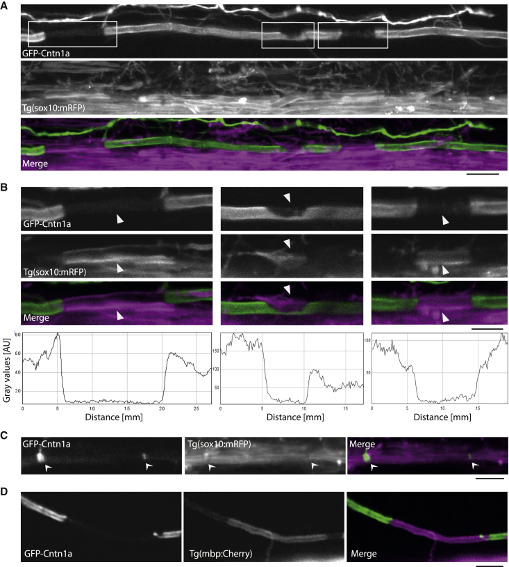
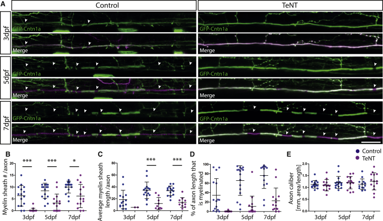
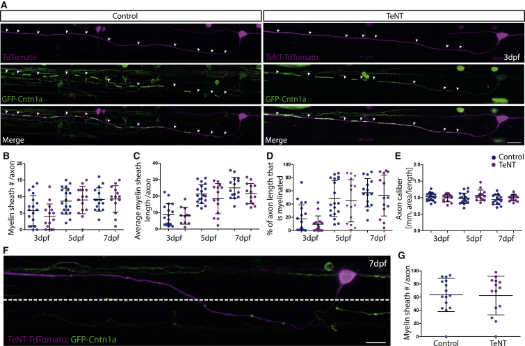
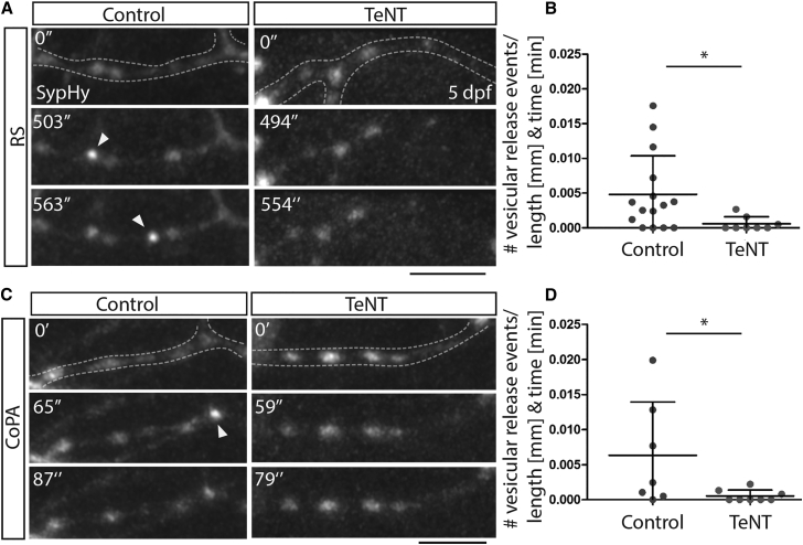
Regulation of myelination by oligodendrocytes in the CNS has important consequences for higher-order nervous system function (e.g., [1-4]), and there is growing consensus that neuronal activity regulates CNS myelination (e.g., [5-9]) through local axon-oligodendrocyte synaptic-vesicle-release-mediated signaling [10-12]. Recent analyses have indicated that myelination along axons of distinct neuronal subtypes can differ [13, 14], but it is not known whether regulation of myelination by activity is common to all neuronal subtypes or only some. This limits insight into how specific neurons regulate their own conduction. Here, we use a novel fluorescent fusion protein reporter to study myelination along the axons of distinct neuronal subtypes over time in zebrafish. We find that the axons of reticulospinal and commissural primary ascending (CoPA) neurons are among the first myelinated in the zebrafish CNS. To investigate how activity regulates myelination by different neuronal subtypes, we express tetanus toxin (TeNT) in individual reticulospinal or CoPA neurons to prevent synaptic vesicle release. We find that the axons of individual tetanus toxin expressing reticulospinal neurons have fewer myelin sheaths than controls and that their myelin sheaths are 50% shorter than controls. In stark contrast, myelination along tetanus-toxin-expressing CoPA neuron axons is entirely normal. These results indicate that while some neuronal subtypes modulate myelination by synaptic vesicle release to a striking degree in vivo, others do not. These data have implications for our understanding of how different neurons regulate myelination and thus their own function within specific neuronal circuits.


Figures

Figure 1
Figure 1
GFP-Cntn1a as a Tool to Visualize Myelin Sheaths along Single Axons (A) Reticulospinal axon labeled with GFP-Cntn1a (top) in a Tg(sox10:mRFP) embryo, which labels oligodendrocytes and their myelin sheaths (middle). The myelin sheaths along the reticulospinal axon are localized to the gaps in GFP expression. Scale bar, 20 μm. (B) High-magnification views of areas outlined in (A) (top). Scale bar, 5 μm. GFP-Cntn1a fluorescent intensity profiles of the insets from (A) (bottom). (C) GFP-Cntn1a expression clustered at putative nodes of Ranvier (left) (arrows), as indicated by gaps in Tg(sox10:mRFP) expression (middle and right). Scale bar, 20 μm. (D) GFP-Cntn1A along a CoPA axon in a Tg(mbp:mCherry-CAAX) embryo at 4 dpf shows expression of the myelin reporter in the gap of GFP-Cntn1A localization. Scale bar, 5 μm. See also Figures S1 and S2 and Table S1.
Figure 2
Figure 2
TeNT Expression in Reticulospinal Neurons Impairs Myelination along Individual Axons (A) Individual reticulospinal axons labeled with GFP-Cntn1a and TdTomato (left) and with GFP-Cntn1a and TeNT-Tdtomato (right) at 3 dpf, 5 dpf, and 7 dpf. Scale bar, 15 μm. (B–E) Quantification of myelin sheath number per axon per 425-μm imaging window (B), average length of myelin sheath per axon (C), percentage of axon length (per 425-μm imaging window) that is myelinated (D), and axon caliber (E) at 3 dpf, 5 dpf, and 7 dpf in control and TeNT-expressing reticulospinal neurons. All error bars indicate ± SD. See also Figure S3.
Figure 3
Figure 3
TeNT Expression in CoPA Neurons Does Not Impair Myelination along Individual Axons (A) Individual CoPA axons labeled with GFP-cntn1a and TdTomato (left) and with GFP-Cntn1a and TeNT-TdTomato (right) at 3 dpf. Scale bar, 10 μm. (B–E) Quantification of myelin sheath number per axon (B), average length of myelin sheath per axon per 425-μm imaging window (C), percentage of axon length (per 425-μm imaging window) that is myelinated (D), and axon caliber (E) at 3 dpf, 5 dpf, and 7 dpf in control and TeNT expressing CoPA neurons. (F) Individual CoPA neuron and axon labeled with GFP-Cntn1a and TeNT-TdTomato. Dashed line indicates dorsoventral cutoff for axonal region analyzed when assessing region of CoPA axons in ventral spinal cord. Scale bar, 15 μm. (G) Percentage of axon length that is myelinated in the ventral spinal cord of control and TeNT expressing CoPA neurons at 7 dpf. All error bars indicate ± SD. See also Figure S3.
Figure 4
Figure 4
Tetanus Toxin Expression in Reticulospinal and CoPA Neurons Impairs Vesicular Release from Their Axons (A) Images from time-lapse movies of sypHy expression in reticulospinal axon collaterals in control (left) and TeNT-expressing (right) neurons at 5 dpf. Dashed lines outline the collateral. Arrowheads point to punctate increases in GFP expression indicative of vesicular release. Scale bar, 5 μm. (B) Quantitation indicates number of GFP events per collateral per micron per minute in control and TeNT-expressing reticulospinal neurons. (C) Images from time-lapse movies of sypHy expression in CoPA axon collaterals in control (left) and TeNT-expressing (right) neurons at 5 dpf. Dashed lines outline the collateral. Arrowheads point to punctate increases in GFP expression indicative of vesicular release. Scale bar, 5 μm. (D) Quantitation indicates number of GFP events per collateral per micron per minute in control and TeNT-expressing CoPA neurons. See also Movies S1, S2, S3, and S4.

Comment in

References

    1. Scholz J., Klein M.C., Behrens T.E.J., Johansen-Berg H. Training induces changes in white-matter architecture. Nat. Neurosci. 2009;12:1370–1371. - PMC - PubMed
    1. Zatorre R.J., Fields R.D., Johansen-Berg H. Plasticity in gray and white: neuroimaging changes in brain structure during learning. Nat. Neurosci. 2012;15:528–536. - PMC - PubMed
    1. McKenzie I.A., Ohayon D., Li H., de Faria J.P., Emery B., Tohyama K., Richardson W.D. Motor skill learning requires active central myelination. Science. 2014;346:318–322. - PMC - PubMed
    1. Makinodan M., Rosen K.M., Ito S., Corfas G. A critical period for social experience-dependent oligodendrocyte maturation and myelination. Science. 2012;337:1357–1360. - PMC - PubMed
    1. Demerens C., Stankoff B., Logak M., Anglade P., Allinquant B., Couraud F., Zalc B., Lubetzki C. Induction of myelination in the central nervous system by electrical activity. Proc. Natl. Acad. Sci. USA. 1996;93:9887–9892. - PMC - PubMed

Publication types