. 2016 May 10:7:11544.
doi: 10.1038/ncomms11544.

Most neutralizing human monoclonal antibodies target novel epitopes requiring both Lassa virus glycoprotein subunits

Affiliations

Most neutralizing human monoclonal antibodies target novel epitopes requiring both Lassa virus glycoprotein subunits

James E Robinson et al. Nat Commun. .

Abstract

Lassa fever is a severe multisystem disease that often has haemorrhagic manifestations. The epitopes of the Lassa virus (LASV) surface glycoproteins recognized by naturally infected human hosts have not been identified or characterized. Here we have cloned 113 human monoclonal antibodies (mAbs) specific for LASV glycoproteins from memory B cells of Lassa fever survivors from West Africa. One-half bind the GP2 fusion subunit, one-fourth recognize the GP1 receptor-binding subunit and the remaining fourth are specific for the assembled glycoprotein complex, requiring both GP1 and GP2 subunits for recognition. Notably, of the 16 mAbs that neutralize LASV, 13 require the assembled glycoprotein complex for binding, while the remaining 3 require GP1 only. Compared with non-neutralizing mAbs, neutralizing mAbs have higher binding affinities and greater divergence from germline progenitors. Some mAbs potently neutralize all four LASV lineages. These insights from LASV human mAb characterization will guide strategies for immunotherapeutic development and vaccine design.


Conflict of interest statement

The Viral Hemorrhagic Fever Consortium (http://www.vhfc.org) is a public-private partnership of academic and industry scientists who are developing diagnostic tests, therapeutic agents and vaccines for Lassa fever, Ebola and other severe diseases. Tulane University and its various academic and industry partners have filed United States and foreign patent applications on behalf of the VHFC for several technologies that have resulted from these efforts. Technical information may also be kept as trade secrets. If commercial products are developed, consortium members may receive royalties or profits. This does not alter our adherence to all policies of the National Institutes of Health and on sharing data and materials.

Figures

Figure 1
Figure 1. Subunit specificity of LASV mAbs.
(a) The LASV GPC is synthesized as a precursor protein. Signal peptidase (SPase) cleaves the small SSP, which remains associated with the complex. SKI protease (SK1/S1P) cleaves GPC into GP1 and GP1. A recombinant LASV (Josiah strain, lineage IV) GPC ectodomain construct (rGPe) was expressed that substituted an uncleavable linker for the SK1 site and deleted the transmembrane and extracellular domains. Constructs expressing recombinant SSP-GP1 (rGP1) and SSP linked to GP2 were also utilized. Molecular weights are indicated in kiloDaltons (kDa). (b) Examples of immunofluorescence assays with unfixed, air-dried cells expressing GP1, GP2 or GPC. All assays were performed two or more times. Scale bar, 10 μm. (c) Distribution of 113 mAbs by subunit specificity, neutralizing activity and reactivity to linear peptides.
Figure 2
Figure 2. LASV neutralization by mAbs and assignment of competition groups.
Neutralizing activity of mAbs was evaluated in two pseudovirus assays and by a PRNT. (a) LASVpp expressing GPC representing the four LASV lineages (I–IV) and the HIV-1 core. (b) LASVpp expressing GPC representing LASV lineage IV and the LCMV core. (c) PRNT. The inset indicates the potency (heat map) for IC50 and IC80 neutralization by each of the mAbs tested in ac. All three pairwise correlation coefficients were significantly different at the 5% significance level according to both Pearson's and Spearman's approaches. (d) Cross-competition analysis was performed pairwise with biotinylated (Biot.) mAbs. The analysis places neutralizing mAbs into four competition groups. 8.9F competes with itself and its binding is blocked by 12.1F. Results in Fig. 2 are representative of at least two experiments performed for each assay.
Figure 3
Figure 3. Immunoprecipitation with anti-GPC mAbs in RIPA buffer, Triton X-100 and Triton X-100±NaSCN.
LASV mAbs indicated were used as capture antibodies in immunoprecipitation studies employing lysates from cells expressing LASV GPs (GPC, rGP1 and rGP2). Immunoprecipitated proteins were resolved by SDS–PAGE, transferred to nitrocellulose. Western blots were probed with mouse mAbs to either GP1 (upper gel image in each panel) or GP2 (lower image). (a) Immunoprecipitation with the indicated mAbs was performed with cell lysates prepared in RIPA buffer. (b) Immunoprecipitation was performed with cell lysates prepared in Triton X-100-containing buffer. (c) mAb-GPC immune complexes bound to magnetic protein A beads were treated or not treated with NaSCN before SDS–PAGE and western blotting. Gel lanes have been reordered for clarity. Uncropped western blot images are presented in Supplementary Fig. 6. Experiments were repeated at least twice.
Figure 4
Figure 4. Cross-reactivity of mAbs isolated from survivors of LASV lineage IV infection with glycoproteins of other arenaviruses.
Cross-reactivity of human mAbs produced after natural infection with LASV lineage IV was investigated by immunofluorescence or by antibody-capture ELISA. (a) Eukaryotic expression vectors encoding GPCs of various OW arenaviruses, including LASV lineages I–IV, LCMV and LUJV were transfected into HEK293T cells. Fixed cell monolayers were then incubated with LASV mAbs and mAb binding was detected by indirect immunofluorescence. An expanded panel of mAbs and OW arenavirus GPCs is presented in Supplementary Fig. 7. Scale bar, 10 μm. (b) GPC from the OW arenaviruses LASV (Josiah strain, lineage IV), LCMV, LUJV and the New World arenavirus MACV were expressed and purified. Reactivity of the indicated mAbs was assessed by ELISA. An expanded panel of mAbs is presented in Supplementary Fig. 8a. Data are pooled from at least four independent experiments.
Figure 5
Figure 5. Mapping of putative epitopes recognized by LASV mAbs by deletion mutagenensis.
Wild-type recombinant LASV GPC was engineered with deletions of amino-acid sequences to map putative B-cell epitopes on LASV glycoproteins. (a) Location of the N-terminal (N-term) deletion of GP1, nested deletions in GP2 and mutations that delete the fusion loop, HR1, T-loop and HR2 are indicated. Alpha helices and beta sheets are labelled according to Hastie et al.. (b) Location of deletions in engineered GPC are mapped to structural models of GP1 and GP2 derived from threading of the LASV GPC ectodomain to the structure of LCMV GPC. Deletions are colour coded as in a. The location of the last amino acid of each of the nested deletions in GP is shown as a black sphere. The inset depicts the LASV GPe structural model and interaction of the N terminus of GP1 with the fusion loop and T-loop of GP2 (see also Supplementary Fig. 2a). (c) ELISA reactivity of selected mAbs to GPC constructs containing deletions of the N terminus of GP1 and indicated regions of GP2. ELISA was performed with GPC in Triton X lysates captured in wells coated with ConA. We normalized the binding of mAbs to each mutant by comparing optical density values observed with each mAb reacted with mutated GPC to that observed with unmutated GPC. Values indicate per cent binding with mutant compared with unmutated control. Mutations that resulted in binding that was <10% of binding to unmutated GPC were considered as having a significant effect on mAb binding. Mutations that reduced binding to 10% or less of control or 11–24% of control are highlighted. Reactivity of GP1-B and GP2-L1, L2 and L3 mAbs with each mutant reflected the degree to which mutations had global effects on glycoprotein conformation. An expanded panel of mAbs is presented in Supplementary Fig. 7b. (d) ELISA reactivity of selected mAbs to GPC constructs containing increasingly larger deletions in GP2. An expanded panel of mAbs is presented in Supplementary Fig. 8c. The results shown are representative of multiple (n>4) independent experiments.
Figure 6
Figure 6. Mapping of putative epitopes recognized by LASV mAbs by site-directed mutagenesis.
Wild-type recombinant LASV GPC was engineered with altered amino-acid sequences to map putative B-cell epitopes on LASV glycoproteins. (a) Location of mutations in engineered LASV glycoproteins are mapped to a structural model of GP1. (b) Binding of mAbs to GPC constructs in which the indicated sets of three amino acids in the amino terminus of GP1 were mutated to three alanines. ConA-capture ELISA performed as in Fig. 5; results expressed as per cent of binding to unmutated control. An expanded panel of mAbs is presented in Supplementary Fig. 8d. (c) 19.7E fails to neutralize LASVpp expressing GPC from lineage III LASV. The sequence of lineage IV GP1 was partially altered to the sequence in lineage III GPC by changing the sequence IIN to LLN. Binding of mAbs was quantified by ConA ELISA as described above. Mutations that reduced binding to 10% or less of control or 11–24% of control are highlighted. (d) Binding of mAbs to GP1 constructs in which the indicated single amino acids in GP1 were mutated to alanine and expressed as D7-tagged proteins. Binding of mAbs to mutant and control GP1-D7 constructs captured in wells coated with mAb JR-52. A mixture of anti-GP1 mAbs was included as a control. Values indicate per cent binding of mutants compared with unmutated GP1. The results shown are representative of multiple (n>4) independent experiments.
Figure 7
Figure 7. Location of putative epitopes on a structural model of LASV GPC.
Antibody epitopes are mapped onto the model of the LASV GP ectodomain monomer. (a) Ribbon view. (b) Surface view. GP1, outside putative epitopes, is coloured yellow. GP2, outside putative epitopes, is coloured white. Epitope colour code: GP1-A and GP1-B, blue; GP2-A and GP2-B, green; GP2-L1-3, purple; GPC-A, red; and GPC-B, orange. The location of the conformational epitope of GPC-C mAb 8.9F is unknown.
Figure 8
Figure 8. LASV mAbs ontogeny and binding kinetics.
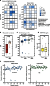
Antibody heavy- and light-chain constructs were sequenced and consensus immunoglobulin sequences spanning framework 1 through 4, and CDRs 1–3 were subjected to in silico analysis using IMGT/V-QUEST and the abYsis integrated antibody analysis and prediction software. (a) Use of heavy and light chains by neutralizing (left) and non-neutralizing mAbs. (b) Dissociation constants of neutralizing and non-neutralizing mAbs. (c) Divergence from presumed germline genes of heavy and light chains of neutralizing and non-neutralizing mAbs. (d) CDR-H3 lengths of neutralizing and non-neutralizing mAbs. (e) Similarity to presumed germline genes of heavy and light chains of human mAbs as a function of time between infection and PBMC collection. Neut., neutralizing; Non neut., non-neutralizing.

References

    1. Monath T. P., Newhouse V. F., Kemp G. E., Setzer H. W. & Cacciapuoti A. Lassa virus isolation from Mastomys natalensis rodents during an epidemic in Sierra Leone. Science 185, 263–265 (1974). - PubMed
    1. Richmond J. K. & Baglole D. J. Lassa fever: epidemiology, clinical features, and social consequences. BMJ 327, 1271–1275 (2003). - PMC - PubMed
    1. Andersen K. G. et al.. Clinical sequencing uncovers origins and evolution of Lassa virus. Cell 162, 738–750 (2015). - PMC - PubMed
    1. Ehichioya D. U. et al.. Current molecular epidemiology of Lassa virus in Nigeria. J. Clin. Microbiol. 49, 1157–1161 (2011). - PMC - PubMed
    1. Shaffer J. G. et al.. Lassa fever in post-conflict Sierra Leone. PLoS Negl. Trop. Dis. 8, e2748 (2014). - PMC - PubMed

Publication types

MeSH terms