. 2016 Jul 10;34(20):2404-15.
doi: 10.1200/JCO.2016.66.6487. Epub 2016 May 9.

Evaluating Cancer of the Central Nervous System Through Next-Generation Sequencing of Cerebrospinal Fluid

Affiliations

Evaluating Cancer of the Central Nervous System Through Next-Generation Sequencing of Cerebrospinal Fluid

Elena I Pentsova et al. J Clin Oncol. .

Erratum in

  • Erratum.
    [No authors listed] [No authors listed] J Clin Oncol. 2017 Jun 10;35(17):1972. doi: 10.1200/JCO.2017.73.8708. J Clin Oncol. 2017. PMID: 28591544 Free PMC article. No abstract available.

Abstract

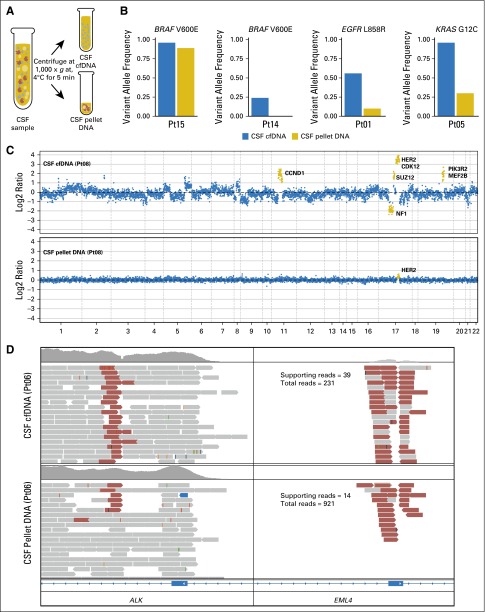
Purpose: Cancer spread to the central nervous system (CNS) often is diagnosed late and is unresponsive to therapy. Mechanisms of tumor dissemination and evolution within the CNS are largely unknown because of limited access to tumor tissue.

Materials and methods: We sequenced 341 cancer-associated genes in cell-free DNA from cerebrospinal fluid (CSF) obtained through routine lumbar puncture in 53 patients with suspected or known CNS involvement by cancer.

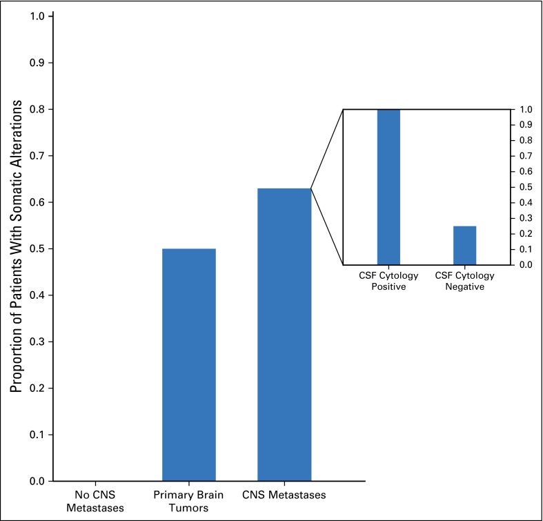
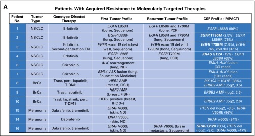
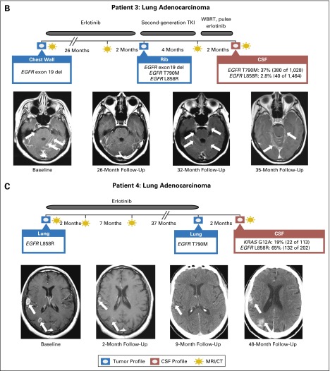
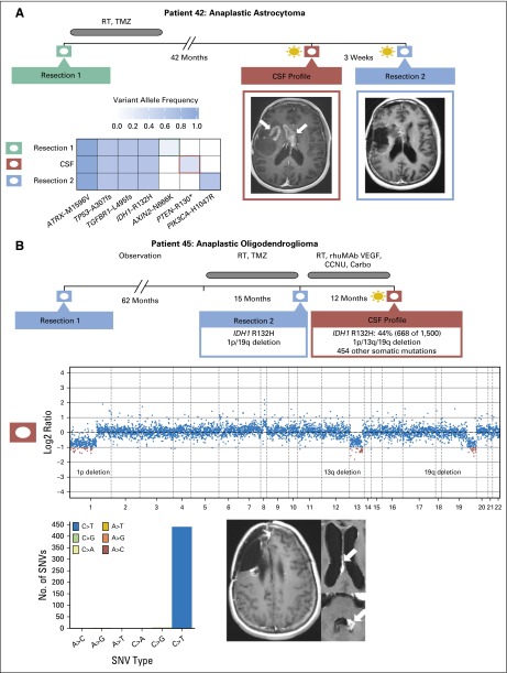
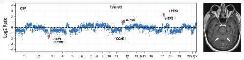
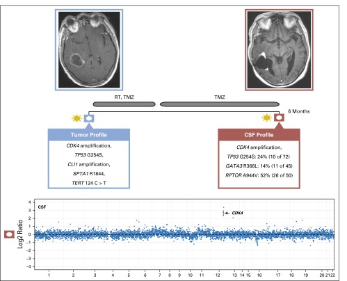
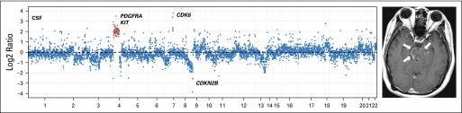
Results: We detected high-confidence somatic alterations in 63% (20 of 32) of patients with CNS metastases of solid tumors, 50% (six of 12) of patients with primary brain tumors, and 0% (zero of nine) of patients without CNS involvement by cancer. Several patients with tumor progression in the CNS during therapy with inhibitors of oncogenic kinases harbored mutations in the kinase target or kinase bypass pathways. In patients with glioma, the most common malignant primary brain tumor in adults, examination of cell-free DNA uncovered patterns of tumor evolution, including temozolomide-associated mutations.

Conclusion: The study shows that CSF harbors clinically relevant genomic alterations in patients with CNS cancers and should be considered for liquid biopsies to monitor tumor evolution in the CNS.


Conflict of interest statement

Authors’ disclosures of potential conflicts of interest are found in the article online at www.jco.org. Author contributions are found at the end of this article.

Figures

Fig 1.
Fig 1.
Comparison of tumor-derived DNA from cerebrospinal fluid (CSF) cell pellet and supernatant. (A) Schematic of separation of CSF pellet and supernatant. Cellular DNA is isolated from the pellet, and cell-free DNA (cfDNA) is isolated from the supernatant. (B) Variant allele frequencies for known mutations in CSF cfDNA and pellet DNA. (C) Log2 ratios of normalized sequence coverage for target exons in CSF cfDNA and pellet DNA for patient 8. Greater than 10-fold amplification of HER2 was observed in CSF cfDNA, whereas HER2 amplification was barely detectable in pellet DNA. (D) Evidence of EML4-ALK gene fusion in CSF cfDNA and pellet DNA for patient 6. Read pairs supporting the fusion (red) were visualized by using the Integrative Genomics Viewer. Pt, patient ID.
Fig 2.
Fig 2.
Detection of tumor-associated mutations in CSF in patients with solid tumors and primary brain tumors. Inset shows the percentage of success in finding somatic alterations in patients with central nervous system (CNS) metastasis with positive and negative cerebrospinal fluid (CSF) cytology.
Fig 3.
Fig 3.
Drug-resistance mutations in patients whose central nervous system (CNS) disease progresses during kinase inhibitor therapy. (A) Summary of genomic profiling results from cerebrospinal fluid (CSF) and other tumor sites in patients in whom progressive CNS disease developed during treatment with the indicated kinase inhibitors. (B) Disease timeline and brain magnet resonance images (MRIs) from a patient with EGFR-mutant NSCLC (patient 3) who presented with leptomeningeal metastasis (baseline MRI, arrows), responded to erlotinib (follow-up MRI at 26 months), was found to have a secondary EGFR mutation (T790M) in a bone metastasis, and developed progressive CNS disease (brain MRIs at 32 and 35 months) that did not respond to second-generation EGFR TKI or pulse erlotinib. CSF cell-free DNA (cfDNA) identified an EGFR T790M mutation. (C) Disease timeline and brain MRIs from a patient with EGFR-mutant NSCLC (patient 4) who presented with brain metastases (baseline MRI), responded to erlotinib (follow-up brain MRI at 2 months and brain CT scan at 9 months), and later developed progressive brain metastases. Molecular profiling of the recurrent lung tumor showed a secondary EGFR mutation (T790M), whereas CSF cfDNA identified an activating KRAS mutation (and the absence of T790M). Sequenom mass spectrometry genotyping was performed for specific mutations in eight genes: AKT1, BRAF, EGFR, ERBB2, KRAS, MEK1 (MAP2K1), NRAS, and PIK3CA. ALK, anaplastic lymphoma kinase; AMP, amplification; BrCa, breast cancer; CT, computed tomography; del, deletion; EGFR, epidermal growth factor receptor; FISH, fluorescent in situ hybridization; HER2, human epidermal growth factor receptor 2; IHC, immunohistochemistry; IMPACT, Integrated Molecular Profiling of Actionable Cancer Targets; ND, not determined; NSCLC, non–small-cell lung cancer; PCR, polymerase chain reaction; pert, pertuzumab; T-DM1, trastuzumab emtansine; TKI, tyrosine kinase inhibitor; trast, trastuzumab; WBRT, whole-brain radiotherapy.
Fig 3.
Fig 3.
Drug-resistance mutations in patients whose central nervous system (CNS) disease progresses during kinase inhibitor therapy. (A) Summary of genomic profiling results from cerebrospinal fluid (CSF) and other tumor sites in patients in whom progressive CNS disease developed during treatment with the indicated kinase inhibitors. (B) Disease timeline and brain magnet resonance images (MRIs) from a patient with EGFR-mutant NSCLC (patient 3) who presented with leptomeningeal metastasis (baseline MRI, arrows), responded to erlotinib (follow-up MRI at 26 months), was found to have a secondary EGFR mutation (T790M) in a bone metastasis, and developed progressive CNS disease (brain MRIs at 32 and 35 months) that did not respond to second-generation EGFR TKI or pulse erlotinib. CSF cell-free DNA (cfDNA) identified an EGFR T790M mutation. (C) Disease timeline and brain MRIs from a patient with EGFR-mutant NSCLC (patient 4) who presented with brain metastases (baseline MRI), responded to erlotinib (follow-up brain MRI at 2 months and brain CT scan at 9 months), and later developed progressive brain metastases. Molecular profiling of the recurrent lung tumor showed a secondary EGFR mutation (T790M), whereas CSF cfDNA identified an activating KRAS mutation (and the absence of T790M). Sequenom mass spectrometry genotyping was performed for specific mutations in eight genes: AKT1, BRAF, EGFR, ERBB2, KRAS, MEK1 (MAP2K1), NRAS, and PIK3CA. ALK, anaplastic lymphoma kinase; AMP, amplification; BrCa, breast cancer; CT, computed tomography; del, deletion; EGFR, epidermal growth factor receptor; FISH, fluorescent in situ hybridization; HER2, human epidermal growth factor receptor 2; IHC, immunohistochemistry; IMPACT, Integrated Molecular Profiling of Actionable Cancer Targets; ND, not determined; NSCLC, non–small-cell lung cancer; PCR, polymerase chain reaction; pert, pertuzumab; T-DM1, trastuzumab emtansine; TKI, tyrosine kinase inhibitor; trast, trastuzumab; WBRT, whole-brain radiotherapy.
Fig 4.
Fig 4.
Tumor evolution in patients with primary brain tumors. (A) Spatial and temporal heterogeneity among samples obtained at diagnosis, at recurrence, and from cerebrospinal fluid (CSF) in patient 42 with recurrent glioblastoma. CSF cell-free DNA harbors a PTEN R130* mutation (variant allele frequency, 0.25), whereas resection 2 harbors a PIK3CA H1047R mutation (variant allele frequency, 0.441). (B) CSF molecular profile for patient 45 with anaplastic oligodendroglioma contains the IDH1 R132H mutation and 1p/19q deletion found in tissue resection 2 as well as 454 nonsilent somatic mutations. Four hundred forty-eight SNVs represent C>T/G>A mutations that demonstrate TMZ-induced mutagenesis. Carbo, carboplatin; CCNU, lomustine; rhuMAB VEGF, bevacuzumab; RT, radiotherapy; SNV, single nucleotide variant; TMZ, temozolomide.
Fig A1.
Fig A1.
Copy number plot for patient 12. cfDNA, cell-free DNA; CSF, cerebrospinal fluid; Pt, patient ID.
Fig A2.
Fig A2.
Detection of EML4-ALK gene fusion in cerebrospinal fluid (CSF) cell-free DNA (cfDNA) and pellet DNA for patient 7. Pt, patient ID.
Fig A3.
Fig A3.
(A) DNA input, (B) library yield, and (C) unique mean sequence coverage.
Fig A4.
Fig A4.
Patient 18: the copy number plot (left panel) showing multiple somatic copy number alterations, including the loci for the receptor tyrosine kinases HER2 and FGFR2 and a brain MRI (right panel) with arrows pointing toward leptomeningeal metastases. CSF, cerebrospinal fluid; FGFR2, fibroblast growth factor receptor 2; HER2, human epidermal growth factor receptor 2.
Fig A5.
Fig A5.
CSF cell-free DNA profiling of glioblastoma for patient 46. CSF, cerebrospinal fluid; RT, radiotherapy; TMZ, temozolomide.
Fig A6.
Fig A6.
Patient 47: the copy number plot (left panel) showing amplification of the PDGFRA gene locus and a brain MRI (right panel) with arrows pointing toward the enhancing tumor in the brainstem.

References

    1. Haber DA, Gray NS, Baselga J. The evolving war on cancer. Cell. 2011;145:19–24. - PubMed
    1. Bettegowda C, Sausen M, Leary RJ, et al. Detection of circulating tumor DNA in early- and late-stage human malignancies. Sci Transl Med. 2014;6:224ra24. - PMC - PubMed
    1. Rhodes CH, Honsinger C, Sorenson GD. PCR-detection of tumor-derived p53 DNA in cerebrospinal fluid. Am J Clin Pathol. 1995;103:404–408. - PubMed
    1. Swinkels DW, de Kok JB, Hanselaar A, et al. Early detection of leptomeningeal metastasis by PCR examination of tumor-derived K-ras DNA in cerebrospinal fluid. Clin Chem. 2000;46:132–133. - PubMed
    1. Shingyoji M, Kageyama H, Sakaida T, et al. Detection of epithelial growth factor receptor mutations in cerebrospinal fluid from patients with lung adenocarcinoma suspected of neoplastic meningitis. J Thorac Oncol. 2011;6:1215–1220. - PubMed

Publication types