. 2016 May 24;113(21):E2925-34.
doi: 10.1073/pnas.1523201113. Epub 2016 May 9.

Loss of RPGR glutamylation underlies the pathogenic mechanism of retinal dystrophy caused by TTLL5 mutations

Affiliations

Loss of RPGR glutamylation underlies the pathogenic mechanism of retinal dystrophy caused by TTLL5 mutations

Xun Sun et al. Proc Natl Acad Sci U S A. .

Abstract

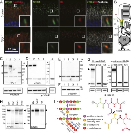
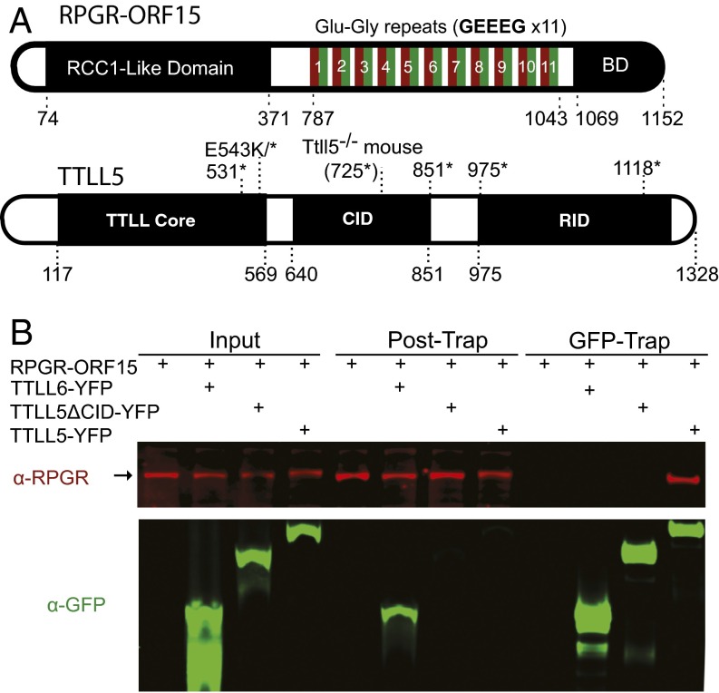
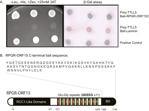
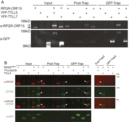
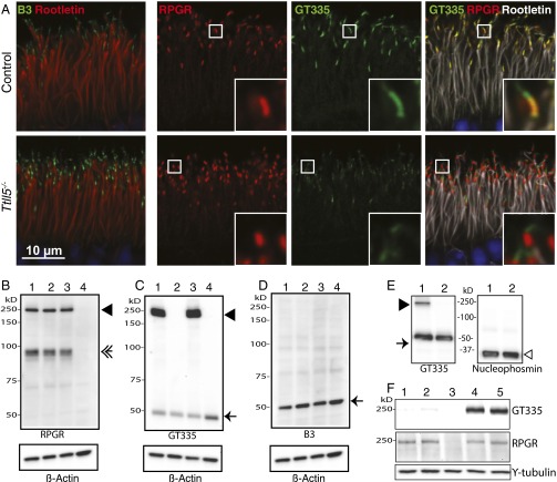
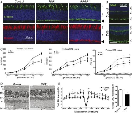
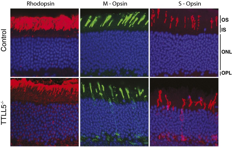
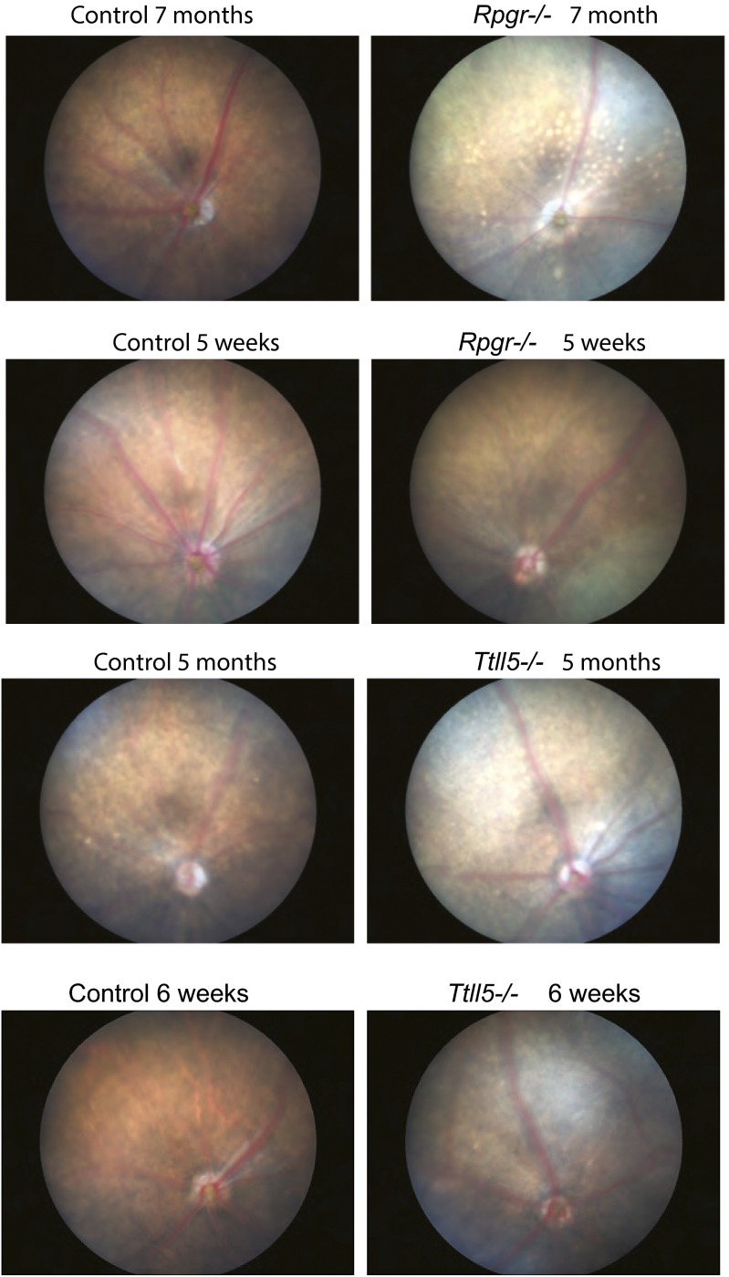
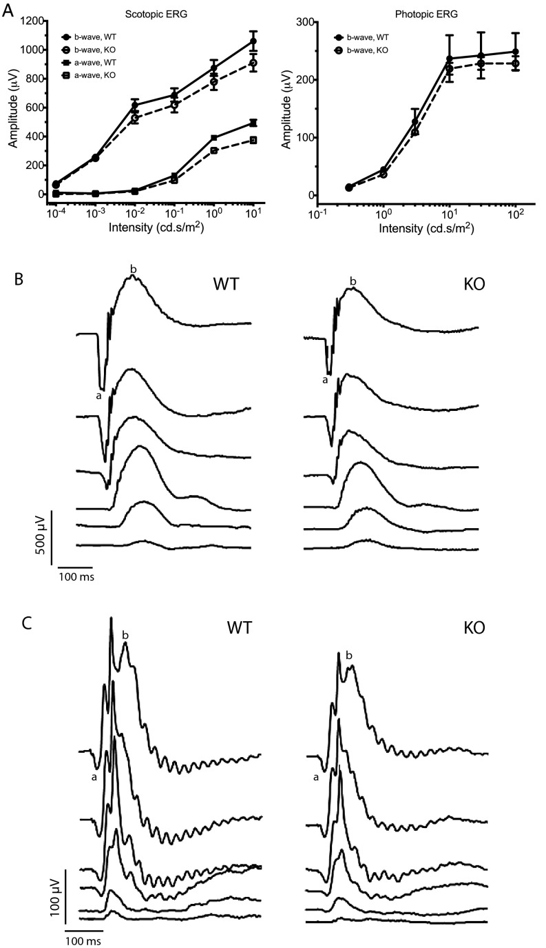
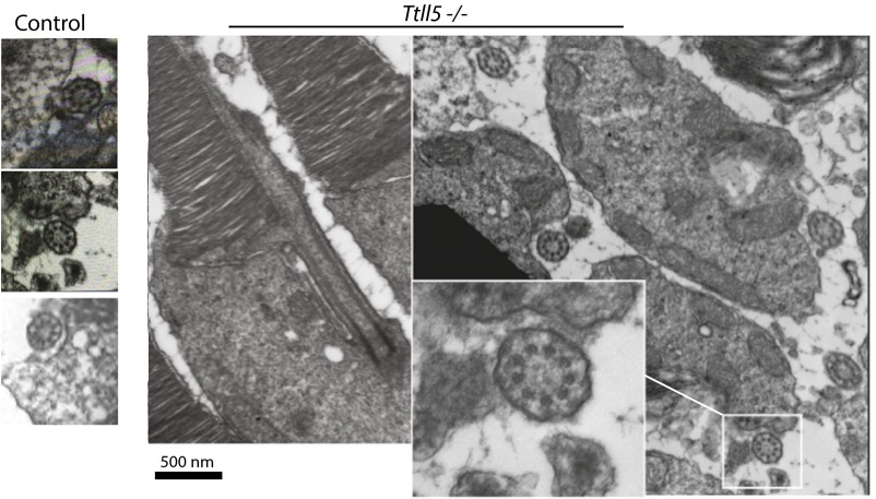
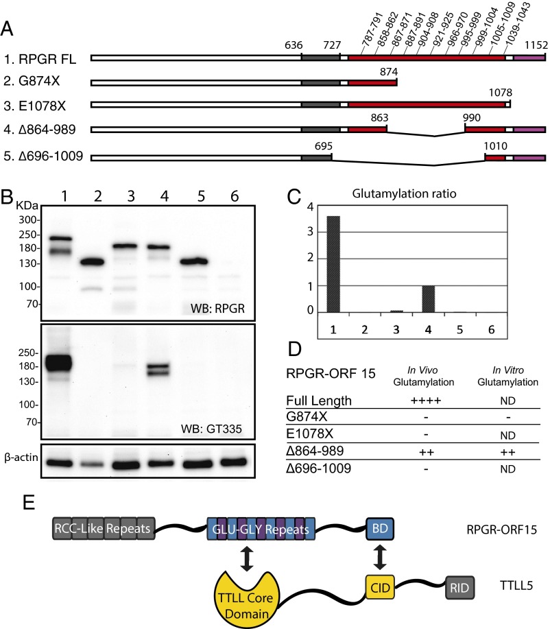
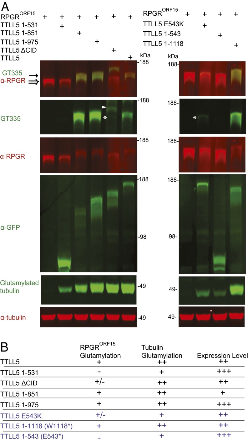
Mutations in the X-linked retinitis pigmentosa GTPase regulator (RPGR) gene are a major cause of retinitis pigmentosa, a blinding retinal disease resulting from photoreceptor degeneration. A photoreceptor specific ORF15 variant of RPGR (RPGR(ORF15)), carrying multiple Glu-Gly tandem repeats and a C-terminal basic domain of unknown function, localizes to the connecting cilium where it is thought to regulate cargo trafficking. Here we show that tubulin tyrosine ligase like-5 (TTLL5) glutamylates RPGR(ORF15) in its Glu-Gly-rich repetitive region containing motifs homologous to the α-tubulin C-terminal tail. The RPGR(ORF15) C-terminal basic domain binds to the noncatalytic cofactor interaction domain unique to TTLL5 among TTLL family glutamylases and targets TTLL5 to glutamylate RPGR. Only TTLL5 and not other TTLL family glutamylases interacts with RPGR(ORF15) when expressed transiently in cells. Consistent with this, a Ttll5 mutant mouse displays a complete loss of RPGR glutamylation without marked changes in tubulin glutamylation levels. The Ttll5 mutant mouse develops slow photoreceptor degeneration with early mislocalization of cone opsins, features resembling those of Rpgr-null mice. Moreover TTLL5 disease mutants that cause human retinal dystrophy show impaired glutamylation of RPGR(ORF15) Thus, RPGR(ORF15) is a novel glutamylation substrate, and this posttranslational modification is critical for its function in photoreceptors. Our study uncovers the pathogenic mechanism whereby absence of RPGR(ORF15) glutamylation leads to retinal pathology in patients with TTLL5 gene mutations and connects these two genes into a common disease pathway.

Keywords: RPGR; cilia; polyglutamylation; retinitis pigmentosa; tubulin tyrosine ligase-like.


Conflict of interest statement

The authors declare no conflict of interest.

Figures

Fig. 1.
Fig. 1.
RPGRORF15, but not RPGRdefault, is glutamylated in vivo. (A) Immunofluorescence staining of retinal cryosections. (Left) RPGR (green) localizes to the connecting cilia as indicated by its position just distal to the ciliary rootlet (red) in the control retina but is absent in the RPGR−/− retina. (Right) GT335 (green) and B3 (red) antibodies both stain the connecting cilia in the control; GT335 signal is greatly diminished in the RPGR−/− retina, whereas B3 is maintained or slightly enhanced. GT335 is a monoclonal antibody that detects monoglutamylation; B3 detects longer glutamate chains on α-tubulin. Insets show images at higher magnification. (B) Schematic representation of a photoreceptor cell. CC, connecting cilia; BB, basal bodies. (C and D) Immunoblotting of mouse tissue extracts with antibodies indicated at the bottom of each blot. (Lower) Same blots probed with γ-tubulin as a loading control. Lane 1, WT retina; 2, RPGRdefault KO (lacking RPGRdefault only) retina; 3, RPGR KO (lacking both RPGRdefault and RPGRORF15 isoforms) retina; 4, rd9 (lacking RPGRORF15) retina; 5, WT brain. (C) An RPGR antibody detected both RPGRORF15 (solid arrowhead) and RPGRdefault (open arrowhead) in the WT retina. RPGRORF15 was retained (solid arrowhead) in the RPGRdefault KO retina but was lost in the full RPGR KO and the rd9 retinas. (D) GT335 reacts with RPGRORF15 (arrowhead) but not with RPGRdefault. Glutamylated tubulin bands (arrow) show comparable intensities in all samples. Brain expresses only RPGRdefault and hence does not have the higher-molecular-weight RPGRORF15 band (lane 5). (E) The B3 antibody shows similar signal intensity in all lanes. (F) α-Tubulin expression levels are comparable in all samples, indicating that tubulin glutamylation is similar in all samples. (G) RPGRORF15 is predominantly monoglutamylated. (Upper) Retinal lysates from a WT mouse or an Rpgr-null mouse expressing human RPGRORF15 (through AAV gene transduction) were run in triplicate and probed with the GT335, polyE, and 1D5 antibodies. Glutamylated mouse and human RPGRORF15 is readily detected with GT335, which recognizes branched glutamate side chains. PolyE, which reacts with the side chains of three or more glutamates, did not detect RPGR. 1D5, which reacts with the chains of two or more glutamates, detects trace amounts of RPGR on prolonged exposure. (H) RPGRORF15 from human photoreceptors is glutamylated. (Left) Three donor retinal lysates (Ret 1, 2, and 3) were run together with recombinant human RPGRORF15 expressed in mouse retina (aav, lane 1) on an immunoblot probed with GT335. Bands corresponding to the position of RPGR were detected (arrowhead). (Right) Following immunoprecipitation with an RPGR antibody, the bound fraction was enriched for the GT335-reactive band (Ret-IP). Ret is lysate before IP (5% input). Note that the tubulin band (arrow) was present in the lysate (Ret) but was lost in the eluate (Ret-IP). Separate blot probed with an RPGR antibody confirmed enrichment of RPGR in the eluate and reduction in the flow-through fraction. (I) (Left) Schematic diagram of glutamylation. The sequence in the diagram can be found in RPGR and the α-tubulin C-terminal tail but does not denote an exact, experimentally mapped modification site. Epitopes for GT335, B3, polyE, and 1D5 antibodies are shown. (Right) Chemical structure of two postranslationally added glutamates (the first one connected to the main chain of the target protein through an isopeptide bond).
Fig. 2.
Fig. 2.
RPGRORF15 interacts with TTLL5 via its C-terminal basic domain (BD). (A) Diagrams showing the domain organization of RPGRORF15 and TTLL5. The RCC1-like domain is shared among all RPGR variants, whereas the Glu-Gly–rich region and the basic C-terminal domain are unique to RPGRORF15. Human RPGRORF15 possesses 11 tandem GEEEG repeats, whereas mouse RPGRORF15 possesses 18. TTLL5 is comprised of a core tubulin tyrosine ligase-like (TTLL) domain, a cofactor interaction domain (CID), and a receptor interaction domain (RID). Point and deletion mutations relevant to this study (see later sections) are denoted on the diagrams. (B) Confirmation of the physical interaction between RPGRORF15 and TTLL5 by GFP-trap precipitation and immunoblotting of transiently transfected HEK293 cell lysates. RPGRORF15 was detected with a polyclonal antibody (red; Upper). All TTLL constructs were YFP-fusions. TTLL5, TTLL5 ΔCID, and TTLL6 (Lower) were detected with a GFP antibody (green; Lower). Input, posttrap (unbound), and GFP-Trap (bound) fractions were analyzed. In the bound (GFP-Trap) fractions, TTLL5, TTLL5 ΔCID, and TTLL6 proteins were efficiently recovered. RPGR was recovered only together with TTLL5. GFP-Trap results were reproduced in more than four independent trials.
Fig. S1.
Fig. S1.
Identification of TTLL5 as a putative interacting partner of RPGR through yeast two-hybrid screens. (A) Confirmation assays. Image on the Left shows growth of yeast colonies. Presence of both TTLL5 and RPGR promoted growth. TTLL5 cotransfected with laminin did not allow growth (negative control). Positive control yeast cells carried SV40 large T antigen and P53, two proteins known to interact. Image on the Right shows lacZ expression in yeast cells where bait and prey plasmids were able to bind each other. (B) Sequence of the human RPGRORF15 C-terminal basic domain used as bait in the screen.
Fig. S2.
Fig. S2.
RPGRORF15 interacts stably with full-length TTLL5, but not with the closely related paralog TTLL 7 or TTLL5 ΔCID. All TTLL constructs were YFP-fusions. (A) RPGRORF15 is recovered together with TTLL5 in the GFP-Trap fraction in cotransfected heterologous cells. TTLL5 also glutamylates and reduces the electrophoretic mobility of RPGRORF15, as indicated by an upward shift (hollow to solid arrow). TTLL5 paralog family member TTLL7 does not interact with nor does it glutamylate RPGRORF15. (B) RPGRORF15 interacts with and is glutamylated by full-length TTLL5 but not TTLL5ΔCID. (Left) RPGRORF15 is recovered from the GFP-trap fraction only when cotransfected with TTLL5-YFP. Glutamylation of RPGRORF15 is detected by the GT335 antibody and is also marked by an upward shift in mobility (white arrowheads). Additional glutamylated (GT335 reactive) bands were also observed. These bands are unrelated to RPGR (they are present also in cells transfected only with TTLL5 or TTLL5 ΔCID) and are presently unknown. (Right) A second round of GFP-Trap binding was performed using Post-Trap supernatants to exclude the possibility that glutamylated RPGRORF15 bound nonspecifically to GFP-Trap in the absence of TTLL5-YFP. Post-Trap2 and GFP-Trap2 are unbound and bound fractions, respectively, from the second round of the GFP-Trap experiment. White arrowheads indicate glutamylated RPGRORF15. The results show that residual RPGRORF15 from the first round Post-Trap fraction, in which TTLL5-YFP fusion proteins were largely depleted, did not bind to GFP-Trap in isolation (in the absence of TTLL5-YFP).
Fig. 3.
Fig. 3.
RPGRORF15 is glutamylated by TTLL5 in vivo. (A) Analysis of WT and Ttll5 mutant retinal sections by immunofluorescence. Polyglutamylation of ciliary tubulin (indicated by staining with the B3 antibody) is not diminished on TTLL5 loss (green; leftmost panels), nor is RPGR itself (red; second column). However, GT335 signal is greatly diminished on TTLL5 loss. Zoomed in images in Insets reveal two domains of GT335 signal: one overlapping with RPGR and the other extending more distally. The GT335 signal in the distal domain, which corresponds to glutamylated tubulin, is unaffected by the Ttll5 mutation. (BD) Immunoblotting analysis of Ttll5+/− (lane 1), Ttll5−/− (lane 2), WT (lane 3), and Rpgr−/− (lane 4) mouse retinal lysates. Ttll5+/−, Ttll5−/− and WT mice were littermates. Antibodies used for immunoblotting are marked under each panel. RPGRORF15 (solid arrowhead) and RPGRdefault (double arrow) expression was unchanged in the Ttll5−/− mutant (B). Glutamylation of RPGRORF15 is abolished in the Ttll5−/− retina (lane 2 of C), whereas tubulin glutamylation (arrow) remains constant. The B3 antibody, which recognizes polyglutamylated tubulin specifically, shows comparable signal intensity among all samples (D). (Lower) Blots reprobed for β-actin as loading controls (BD). Similar results were obtained from two independent experiments. (E) Nucleophosmin is not glutamylated. Mouse retinal lysates probed with GT335 show signal for tubulin (arrow) in both WT (1) and Ttll5−/− mutant (2) samples, and RPGRORF15 (solid arrowhead) only in the WT sample. No GT335-reactive bands at the molecular weight of nucleophosmin (∼33 kDa) were detected even after prolonged exposure (Left). Nucleophosmin (open arrowhead) was readily detected in both samples (Right). (F) Glutamylation of RPGRORF15 is greatly diminished in the Rpgrip1−/− retina. (Top) GT335 immunoblot of retinal extracts from Rpgrip1−/− retinas (lanes 1 and 2) at age postnatal day 20 and age-matched WT controls (lanes 4 and 5). Four mice of each genotype were analyzed in this experiment. Lane 3: Rpgr−/− as a negative control. (Middle) Immunoblot of the same retinal lysates probed with an RPGRORF15 C-terminal antibody. (Bottom) A γ-tubulin immunoblot is shown as a loading control.
Fig. 4.
Fig. 4.
Ttll5−/− mice develop late-onset, slowly progressive photoreceptor degeneration that phenocopies Rpgr−/− mice. (A) Immunofluorescence analysis of cone opsins at a young age (20–40 d). Comparison with the control shows both the S- and M-opsins mislocalized to the cell body and the synaptic layer (arrowhead) in the Ttll5−/− and Rpgr−/− retinas. (B) GFAP is up-regulated in the Ttll5−/− retinas at older ages (20–22 mo old). (C) ERG stimulus intensity–amplitude functions in aged mice. ERG amplitudes in Ttll5−/− mice are significantly lower than control mice of this age (20–22 mo; P < 0.0001 for scotopic ERG a- and b- waves; P = 0.0013 for photopic ERG b-wave in two-way ANOVA). (D) Light microscopy of retina sections show mild thinning of the photoreceptor layers. (E) Measurements of ONL thickness at different positions from the optic nerve head (ONH) to the far peripheral retinas (Left) shows thinning of ONL in the Ttll5−/− mice (n = 11) compared with the controls (n = 9). Overall, the mean ONL thickness (Right) in Ttll5−/− retinas was significantly reduced compared with controls (P < 0.05). Mice were 20–22 mo old in D and E.
Fig. S3.
Fig. S3.
Immunofluorescence analysis of rhodopsin and cone opsins at 20–22 mo. Rhodopsin staining reveals outer segments are shorter in the mutant with some mislocalization of rhodopsin in the inner segments and nuclear layer. Both the S- and M-opsins mislocalize to the cell body and the synaptic layer in the Ttll5−/− retinas, but not in that of controls.
Fig. S4.
Fig. S4.
Representative fundus photographs from Rpgr and Ttll5−/− mice and age-matched controls. The controls for Rpgr-null mice were age-matched C57BL/6J and, for Ttll5 mice, WT littermates were used.
Fig. S5.
Fig. S5.
ERG analysis of Ttll5−/− (KO) mice and littermate WT mice at 4 mo of age. (A) ERG stimulus intensity-amplitude functions. (B) Representative dark-adapted ERG waveforms. (C) Representative light-adapted ERG waveforms.
Fig. S6.
Fig. S6.
The 9+0 microtubule doublet array appeared normal in the photoreceptor connecting cilia of Ttll5−/− mice. Transmission electron micrographs showing cross-sectional views of the connecting cilia. Mice were at 20 mo of age. Controls and mutant mice were littermates. Two animals of each genotype were examined.
Fig. 5.
Fig. 5.
RPGR glutamylation in vivo requires both the C-terminal basic domain and the Glu-Gly–rich region. (A) Diagrams of human RPGRORF15 expression constructs packaged into AAV vectors. The Glu-Gly–rich region is marked in red and the C-terminal basic domain in magenta. The position of glutamylation consensus motifs is shown in the schematic for the full-length (FL) construct. (B) Immunoblots of retinal extracts from Rpgr−/− mice injected with RPGR expression constructs. Lanes 1–5 match the construct numbers shown in A, and lane 6 is an uninjected control. Full-length RPGR and to a lesser extent RPGRΔ864–989 are glutamylated as indicated by detection with GT335 (Middle). Probing with an RPGR antibody shows expression levels for recombinant RPGR (Top). Reprobing blots with β-actin provides a loading control (Bottom). (C) Quantification of glutamylation levels by densitometry after normalizing for RPGR levels, with sample 4 level set arbitrarily at 1. These results are summarized in D. ND, not determined. Similar results were obtained from two independent experiments. (E) Schematic diagram illustrating mapped interactions between RPGR and TTLL5.
Fig. 6.
Fig. 6.
Effect of TTLL5 domain deletion and disease mutations on RPGRORF15 glutamylation in transient cotransfection assays. (A) Coexpression of RPGRORF15 and WT TTLL5 in HEK293 cells leads to RPGRORF15 glutamylation as detected by GT335 staining. An early truncation mutant TTLL5 1–531 shows no activity toward RPGRORF15. Deletion of the CID or mutation of a conserved residue E543K in the TTLL core dramatically reduce RPGRORF15 glutamylation as indicated by only residual GT335 staining (denoted by a star). Truncation downstream from CID generally preserves TTLL5 activity toward RPGRORF15 in the in vitro assay. White arrowhead indicates the unknown high-molecular-weight protein that is also gluatmylated. Note an upward shift in molecular weight of RPGRORF15 on glutamylation as denoted by hollow (unmodified) and solid (modified) arrows, consistent with the glutamate addition at multiple Glu-Gly–rich motifs. Expression of YFP-TTLL5 constructs was confirmed with a GFP antibody. This experiment is representative for more than three independent cotransfections. (B) Summary table of RPGR-ORF15 glutamylation by mutant TTLL5 constructs in heterologous cells (disease-causing mutations are shown in blue).

Similar articles

Cited by

References

    1. Berson EL. Retinitis pigmentosa. The Friedenwald Lecture. Invest Ophthalmol Vis Sci. 1993;34(5):1659–1676. - PubMed
    1. Bader I, et al. X-linked retinitis pigmentosa: RPGR mutations in most families with definite X linkage and clustering of mutations in a short sequence stretch of exon ORF15. Invest Ophthalmol Vis Sci. 2003;44(4):1458–1463. - PubMed
    1. Pelletier V, et al. Comprehensive survey of mutations in RP2 and RPGR in patients affected with distinct retinal dystrophies: Genotype-phenotype correlations and impact on genetic counseling. Hum Mutat. 2007;28(1):81–91. - PubMed
    1. Branham K, et al. Mutations in RPGR and RP2 account for 15% of males with simplex retinal degenerative disease. Invest Ophthalmol Vis Sci. 2012;53(13):8232–8237. - PMC - PubMed
    1. Churchill JD, et al. Mutations in the X-linked retinitis pigmentosa genes RPGR and RP2 found in 8.5% of families with a provisional diagnosis of autosomal dominant retinitis pigmentosa. Invest Ophthalmol Vis Sci. 2013;54(2):1411–1416. - PMC - PubMed

Publication types