. 2016 May 24;113(21):5946-51.
doi: 10.1073/pnas.1601918113. Epub 2016 May 9.

Enhancement of β-catenin activity by BIG1 plus BIG2 via Arf activation and cAMP signals

Affiliations

Enhancement of β-catenin activity by BIG1 plus BIG2 via Arf activation and cAMP signals

Chun-Chun Li et al. Proc Natl Acad Sci U S A. .

Abstract

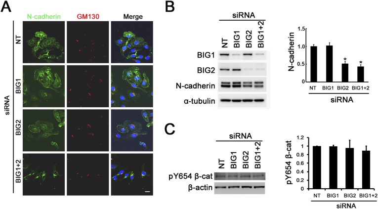
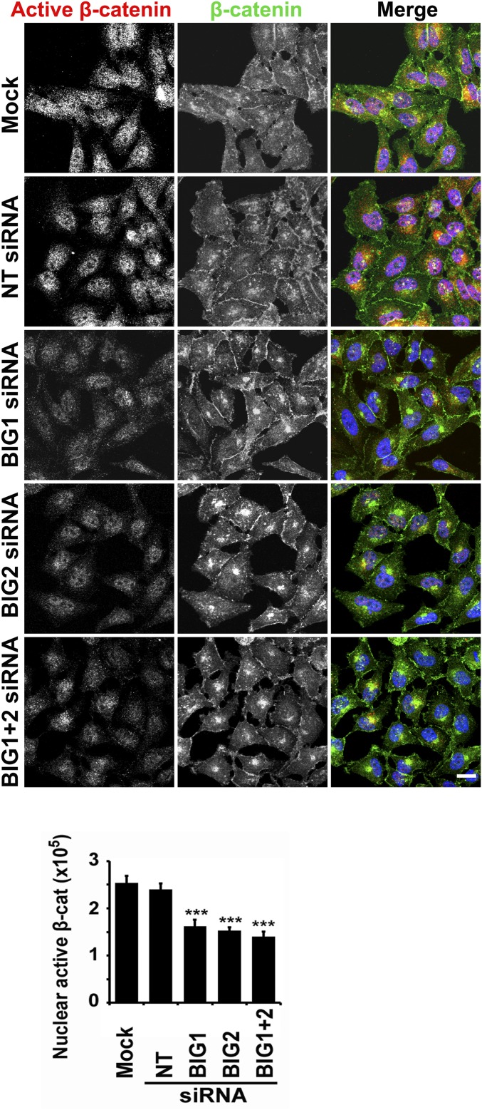
Multifunctional β-catenin, with critical roles in both cell-cell adhesion and Wnt-signaling pathways, was among HeLa cell proteins coimmunoprecipitated by antibodies against brefeldin A-inhibited guanine nucleotide-exchange factors 1 and 2 (BIG1 or BIG2) that activate ADP-ribosylation factors (Arfs) by accelerating the replacement of bound GDP with GTP. BIG proteins also contain A-kinase anchoring protein (AKAP) sequences that can act as scaffolds for multimolecular assemblies that facilitate and limit cAMP signaling temporally and spatially. Direct interaction of BIG1 N-terminal sequence with β-catenin was confirmed using yeast two-hybrid assays and in vitro synthesized proteins. Depletion of BIG1 and/or BIG2 or overexpression of guanine nucleotide-exchange factor inactive mutant, but not wild-type, proteins interfered with β-catenin trafficking, leading to accumulation at perinuclear Golgi structures. Both phospholipase D activity and vesicular trafficking were required for effects of BIG1 and BIG2 on β-catenin activation. Levels of PKA-phosphorylated β-catenin S675 and β-catenin association with PKA, BIG1, and BIG2 were also diminished after BIG1/BIG2 depletion. Inferring a requirement for BIG1 and/or BIG2 AKAP sequence in PKA modification of β-catenin and its effect on transcription activation, we confirmed dependence of S675 phosphorylation and transcription coactivator function on BIG2 AKAP-C sequence.

Keywords: ADP-ribosylation factor; AKAP; cell migration; phospholipase D.


Conflict of interest statement

The authors declare no conflict of interest.

Figures

Fig. 1.
Fig. 1.
Interaction of β-catenin with BIG1 and/or BIG2. (A) Similar six-domain structures of BIG1 and BIG2 molecules include dimerization and cyclophilin binding (DCB, blue), homology upstream of Sec7 (HUS, green), Sec7 (BFA-inhibited Arf activation, yellow), and three homology downstream of Sec7 (HDS, pink). BIG1 contains one AKAP sequence and BIG2 three (AKAP-A, -B, -C, red). (B and C) Samples (5%, input) of endogenous HeLa cell proteins for IP (100 μg) and of proteins collected by IP (50%) with antibodies against BIG1, BIG2, or β-catenin (β-cat), or control IgG were analyzed by Western blotting with indicated antibodies. Exposure times differed in B and C to optimize images. (D) HA-BIG1 and FLAG–β-catenin, synthesized singly (wheat germ extract), were incubated together as indicated before IP with antitag antibodies. Samples of in vitro synthesized proteins before (5%, input) and from IP (100%) with antibodies against HA or FLAG were subjected to Western blotting, respectively, with the same antibodies. (E) Samples of in vitro synthesized BIG2 and FLAG–β-catenin were mixed before IP and analysis of collected proteins. (F) Yeast transformed with N terminus of Gal4p-activation domain fusion constructs of GSK-3β (3β) or indicated BIG1 amino acid sequences [1–1849 full length (F), 1–887 (N), or 888–1849 (C)], together with pGBKT7 vector with N terminus of full-length β-catenin fused to the DNA-binding domain of Gal4p were grown (3 d, 30 °C) on synthetic histidine-containing medium lacking leucine and tryptophan (LW); colonies from LW plates were grown (3 d, 30 °C) on selective medium lacking also histidine (LWH) with or without adenine (LWHAde).
Fig. S1.
Fig. S1.
Density gradient distribution of endogenous HeLa cell proteins: BIG1, BIG2, and β-catenin. (A) Proteins in postnuclear supernatants of cells separated in iodixanol gradients as described in Materials and Methods. Lower gradient numbers correspond to lower and higher to greater densities. Amounts of protein are percentage in each fraction of the total amount of that protein recovered. (B) Cells were fixed and reacted with rabbit antibodies against BIG1 or BIG2 and mouse anti–β-catenin antibodies. (Scale bar, 10 μm.) (C) Quantification of Fig. 2A. β-Catenin in cells was quantified as the ratio of mean β-catenin staining intensity of Golgi to mean β-catenin staining intensity of cytosol (Golgi/cytosol). At least 20 cells were analyzed in each of three experiments. ***P < 0.005; *P < 0.05 vs. NT siRNA.
Fig. 2.
Fig. 2.
Effects of BIG1 and/or BIG2 depletion on intracellular distribution of endogenous β-catenin. After a 72-h incubation with vehicle alone (mock) or siRNA, nontargeted (NT) or specific for BIG1 or BIG2 or both, cells were fixed, reacted with rabbit anti–β-catenin and mouse anti-GM130 antibodies (A), or mouse anti–syntaxin-6 antibodies (B), before preparation for confocal immunofluorescence microscopy. (Scale bar, 10 μm.)
Fig. S2.
Fig. S2.
Effects of BIG1 and/or BIG2 depletion on amounts of N-cadherin and Y654-phosphorylated (pY654) β-catenin. (A) After a 72-h incubation with siRNA, nontarget (NT) or specific for BIG1 or BIG2, or both, cells were fixed, reacted with antibodies against N-cadherin and GM130, and prepared for confocal immunofluorescence microscopy. (Scale bar, 10 μm.) (B) Samples (20 μg) of total lysate proteins were separated in 4–12% NuPAGE Bis⋅Tris gels, reacted with indicated antibodies, and quantified by densitometry; values were normalized to that of control (NT siRNA) cells = 1.0. Data are means ± SEM (n = 3), *P < 0.05 vs. NT siRNA. (C) Cells were incubated 72 h with NT or specific BIG1 and/or BIG2 siRNA before analyses of indicated proteins by Western blotting with antibodies against pY654 β-catenin (pY654–β-cat), densitometric quantification, and statistical analysis of data from three experiments as in B.
Fig. S3.
Fig. S3.
Effects of overexpressed GEF-inactive BIG1 or BIG2 mutants, but not BIG1 AKAP mutant, on cellular distribution of β-catenin. After a 24-h transfection with cDNA encoding HA-tagged BIG1 WT or mutants E793K, or AKAP (A), or GFP-tagged BIG2 WT or mutant E738K (B), cells were fixed, reacted with antibodies against β-catenin or GM130 (cis-Golgi marker), and HA or GFP, and prepared for confocal immunofluorescence microscopy. Perinuclear accumulation of β-catenin in cells expressing BIG1 (E793K) or BIG2 (E738K), but not WT BIG1 or BIG2 or BIG1 AKAP mutant, is consistent with blockage of Arf activation at membranes in a β-catenin translocation pathway. (Scale bar, 10 μm.)
Fig. 3.
Fig. 3.
Effects of BIG1 and/or BIG2 depletion on levels of pS675 β-catenin, active β-catenin (unreactive with antibodies to phosphorylated N terminus), total β-catenin, and interaction between β-catenin and PKA Cα. (A) Cells were incubated 72 h with vehicle alone (mock) or with nontarget (NT) or specific BIG1 and/or BIG2 siRNA before analysis of indicated proteins by Western blotting and densitometric quantification. **P < 0.005 vs. NT siRNA. (B and C) CoIP of endogenous β-catenin with PKA catalytic subunit Cα and BIG1 or BIG2. Samples (5%, input) of total cell proteins used for IP (200 μg) and 50% of proteins collected by IP with antibodies against BIG1, BIG2, or control IgG (B) or Cα antibodies (C) were analyzed. (D) Proteins from Cα IP of extracts of cells incubated 72 h without (none) or with indicated siRNA, were reacted with antibodies against Cα, β-catenin, BIG1, BIG2, and β-actin. *P < 0.05 vs. NT siRNA.
Fig. S4.
Fig. S4.
Effects of BIG1 and/or BIG2 depletion on levels of specifically serine-phosphorylated and total β-catenin or Akt. (A and B) Cells were incubated 72 h with vehicle alone (mock) or with NT or specific BIG1 and/or BIG2 siRNA before analyses of indicated proteins by Western blotting and densitometric quantification. *P < 0.05; **P < 0.01 vs. NT siRNA.
Fig. 4.
Fig. 4.
Overexpression of BIG1 WT, BIG1 AKAP, or BIG2 AKAP-A or -B mutants, but not BIG2 AKAP-C mutant, reversed effects of BIG1 or BIG2 depletion on levels of pS675 β-catenin. (A) After a 72-h depletion of BIG1, cells were incubated 24 h with 4 μg of empty vector (EV) or 4 or 2 μg of HA-BIG1 WT before analysis of proteins. *P < 0.01 vs. NT siRNA, **P < 0.05 vs. BIG1 siRNA + EV. (B and C) After 72 h with BIG1 (B) or BIG2 (C) siRNA, cells were incubated for 48 h with 4 or 2 μg of WT or HA-tagged BIG1, or 4 μg of BIG2 WT or AKAP-A (A), -B (B), -C (C) mutants before analysis of proteins. *P < 0.01 vs. NT siRNA; **P < 0.01 vs. BIG1 or BIG2 siRNA.
Fig. S5.
Fig. S5.
Overexpression of BIG1-F, BIG1-N, but not BIG1 (E793K) mutant, or BIG1-S, or -C, reversed effects of BIG1 or BIG2 depletion on levels of pS675 β-catenin. (A) After a 72-h depletion of BIG1, cells were incubated 24 h with 4 μg of empty vector (EV) or 4 or 2 μg of HA-BIG1 E793K mutant before analysis of proteins. *P < 0.01 vs. NT siRNA. (B) Western blots of proteins from cells after 72 h with BIG1 siRNA followed by 24 h with plasmids (4 μg) for expression of, respectively, BIG1-full length (F), -N terminus (N), -Sec7 domain (S), or -C terminus. *P < 0.05 vs. NT siRNA; **P < 0.05 vs. BIG1 siRNA.
Fig. 5.
Fig. 5.
Effects of FIPI or PKA (H-89) on levels of pS675 β-catenin. (A and B) After a 72-h depletion of BIG1, cells were incubated 24 h with 4 μg of EV or WT HA-BIG1 plus 1 h without or with vehicle, 750 nM FIPI, or 10 μM H-89 before analyses of proteins. *P < 0.01 vs. NT siRNA; **P < 0.05 vs. BIG1 siRNA + EV.
Fig. S6.
Fig. S6.
Effects of BIG1 and/or BIG2 depletion on cellular localization of active β-catenin. After a 72-h incubation with vehicle (mock) or siRNA, NT, or specific for BIG1 or BIG2, or both, HeLa cells were fixed, reacted with antibodies against β-catenin (total) or active β-catenin (without phosphorylation of Ser37 and Thr41), and prepared for confocal immunofluorescence microscopy. (Scale bar, 10 μm.) To quantify active β-catenin in nuclei of HeLa cells, nuclear perimeter was demarcated and fluorescence intensity quantified in pixels using ImageJ software. In each group, >50 transfected cells were counted. ***P < 0.005 vs. NT siRNA.
Fig. 6.
Fig. 6.
Effects of BIG1 and/or BIG2 depletion on β-catenin–induced transcriptional activity. (A) HeLa cells were incubated with vehicle (mock) or siRNA, NT or specific for BIG1 or BIG2 or both for 72 h before mRNA was extracted, cDNA synthesized, and level of cyclin D1 or c-myc mRNA quantified by RT-PCR. *P < 0.05 vs. NT siRNA. (B) After a 48-h depletion of BIG1 and/or BIG2, cells were assayed for reporter activity. *P < 0.05 vs. NT siRNA. (C) Proposed model for enhancement of β-catenin S675 phosphorylation and transcriptional activity that requires presence of β-catenin bound to BIG1 that is interacting with BIG2. AKAP-C sequence of BIG2 supports a PKA assembly that phosphorylates β-catenin S675. Such dynamic macromolecular machines would be situated appropriately for nuclear translocation of pS675 β-catenin to promote transcriptional activity of TCF/LEF-1 family genes. Identification of other molecules, which may accompany β-catenin into nuclei, could also be important.
Fig. S7.
Fig. S7.
Effects of BIG1 and/or BIG2 depletion on Wnt3a-induced transcriptional activity and amounts of cell-surface receptor proteins. (A) After a 48-h depletion of BIG1 and/or BIG2, cells were incubated for 24 h with TOP-Flash or FOP-Flash, together with pRL-TK reporter plasmids followed by a 1-h incubation with 100 ng/mL Wnt3a, before TCF/LEF reporter activity was measured as described in Materials and Methods. *P < 0.01 vs. NT siRNA, **P < 0.05 vs. NT siRNA + Wnt3a. (B) Cells were incubated (72 h) with vehicle (mock) or siRNA, NT, or specific for BIG1 or BIG2 or both. Samples (20 μg) of total cell lysate or biotinylated cell-surface proteins from replicate samples were separated in 4–12% Bolt Bis⋅Tris Plus gels (Invitrogen), followed by reaction with indicated antibodies. (C and D) Amounts of LRP6, Frizzled 6, and integrin β1 in total cell lysate (C) or at cell surface (D) from three experiments like that in B were quantified by densitometry and values expressed relative to that of control (mock) cells = 1.0. Data are means ± SEM, *P < 0.05, **P < 0.01 vs. NT siRNA.
Fig. S8.
Fig. S8.
Effects of BIG1 and/or BIG2 depletion on β-catenin–induced transcriptional activity and BIG2 AKAP-C mutant on phosphorylation of β-catenin S675. After a 48-h depletion of BIG1 or BIG2, cells were transfected (24 h) without or with 4 μg of EV or β-catenin (S675D) phosphomimetic mutant before measurement of TCF/LEF reporter activity (A). *P < 0.05 vs. NT siRNA, **P < 0.05 vs. BIG1 siRNA + EV or BIG2 siRNA + EV. Expression of β-catenin mutant (S675A) that cannot be phosphorylated was, however, ineffective (B). *P < 0.05 vs. EV. (C and D) After a 48-h depletion of BIG1 or BIG2, cells were transfected (24 h) with EV or appropriate WT BIG1 or BIG2 followed by 24-h incubation with TOP-Flash or FOP-Flash together with pRL-TK reporter plasmids. During a final 1-h incubation, 10 μM H-89 protein kinase inhibitor or DMSO (vehicle) was present, as indicated, before measurement of TCF/LEF reporter activity as in A. *P < 0.05 vs. NT siRNA, **P < 0.05 vs. BIG1 siRNA + EV or BIG2 siRNA + EV. (E and F) After a 48-h depletion of BIG1 or BIG2, cells were transfected (24 h) with 4 μg of EV, BIG1 AKAP mutant (E), or indicated BIG2 AKAP mutant (F), and TCF/LEF reporter activity was measured as in A. *P < 0.05 vs. NT siRNA, **P < 0.05 vs. BIG1 siRNA + EV or BIG2 siRNA + EV.

Similar articles

Cited by

References

    1. Valenta T, Hausmann G, Basler K. The many faces and functions of β-catenin. EMBO J. 2012;31(12):2714–2736. - PMC - PubMed
    1. Kikuchi A, Yamamoto H, Sato A, Matsumoto S. New insights into the mechanism of Wnt signaling pathway activation. Int Rev Cell Mol Biol. 2011;291:21–71. - PubMed
    1. Clevers H, Nusse R. Wnt/β-catenin signaling and disease. Cell. 2012;149(6):1192–1205. - PubMed
    1. Taelman VF, et al. Wnt signaling requires sequestration of glycogen synthase kinase 3 inside multivesicular endosomes. Cell. 2010;143(7):1136–1148. - PMC - PubMed
    1. Zeng X, et al. A dual-kinase mechanism for Wnt co-receptor phosphorylation and activation. Nature. 2005;438(7069):873–877. - PMC - PubMed

MeSH terms

LinkOut - more resources