. 2016 May 24;113(21):6011-6.
doi: 10.1073/pnas.1524198113. Epub 2016 May 9.

Glutathione adducts induced by ischemia and deletion of glutaredoxin-1 stabilize HIF-1α and improve limb revascularization

Affiliations

Glutathione adducts induced by ischemia and deletion of glutaredoxin-1 stabilize HIF-1α and improve limb revascularization

Yosuke Watanabe et al. Proc Natl Acad Sci U S A. .

Abstract

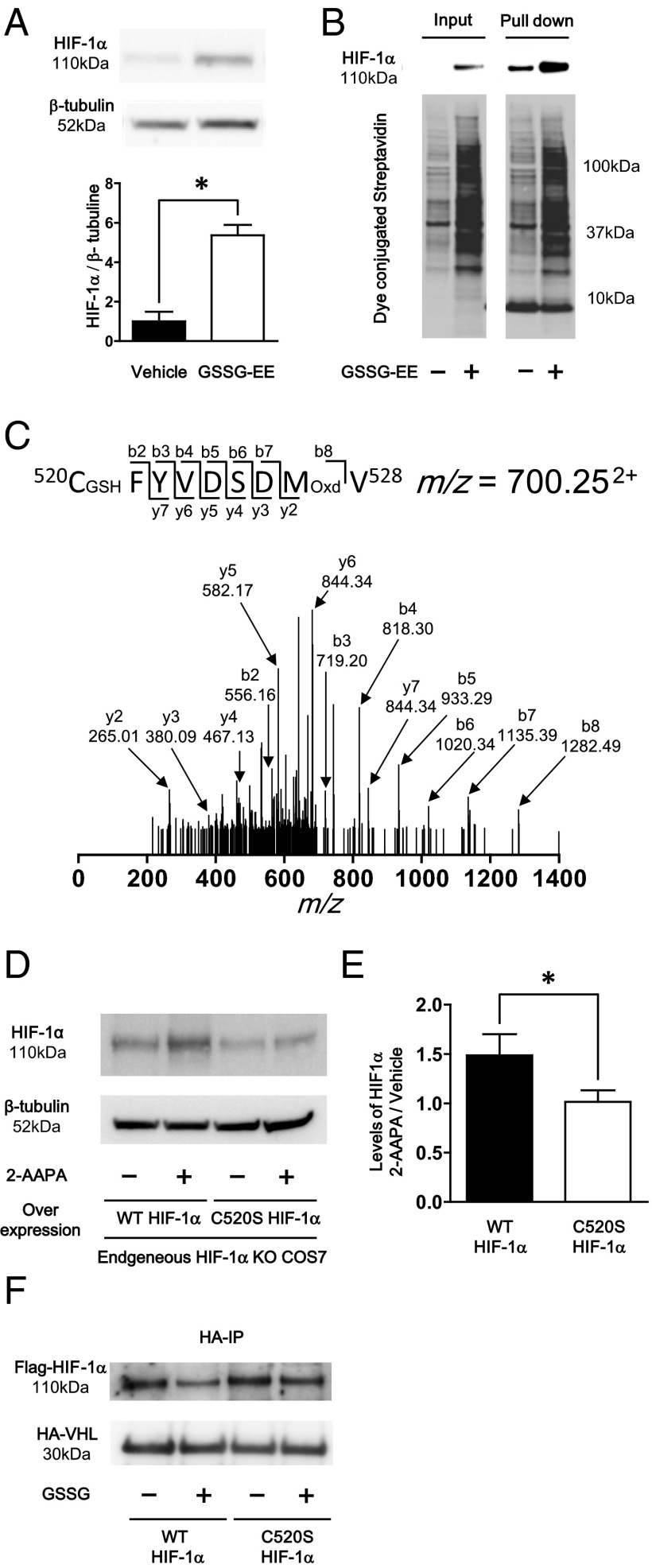
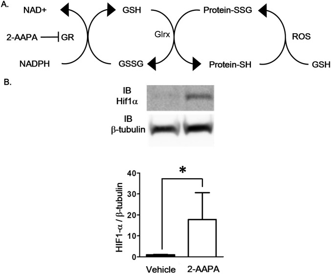
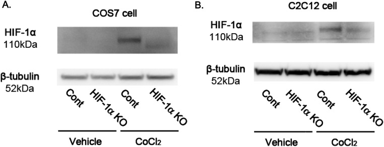
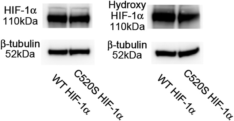
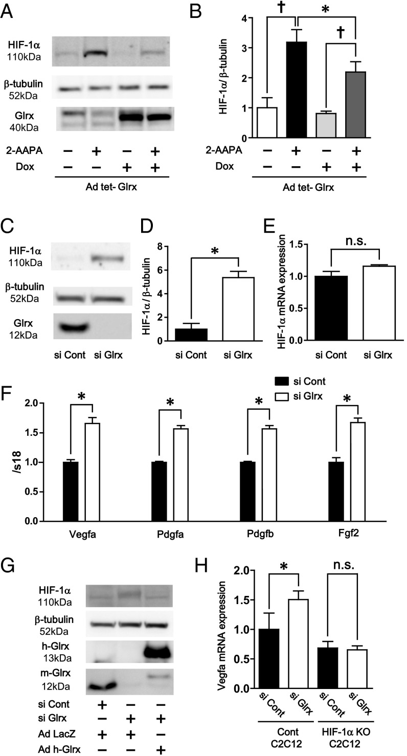
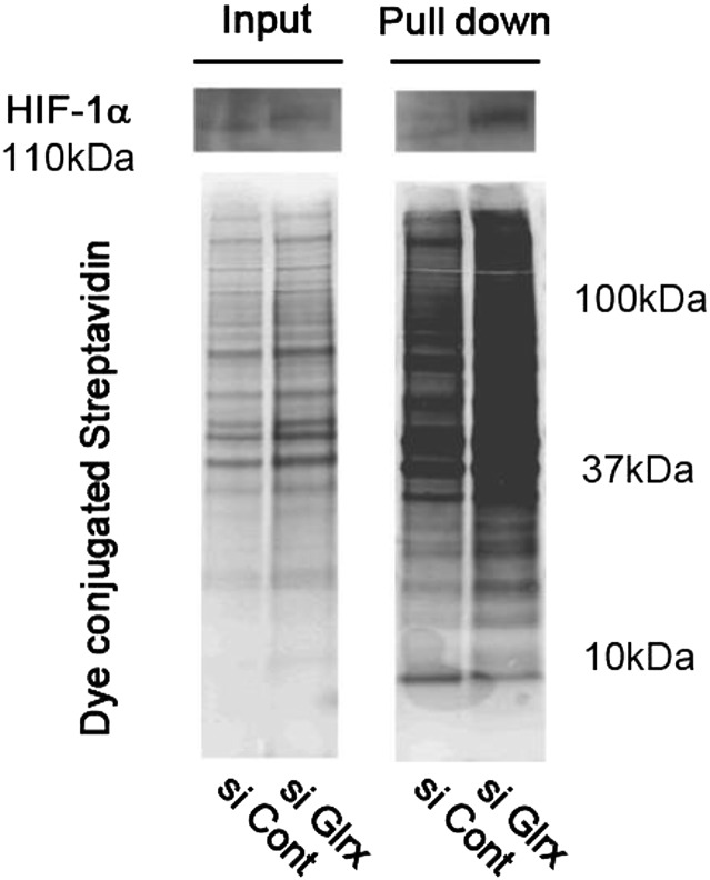
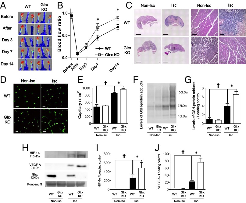
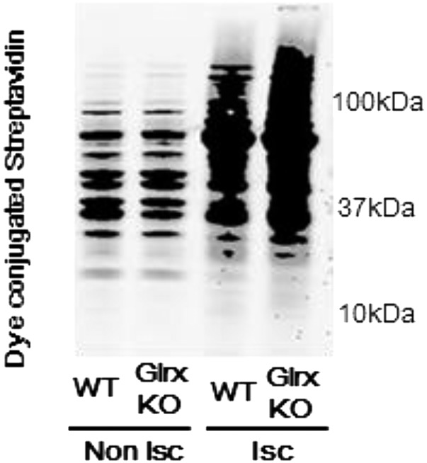
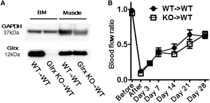
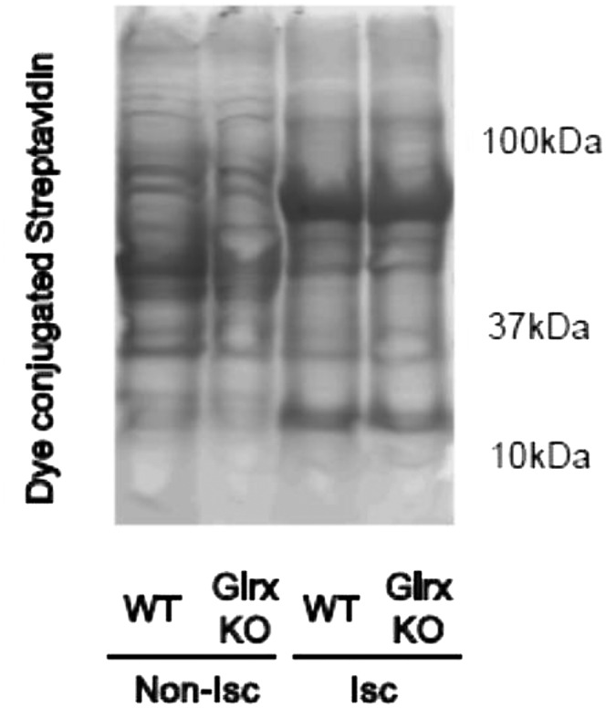
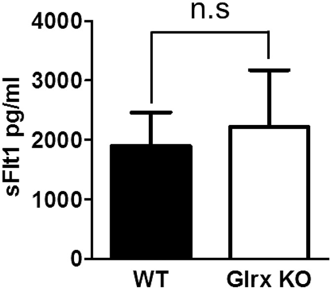
Reactive oxygen species (ROS) are increased in ischemic tissues and necessary for revascularization; however, the mechanism remains unclear. Exposure of cysteine residues to ROS in the presence of glutathione (GSH) generates GSH-protein adducts that are specifically reversed by the cytosolic thioltransferase, glutaredoxin-1 (Glrx). Here, we show that a key angiogenic transcriptional factor hypoxia-inducible factor (HIF)-1α is stabilized by GSH adducts, and the genetic deletion of Glrx improves ischemic revascularization. In mouse muscle C2C12 cells, HIF-1α protein levels are increased by increasing GSH adducts with cell-permeable oxidized GSH (GSSG-ethyl ester) or 2-acetylamino-3-[4-(2-acetylamino-2-carboxyethylsulfanyl thiocarbonylamino) phenylthiocarbamoylsulfanyl] propionic acid (2-AAPA), an inhibitor of glutathione reductase. A biotin switch assay shows that GSSG-ester-induced HIF-1α contains reversibly modified thiols, and MS confirms GSH adducts on Cys(520) (mouse Cys(533)). In addition, an HIF-1α Cys(520) serine mutant is resistant to 2-AAPA-induced HIF-1α stabilization. Furthermore, Glrx overexpression prevents HIF-1α stabilization, whereas Glrx ablation by siRNA increases HIF-1α protein and expression of downstream angiogenic genes. Blood flow recovery after femoral artery ligation is significantly improved in Glrx KO mice, associated with increased levels of GSH-protein adducts, capillary density, vascular endothelial growth factor (VEGF)-A, and HIF-1α in the ischemic muscles. Therefore, Glrx ablation stabilizes HIF-1α by increasing GSH adducts on Cys(520) promoting in vivo HIF-1α stabilization, VEGF-A production, and revascularization in the ischemic muscles.

Keywords: GSH-protein adducts; S-glutathionylation; glutaredoxin-1; hypoxia-inducible factor-1; ischemic limb revascularization.


Conflict of interest statement

The authors declare no conflict of interest.

Figures

Fig. 1.
Fig. 1.
Effect of GSH adduct on HIF-1α stabilization. (A) Effect of GSSG-ethyl-ester on HIF-1α stabilization in C2C12 cells. After differentiation, C2C12 cells were treated with 50 μg GSSG-ethyl ester or PBS for 10 h. Representative Western blot of HIF-1α and β-tubulin (Upper) and densitometry analysis of HIF-1α normalized by β-tubulin (Lower) (n = 8 each group). (B) Biotin switching assay for detection of HIF-1α reversible oxidative modification. DTT-dependent oxidative modified cysteines were labeled with biotin. Then biotin labeled protein was pull-downed using streptavidin beads. (B) Immunoblot of HIF-1α (Upper Left) and total reversible oxidative modified proteins detected by dye conjugated streptavidin (Lower Left) in a total sample. Immunoblot of HIF-1α (Upper Right) and total reversible oxidative modified proteins (Lower Right) in pulled down proteins. (C) Identification of Cys520 GSH adduct by MS. GSH adduct of Cys520 was detected from elastase fragment 520CFYVDSDMV528. The actual mass of this fragment was 1,939.48 (m/z = 700.252+), which was 321 Da more than the original MW. The MS/MS analysis showed that this fragment modified at Cys520 by GSH adduct (+305 Da) and Met527 by oxidation (+16 Da). (D and E) C520S mutation decreased 2-AAPA–dependent stabilization of HIF-1α. Plasmids that included WT and C520S mutant HIF-1α were transfected to COS7 cells in which endogenous HIF-1α was deleted by CRISPR/Cas9. These cells were treated with 20 μmol/L 2-AAPA for 3 h. (D) Representative Western blot of HIF-1α and β-tubulin. (E) Densitometry analysis, data were expressed as HIF-1α induction ratio of 2-AAPA–treated cells to respective vehicle-treated cells (n = 4 each group). (F) Western blotting analysis following Co IP of HA-VHL overexpressed cell lysate and GSSG-treated Flag-tagged WT or C520S mutant HIF-1α–overexpressed cell lysate by anti-HA antibody. Detection of Flag-tagged HIF-1α and HA-tagged VHL was performed by anti-Flag antibody and anti-VHL antibody, respectively. Experiments were repeated three times with similar results. *P < 0.05.
Fig. S1.
Fig. S1.
Effects of 2-AAPA on HIF-1α induction. (A) GSH-protein adduct (Protein-SSG) formation by reactive oxygen species (ROS) and oxidized glutathione (GSSG) and removal by glutaredoxin (Glrx) system are shown. GR, glutathione reductase. (B) HIF-1α stabilization after 2-AAPA treatment. Differentiated C2C12 cells were treated with the GR inhibitor 2-AAPA (20 mmol/L) for 3 h. Representative Western blotting of HIF-1α and β-tubulin in cell lysate (Upper) and densitometry analysis (Lower) (n = 8 each group, *P < 0.05).
Fig. S2.
Fig. S2.
HIF-1α knockdown in COS7 and C2C12 cells by CRISPR/Cas9. pSpCas9(BB)-2A-Puro (PX459) plasmids, which contain a guiding RNA sequence for green monkey or mouse HIF-1α, were transfected to COS7 cells or C2C12 cells. After selection by Puromycin, cells were expanded. To analyze the difference of HIF-1α protein expression in control plasmid-transfected cells and gRNA-inserted plasmid-transfected cells, HIF-1α was inducted by CoCl2 in COS7 cells (A) or C2C12 cells (B), and levels of protein were analyzed by Western blotting.
Fig. S3.
Fig. S3.
Levels of HIF-1α and hydoxy-HIF-1α in WT or C520S HIF-1α–expressed cell lysate. WT or C520S HIF-1α plasmid was transfected to HEK293T cells. These transfected cells were lysed 2 d after transfection, and levels of total HIF-1α and hydroxy HIF-1α were analyzed by Western blotting.
Fig. 2.
Fig. 2.
Regulation of HIF-1α and angiogenic genes by Glrx. (A and B) Glrx overexpression decreased 2-AAPA–dependent HIF-1α stabilization. Ad tet-Glrx was transfected to COS7 cells. After Glrx overexpression was induced by 1 μg doxycycline, these cells were treated with 2-AAPA. (A) Representative Western blot of HIF-1α, β-tubulin, and Glrx. (B) Densitometry analysis of HIF-1α normalized by β-tubulin (n = 4 each group). Glrx knockdown increased HIF-1α and angiogenic genes expression. (C and D) Glrx knockdown by siGlrx increased HIF-1α and angiogenic genes in C2C12 cells. (C) Representative Western blot of HIF-1α, β-tubulin, and Glrx. (D) Densitometry analysis of HIF-1α (n = 8 each group). (E) Relative mRNA expression of HIF-1α, (F) mRNA of Vegfa, Pdgfa, Pdgfb, and Fgf2 (n = 6 each group) in siGlrx-treated C2C12 cells. (G) Overexpression of human Glrx effect on HIF-1α stabilization by Glrx knockdown. Glrx knockdowned C2C12 cells by siGlrx were transfected with ad human Glrx, which is resistant to siGlrx. Levels of HIF-1α, human, and mouse Glrx were analyzed by Western blotting. (H) Effect of HIF-1α knockdown on increment of mRNA levels of Vegfa by siGlrx. HIF-1α was knocked down by CRISPR/Cas9 in C2C12 cells. Then effect of siGlrx on mRNA expression of Vegfa was analyzed by qPCR. P < 0.05, compared with respective vehicle-treated cells, *P < 0.05.
Fig. S4.
Fig. S4.
Biotin switch assay of the Glrx knockdown cell. Glrx knockdown in the C2C12 cell was performed by siGlrx. After C2C12 cells were differentiated, these cells were lysed, and the biotin switch assay was performed. Experiments were repeated three times with similar results. Biotin-labeled proteins were separated on SDS/PAGE in a nonreducing condition and detected by dye-conjugated streptavidin or anti–HIF-1α antibody.
Fig. 3.
Fig. 3.
Lack of Glrx improved ischemic revascularization after hind limb ischemia in mice. (A and B) Blood flow after hind limb ischemia was analyzed by LASER Doppler. (A) Representative images and (B) quantitative serial assessment (n = 8 each group). (C) Representative H&E staining of nonischemic (Non-Isc) and ischemic (Isc) gastrocnemius muscles of WT and Glrx KO mice. Squire area in low-power field images (Left) are shown by high-power images (Right). Black bar is 1 mm for low-power images and 100 μm for high-power field images. (D and E) Capillary density measurement in gastrocnemius muscles of WT and Glrx KO mice 7 d after HLI. Capillaries were stained with fluorescein-conjugated isolectin B4. (D) Representative images. (Scale bar, 50 μm.) (E) Graphical analysis (n = 8 each group). (F and G) Levels of protein GSH adducts in nonischemic (Non-Isc) and ischemic (Isc) gastrocnemius muscles of WT and Glrx KO mice were detected by anti-GSH antibody. (F) Representative blot and (G) densitometry analysis. (HJ) Western blot for HIF-1α, VEGF-A, and Glrx expression in muscles. (H) Representative blot, (I) densitometry analysis of HIF-1α, and (J) densitometry analysis of VEGF-A. Ponceau S staining showed equal loading of proteins and used as loading control to normalize protein expression (n = 4 each group). P < 0.05 compared with nonischemic limbs; *P < 0.05.
Fig. S5.
Fig. S5.
Biotin switch assay of gastrocnemius muscles. The muscles were removed 3 d after hind limb ischemia surgery from WT and Glrx KO mice and processed for the biotin switch assay as described in Materials and Methods. Biotin-labeled proteins were separated on SDS/PAGE in a nonreducing condition and detected by dye-conjugated streptavidin. Nonischemic muscles (Non-Isc) and ischemic muscles (Isc) from each mouse are shown. Experiments were repeated three times with similar results.
Fig. S6.
Fig. S6.
Effects of Glrx deletion in myeloid cells. (A) Western blot showing Glrx expression in bone marrow (BM) and skeletal muscle 7 wk after bone marrow transplantation. (B) Blood flow recovery after hind limb ischemia in recipient mice. Femoral artery ligation surgery was performed 3 wk after bone marrow transplant (n = 8, each group). For bone marrow transplantation, Glrx KO (8 wk old) mice and WT littermate controls were euthanized, and bone marrow was harvested from tibias, femurs, and humeri. Recipient WT mice (9 wk old) were irradiated with 10 Gy. After irradiation, 0.5 × 10⁷ marrow cells were injected through the tail vein of the recipient mice.
Fig. S7.
Fig. S7.
Ascorbate-dependent biotin switch assay after hind limb ischemia. The muscles were removed 3 d after hind limb ischemia surgery from WT and Glrx KO mice and processed for the ascorbate-dependent biotin switch assay for detection of S-nitrosylation. Biotin-labeled proteins were separated on SDS/PAGE and detected by dye-conjugated streptavidin. Nonischemic muscles (Non-Isc) and ischemic muscles (Isc) from each mouse are shown. Experiments were repeated three times with similar results.
Fig. S8.
Fig. S8.
Levels of soluble VEGFR1 (sFlt1) in mouse plasma. Plasma was collected 3 d after hind limb ischemia from WT and Glrx KO mice. Plasma sFlt1 was measured by ELISA (n = 6 in each group).

Similar articles

Cited by

References

    1. Kris-Etherton PM, Lichtenstein AH, Howard BV, Steinberg D, Witztum JL. Nutrition Committee of the American Heart Association Council on Nutrition, Physical Activity, and Metabolism Antioxidant vitamin supplements and cardiovascular disease. Circulation. 2004;110(5):637–641. - PubMed
    1. Tojo T, et al. Role of gp91phox (Nox2)-containing NAD(P)H oxidase in angiogenesis in response to hindlimb ischemia. Circulation. 2005;111(18):2347–2355. - PubMed
    1. Urao N, et al. Critical role of endothelial hydrogen peroxide in post-ischemic neovascularization. PLoS One. 2013;8(3):e57618. - PMC - PubMed
    1. Craige SM, et al. NADPH oxidase 4 promotes endothelial angiogenesis through endothelial nitric oxide synthase activation. Circulation. 2011;124(6):731–740. - PMC - PubMed
    1. Ichihara S, et al. Ablation of the transcription factor Nrf2 promotes ischemia-induced neovascularization by enhancing the inflammatory response. Arterioscler Thromb Vasc Biol. 2010;30(8):1553–1561. - PubMed

Publication types

MeSH terms