. 2016 May 24;113(21):5904-5909.
doi: 10.1073/pnas.1604984113. Epub 2016 May 9.

Endogenous B-ring oxysterols inhibit the Hedgehog component Smoothened in a manner distinct from cyclopamine or side-chain oxysterols

Affiliations

Endogenous B-ring oxysterols inhibit the Hedgehog component Smoothened in a manner distinct from cyclopamine or side-chain oxysterols

Navdar Sever et al. Proc Natl Acad Sci U S A. .

Abstract

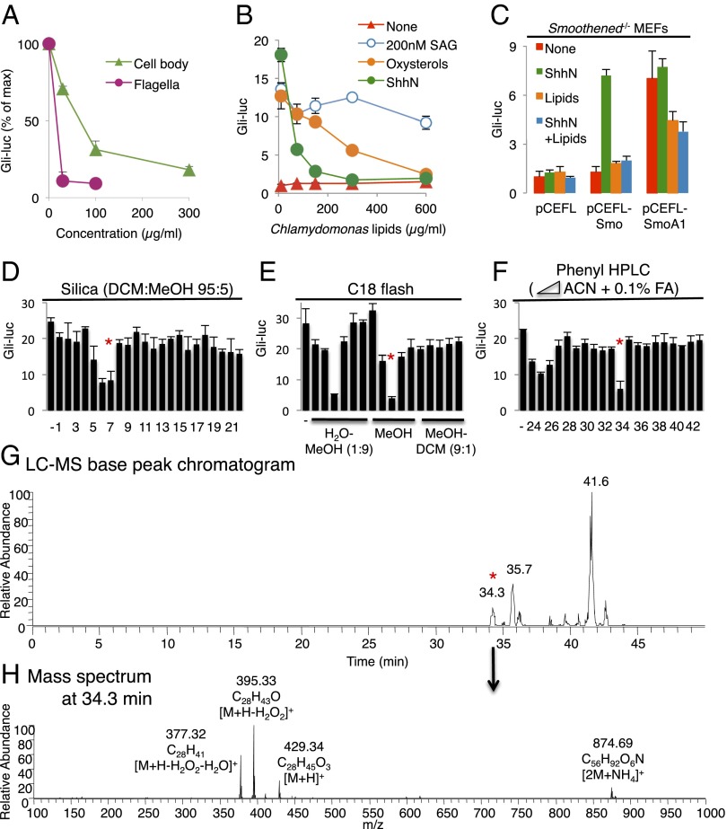
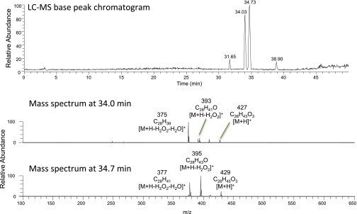
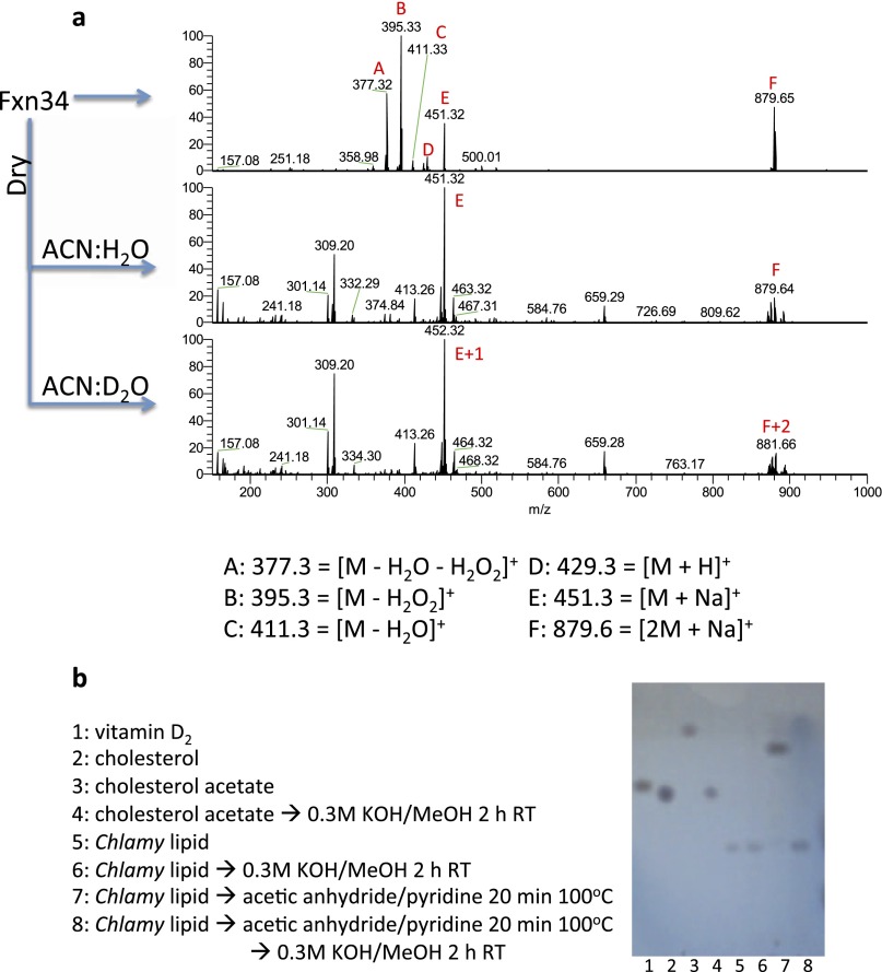
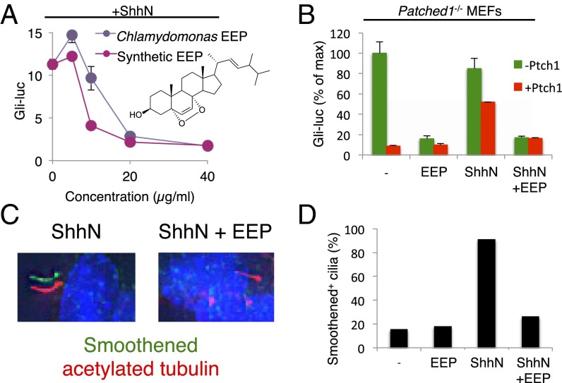
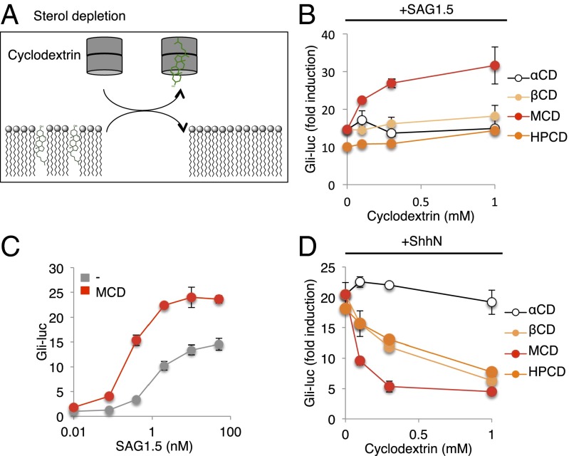
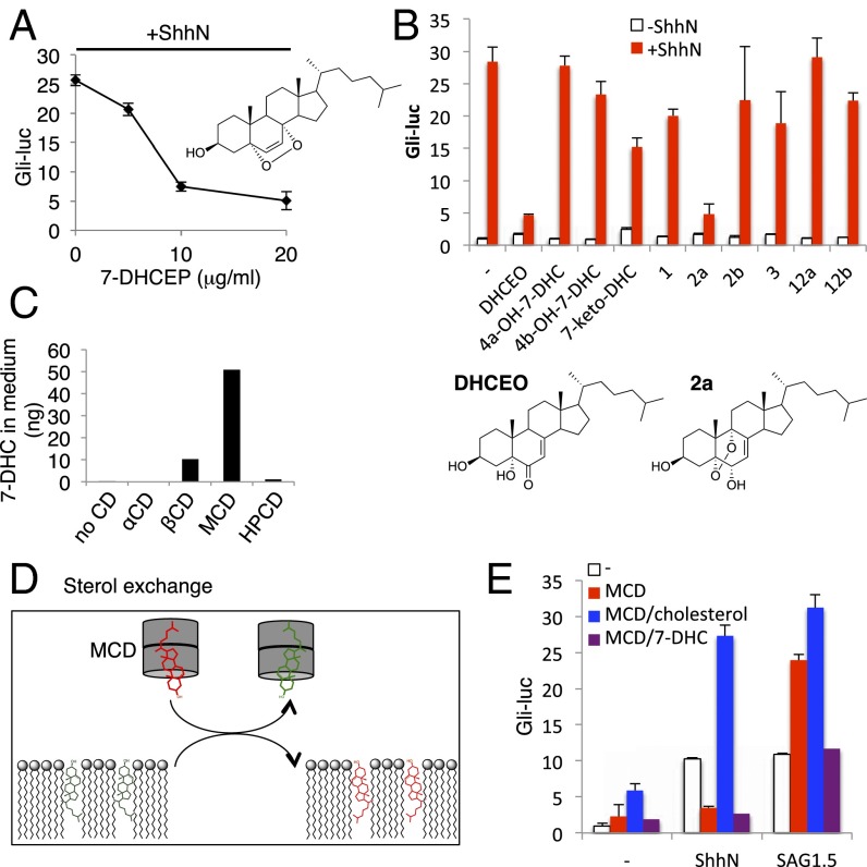
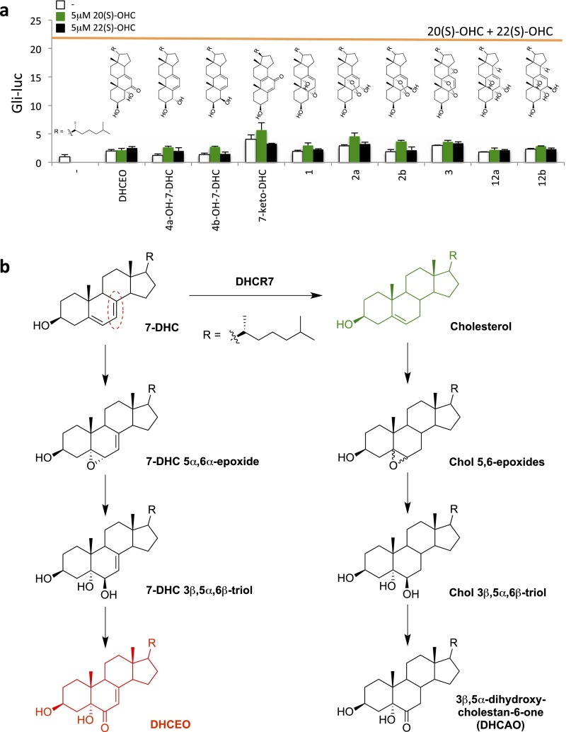
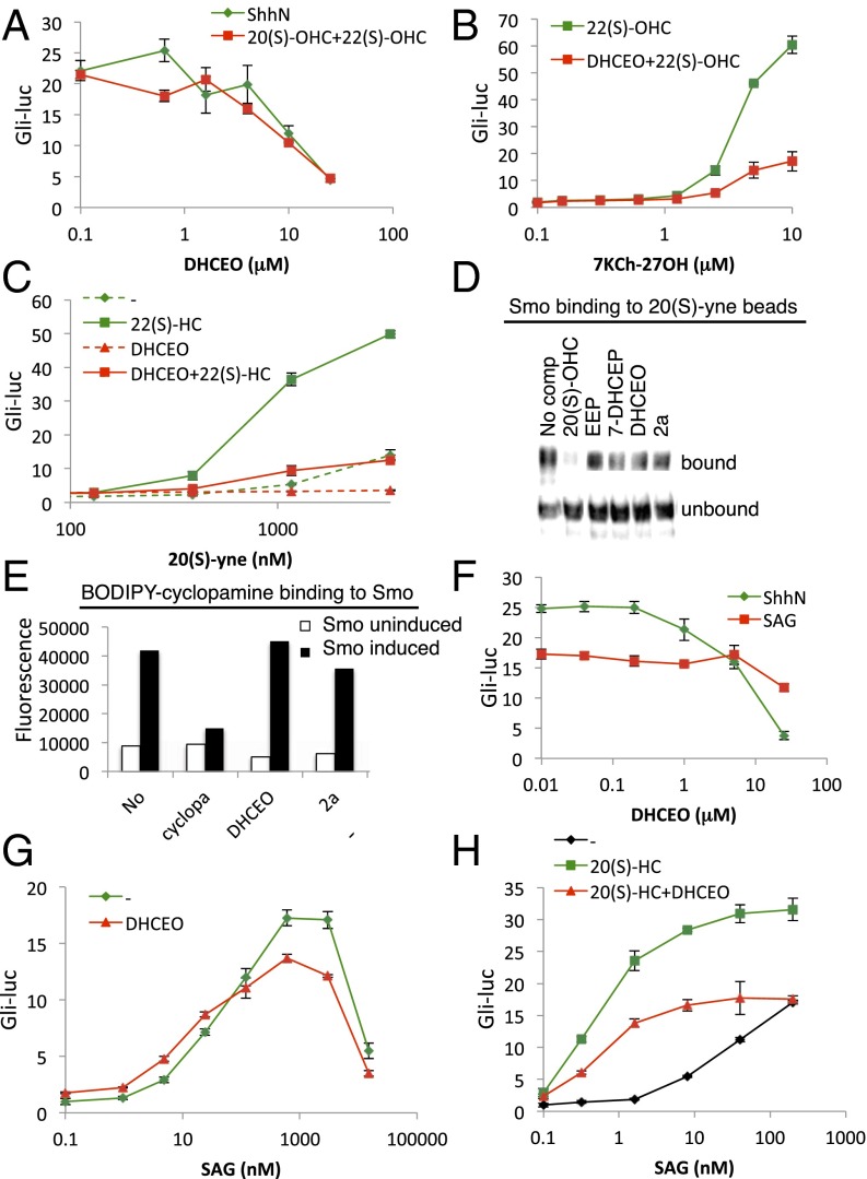
Cellular lipids are speculated to act as key intermediates in Hedgehog signal transduction, but their precise identity and function remain enigmatic. In an effort to identify such lipids, we pursued a Hedgehog pathway inhibitory activity that is particularly abundant in flagellar lipids of Chlamydomonas reinhardtii, resulting in the purification and identification of ergosterol endoperoxide, a B-ring oxysterol. A mammalian analog of ergosterol, 7-dehydrocholesterol (7-DHC), accumulates in Smith-Lemli-Opitz syndrome, a human genetic disease that phenocopies deficient Hedgehog signaling and is caused by genetic loss of 7-DHC reductase. We found that depleting endogenous 7-DHC with methyl-β-cyclodextrin treatment enhances Hedgehog activation by a pathway agonist. Conversely, exogenous addition of 3β,5α-dihydroxycholest-7-en-6-one, a naturally occurring B-ring oxysterol derived from 7-DHC that also accumulates in Smith-Lemli-Opitz syndrome, blocked Hedgehog signaling by inhibiting activation of the essential transduction component Smoothened, through a mechanism distinct from Smoothened modulation by other lipids.

Keywords: DHCEO; Hedgehog signaling; SLOS; Smoothened; cyclodextrin.


Conflict of interest statement

The authors declare no conflict of interest.

Figures

Fig. 1.
Fig. 1.
Identification of ergosterol endoperoxide as a Chlamydomonas lipid that inhibits mammalian Hedgehog signaling. (A) Gli-luciferase activity in Shh-LIGHT2 cells was measured following treatment with ShhN-conditioned medium in combination with the indicated concentrations of organic extracts from Chlamydomonas cell bodies or flagella. (B) Dose–response analysis of Chlamydomonas whole-cell lipid extract in Shh-LIGHT2 cells with no stimulation (red), stimulation with ShhN-conditioned medium (green), 200 nM SAG (blue) or 5 μM 20(S)-OHC and 5 μM 22(S)-OHC (oxysterols, orange). (C) Gli-luciferase activity in Smoothened−/− MEFs transfected with the indicated Smoothened (Smo) constructs and stimulated with ShhN-conditioned medium and Chlamydomonas lipids as indicated. (D–F) Activity profiles following fractionation of Chlamydomonas lipids using the indicated columns and solvents. (G and H) In-line electrospray ionization–Fourier transform MS analysis, showing masses and formulas of in-source fragmentation products. Red asterisks in D and E indicate fractions that were used in subsequent chromatography steps. The asterisks in F and G indicate co-incidence of activity with a chromatographic peak.
Fig. S1.
Fig. S1.
Column fractionation of Drosophila larval extract yielded a partially purified inhibitory fraction containing two related species with the indicated masses of in-source fragmentation products.
Fig. S2.
Fig. S2.
Chemical characterization of the purified Chlamydomonas lipid. (A) Chlamydomonas lipid has a single exchangeable proton. HPLC-purified Chlamydomonas lipid was infused directly into the mass spectrometer (Top), dried and redissolved in 1:1 acetonitrile:water (ACN:H2O) (Middle), or acetonitrile:deuterium oxide (ACN:D2O) (Bottom). Note that the formic acid from the HPLC eluent that promotes formation of the protonated species evaporates after drying, which leaves the sodium adduct as the only means of ionization. Fragmentation of m/z 452 species first caused a neutral loss of 19, confirming that a hydroxyl proton had exchanged with a deuterium (not shown). (B) Chlamydomonas lipid is not an ester. Even though conventional saponification at 80 °C destroyed the purified species and Hedgehog pathway-inhibitory activity (not shown), room temperature saponification under the indicated conditions did not (lanes 5 and 6). As a control, cholesterol acetate (lanes 3 and 4) and preacetylated Chlamydomonas lipid (lanes 7 and 8) were saponified and analyzed by TLC.
Fig. 2.
Fig. 2.
Ergosterol endoperoxide (EEP) inhibits Hedgehog pathway downstream of Patched1 and at the level of Smoothened. (A) Dose–response analysis of EEP purified from Chlamydomonas (purple) or synthesized by photooxidation (red). (B) Gli-luciferase activity in Patched1−/− MEFs transfected with or without Patched1 and treated with ShhN-conditioned medium and 25 μM EEP as indicated. (C and D) Ciliary accumulation of endogenous Smoothened in NIH 3T3 cells treated with ShhN-conditioned medium and 25 μM EEP as indicated. Fixed cells were analyzed by immunofluorescence using antibodies against Smoothened (green) and acetylated tubulin (red) with DAPI counterstain (blue). Representative images for selected conditions are displayed as shifted overlays of Smoothened and acetylated tubulin stains. (Magnification: 63×.)
Fig. S3.
Fig. S3.
Synthesis and purification of EEP and 7-DHCEP. (A) TLC analysis of ergosterol (lane 1), 7-DHC (lane 2), photooxidation products of ergosterol (lane 3), and 7-DHC (lane 4). (B) Photooxidation products of ergosterol and 7-DHC were fractionated using flash chromatography and analyzed by TLC. Major reaction products were found in pure forms in fractions 3 (shown in red) and were analyzed further (Figs. S4 and S5).
Fig. S4.
Fig. S4.
LC-MS analyses of purified EEP (A) and 7-DHCEP (B).
Fig. S5.
Fig. S5.
1H NMR spectra of purified EEP (A) and 7-DHCEP (B).
Fig. S6.
Fig. S6.
Chlamydomonas mutants have sterol endoperoxides with side chains distinct from wild-type. Extracted ion chromatograms of partially purified lipid extracts for the indicated m/z values (Top), mass spectra at the indicated retention times (Middle), and predicted structures of the active species (Bottom). AD indicate spectra corresponding to each of the proposed side chain structures.
Fig. 3.
Fig. 3.
MCD potentiates SAG. (A) Scheme describing use of cyclodextrins to remove cellular sterols. Gli-luciferase activity in Shh-LIGHT2 cells was measured following treatment with (B) 10 nM SAG1.5 or (D) ShhN-conditioned medium and increasing concentrations of α-cyclodextrin (αCD), β-cyclodextrin (βCD), MCD, or hydroxypropyl-β-cyclodextrin (HPCD). (C) Gli-luciferase activity in Shh-LIGHT2 cells was measured following treatment with increasing concentrations of SAG1.5 alone (gray) or together with 1 mM MCD (red).
Fig. 4.
Fig. 4.
Endogenous 7-DHC derivatives inhibit Hedgehog signaling. (A) Dose–response analysis of 7-DHC endoperoxide (7-DHCEP) in Shh-LIGHT2 cells. (B) 7-DHC–derived oxysterols inhibit Hedgehog pathway. Gli-luciferase activity in Shh-LIGHT2 cells was measured following treatment with the indicated sterols either alone (white) or in combination with ShhN-conditioned medium (red). (C) 7-DHC levels in the medium after treatment with the indicated cylodextrins were quantified by tandem MS. (D) Scheme describing use of MCD to exchange cellular sterols. (E) Gli-luciferase activity in Shh-LIGHT2 cells was measured following treatment with ShhN-conditioned medium or 10 nM SAG1.5 and 1 mM MCD complexed with 100 μM cholesterol or 7-DHC.
Fig. S7.
Fig. S7.
Lack of stimulatory activity of 7-DHC derivatives and formation mechanism of DHCEO. (A) Gli-luciferase activity in Shh-LIGHT2 cells was measured following treatment with the indicated sterols (described in ref. 33) alone (white) or in combination with 5 μM 20(S)-OHC (green) or 5 μM 22(S)-OHC (black). For comparison, luciferase induction obtained with 5 μM 20(S)-OHC plus 5 μM 22(S)-OHC (21.4-fold) is shown with the orange line. (B) DHCEO is proposed to form in multiple steps from 7-DHC analogous to a known biotransformation of cholesterol. Note that the double bond reduced by DHCR7 (circled in red) does not undergo a net reaction during DHCEO formation.
Fig. 5.
Fig. 5.
DHCEO can block the action of CRD agonists but not SAG. (A–C) Gli-luciferase activity in Shh-LIGHT2 cells was measured following treatment with: (A) increasing concentrations of DHCEO in combination with ShhN-conditioned medium (green) or 5 μM 20(S)-OHC plus 5 μM 22(S)-OHC (red); (B) increasing concentrations of 7KCh-27OH in combination with 5 μM 22(S)-OHC (green) or 5 μM 22(S)-OHC plus 25 μM DHCEO (red); or (C) increasing concentrations of 20(S)-yne alone (dashed green) or in combination with 5 μM 22(S)-OHC (solid green), 25 μM DHCEO (dashed red), or 5 μM 22(S)-OHC plus 25 μM DHCEO (solid red). (D) Detergent-solubilized membranes from HEK293S-Smo cells were incubated with 20(S)-yne affinity resin in the presence of 50 μM 20(S)-OHC, 250 μM ergosterol endoperoxide (EEP), 250 μM 7-DHC endoperoxide (7-DHCEP), 250 μM DHCEO, or 100 μM 2a. After washing, bound protein was eluted and analyzed by immunoblotting. (E) Binding of 5 nM BODIPY-cyclopamine to membranes from tetracycline-inducible HEK293S cells expressing Smoothened was measured in the presence of 3 μM cyclopamine, 25 μM DHCEO, or 10 μM 2a. (F–H) Gli-luciferase activity in Shh-LIGHT2 cells was measured following treatment with increasing concentrations of DHCEO in combination with ShhN-conditioned medium (green) or 200 nM SAG (red); (G) increasing concentrations of SAG alone (green) or in combination with 25 μM DHCEO (red); or (H) increasing concentrations of SAG alone (black) or in combination with the indicated sterols.

Similar articles

Cited by

References

    1. Briscoe J, Thérond PP. The mechanisms of Hedgehog signalling and its roles in development and disease. Nat Rev Mol Cell Biol. 2013;14(7):416–429. - PubMed
    1. Chiang C, et al. Cyclopia and defective axial patterning in mice lacking Sonic hedgehog gene function. Nature. 1996;383(6599):407–413. - PubMed
    1. Roessler E, et al. Mutations in the human Sonic hedgehog gene cause holoprosencephaly. Nat Genet. 1996;14(3):357–360. - PubMed
    1. Belloni E, et al. Identification of Sonic hedgehog as a candidate gene responsible for holoprosencephaly. Nat Genet. 1996;14(3):353–356. - PubMed
    1. Taipale J, Cooper MK, Maiti T, Beachy PA. Patched acts catalytically to suppress the activity of Smoothened. Nature. 2002;418(6900):892–897. - PubMed

Publication types

MeSH terms