

Deformation of a Titanium Calvarial Implant following Trauma: A Case Report
- PMID: 27162574
- PMCID: PMC4858418
- DOI: 10.1055/s-0035-1567810
Deformation of a Titanium Calvarial Implant following Trauma: A Case Report
Abstract
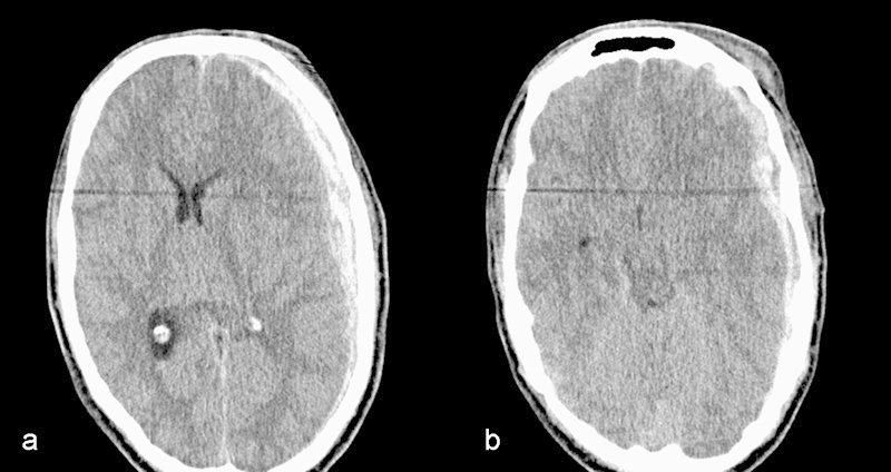
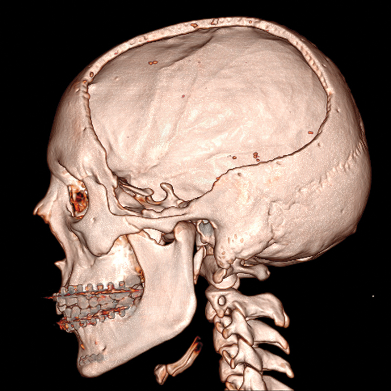
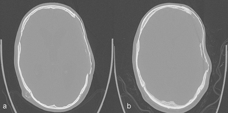
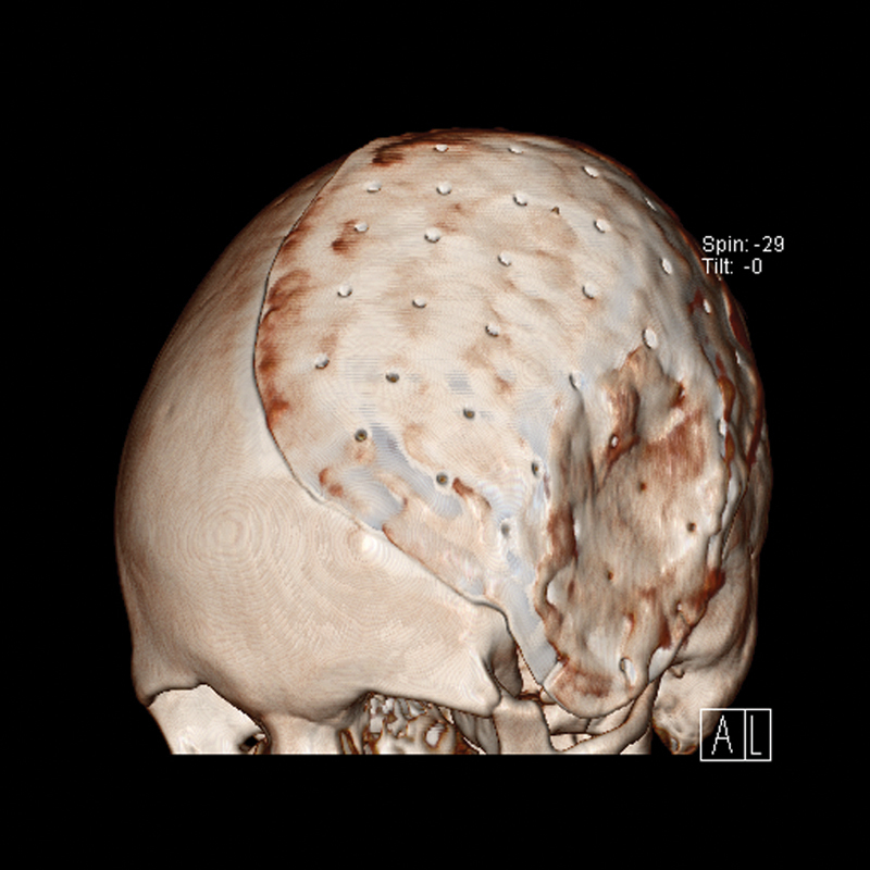
Alloplastic material is widely used for the reconstruction of calvarial defects. The objective of this article is to describe the effect of mechanical impact on a titanium calvarial implant and to discuss mechanical properties of alternative implant materials. The patient is a 19-year-old man who was involved in a traffic accident and underwent decompressive craniectomy for an extensive subdural hematoma. Reimplantation of the skull flap was complicated by infection and the flap had to be removed. The remaining cranial defect was closed with a titanium plate. The recovery was without complications. One year later, the patient was hit on the titanium plate, during a soccer match, by the elbow of a fellow player. The implant deflected inward, leaving a visible indentation of the cranial vault. Fortunately, there were no significant neurological symptoms and radiography did not show any signs of damage or pressure on the brain parenchyma. The patient had no aesthetic complaints regarding the shape. Thus, there was no indication to remove the plate. This case illustrates the limits of the protection offered by titanium cranioplasty.
Keywords: alloplastic material; calvarial defect; cranioplasty; implant failure.
Figures
Similar articles
-
Efficacy and Versatility of the 3-D Titanium Mesh Implant in the Closure of Large Post-Craniectomy Osseous Defects, and its Therapeutic Role in Reversing the Syndrome of the Trephined: Clinical Study of a Case Series and Review of Literature.J Maxillofac Oral Surg. 2016 Mar;15(1):82-92. doi: 10.1007/s12663-015-0807-0. Epub 2015 May 26. J Maxillofac Oral Surg. 2016. PMID: 26929558 Free PMC article.
-
Calvarial reconstruction using high-density porous polyethylene cranial hemispheres.Indian J Plast Surg. 2011 Sep;44(3):422-31. doi: 10.4103/0970-0358.90812. Indian J Plast Surg. 2011. PMID: 22279274 Free PMC article.
-
Reconstruction of Large Calvarial Defects Using Titanium Mesh Versus Autologous Split Thickness Calvarial Bone Grafts: A Comprehensive Comparative Evaluation of the Two Major Cranioplasty Techniques.J Maxillofac Oral Surg. 2018 Sep;17(3):308-323. doi: 10.1007/s12663-017-1047-2. Epub 2017 Sep 27. J Maxillofac Oral Surg. 2018. PMID: 30034149 Free PMC article.
-
Titanium cranioplasty in children and adolescents.J Craniomaxillofac Surg. 2016 Jul;44(7):789-94. doi: 10.1016/j.jcms.2016.03.010. Epub 2016 Apr 4. J Craniomaxillofac Surg. 2016. PMID: 27174495 Review.
-
Custom-made titanium cranioplasty: early and late complications of 151 cranioplasties and review of the literature.Int J Oral Maxillofac Surg. 2015 May;44(5):599-608. doi: 10.1016/j.ijom.2014.09.006. Epub 2014 Dec 5. Int J Oral Maxillofac Surg. 2015. PMID: 25482456 Review.
Cited by
-
Design and Additive Manufacturing of a Biomimetic Customized Cranial Implant Based on Voronoi Diagram.Front Physiol. 2021 Apr 9;12:647923. doi: 10.3389/fphys.2021.647923. eCollection 2021. Front Physiol. 2021. PMID: 33897455 Free PMC article.
-
Deformation of cranioplasty titanium mesh in a paediatric patient following head trauma.BMJ Case Rep. 2019 Jun 11;12(6):e230421. doi: 10.1136/bcr-2019-230421. BMJ Case Rep. 2019. PMID: 31189547 Free PMC article.
References
-
- Honeybul S, Ho K M. Long-term complications of decompressive craniectomy for head injury. J Neurotrauma. 2011;28(6):929–935. - PubMed
-
- Akins P T, Guppy K H. Sinking skin flaps, paradoxical herniation, and external brain tamponade: a review of decompressive craniectomy management. Neurocrit Care. 2008;9(2):269–276. - PubMed
-
- Dujovny M, Aviles A, Agner C, Fernandez P, Charbel F T. Cranioplasty: cosmetic or therapeutic? Surg Neurol. 1997;47(3):238–241. - PubMed
-
- Honeybul S. Complications of decompressive craniectomy for head injury. J Clin Neurosci. 2010;17(4):430–435. - PubMed
Publication types
LinkOut - more resources
Full Text Sources
Other Literature Sources