

Mucin-Inspired Thermoresponsive Synthetic Hydrogels Induce Stasis in Human Pluripotent Stem Cells and Human Embryos
- PMID: 27163030
- PMCID: PMC4827554
- DOI: 10.1021/acscentsci.5b00370
Mucin-Inspired Thermoresponsive Synthetic Hydrogels Induce Stasis in Human Pluripotent Stem Cells and Human Embryos
Abstract
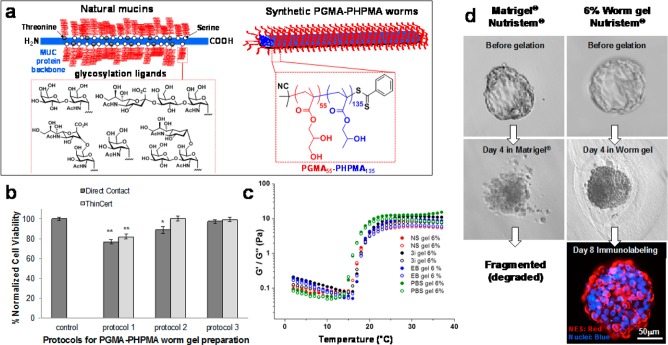
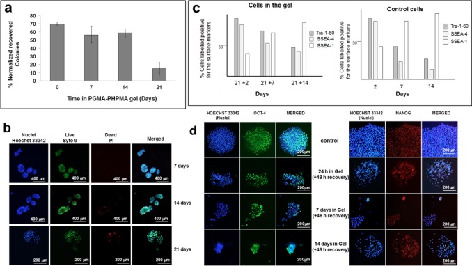
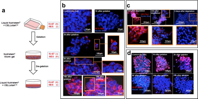
Human pluripotent stem cells (hPSCs; both embryonic and induced pluripotent) rapidly proliferate in adherent culture to maintain their undifferentiated state. However, for mammals exhibiting delayed gestation (diapause), mucin-coated embryos can remain dormant for days or months in utero, with their constituent PSCs remaining pluripotent under these conditions. Here we report cellular stasis for both hPSC colonies and preimplantation embryos immersed in a wholly synthetic thermoresponsive gel comprising poly(glycerol monomethacrylate)-poly(2-hydroxypropyl methacrylate) [PGMA55-PHPMA135] diblock copolymer worms. This hydroxyl-rich mucin-mimicking nonadherent 3D gel maintained PSC viability and pluripotency in the quiescent G0 state without passaging for at least 14 days. Similarly, gel-coated human embryos remain in a state of suspended animation (diapause) for up to 8 days. The discovery of a cryptic cell arrest mechanism for both hPSCs and embryos suggests an important connection between the cellular mechanisms that evoke embryonic diapause and pluripotency. Moreover, such synthetic worm gels offer considerable utility for the short-term (weeks) storage of either pluripotent stem cells or human embryos without cryopreservation.
Conflict of interest statement
The authors declare no competing financial interest.
Figures
Similar articles
-
Probing the mechanism for hydrogel-based stasis induction in human pluripotent stem cells: is the chemical functionality of the hydrogel important?Chem Sci. 2019 Nov 11;11(1):232-240. doi: 10.1039/c9sc04734d. Chem Sci. 2019. PMID: 34040716 Free PMC article.
-
Site-Directed Differentiation of Human Adipose-Derived Mesenchymal Stem Cells to Nucleus Pulposus Cells Using an Injectable Hydroxyl-Functional Diblock Copolymer Worm Gel.Biomacromolecules. 2021 Feb 8;22(2):837-845. doi: 10.1021/acs.biomac.0c01556. Epub 2021 Jan 20. Biomacromolecules. 2021. PMID: 33470795
-
Engineered peptide modified hydrogel platform for propagation of human pluripotent stem cells.Acta Biomater. 2020 Sep 1;113:228-239. doi: 10.1016/j.actbio.2020.06.034. Epub 2020 Jun 27. Acta Biomater. 2020. PMID: 32603868
-
Molecular Regulation of Paused Pluripotency in Early Mammalian Embryos and Stem Cells.Front Cell Dev Biol. 2021 Jul 27;9:708318. doi: 10.3389/fcell.2021.708318. eCollection 2021. Front Cell Dev Biol. 2021. PMID: 34386497 Free PMC article. Review.
-
X chromosome inactivation in human pluripotent stem cells as a model for human development: back to the drawing board?Hum Reprod Update. 2017 Sep 1;23(5):520-532. doi: 10.1093/humupd/dmx015. Hum Reprod Update. 2017. PMID: 28582519 Review.
Cited by
-
Mucus-Inspired Dynamic Hydrogels: Synthesis and Future Perspectives.J Am Chem Soc. 2022 Nov 9;144(44):20137-20152. doi: 10.1021/jacs.1c13547. Epub 2022 Sep 8. J Am Chem Soc. 2022. PMID: 36074739 Free PMC article. Review.
-
Proline pre-conditioning of Jurkat cells improves recovery after cryopreservation.RSC Med Chem. 2023 Jul 27;14(9):1704-1711. doi: 10.1039/d3md00274h. eCollection 2023 Sep 19. RSC Med Chem. 2023. PMID: 37731697 Free PMC article.
-
Polymer Self-Assembly Induced Enhancement of Ice Recrystallization Inhibition.J Am Chem Soc. 2021 May 19;143(19):7449-7461. doi: 10.1021/jacs.1c01963. Epub 2021 May 4. J Am Chem Soc. 2021. PMID: 33944551 Free PMC article.
-
Can percolation theory explain the gelation behavior of diblock copolymer worms?Chem Sci. 2018 Aug 2;9(35):7138-7144. doi: 10.1039/c8sc02406e. eCollection 2018 Sep 21. Chem Sci. 2018. PMID: 30310636 Free PMC article.
-
Poly(l-proline)-Stabilized Polypeptide Nanostructures via Ring-Opening Polymerization-Induced Self-Assembly (ROPISA).ACS Macro Lett. 2024 Aug 20;13(8):1031-1036. doi: 10.1021/acsmacrolett.4c00400. Epub 2024 Jul 29. ACS Macro Lett. 2024. PMID: 39074359 Free PMC article.
References
LinkOut - more resources
Full Text Sources
Other Literature Sources
