

Distal radius anatomy applied to the treatment of wrist fractures by plate: a review of recent literature
- PMID: 27163070
- PMCID: PMC4849245
- DOI: 10.1051/sicotj/2015012
Distal radius anatomy applied to the treatment of wrist fractures by plate: a review of recent literature
Abstract
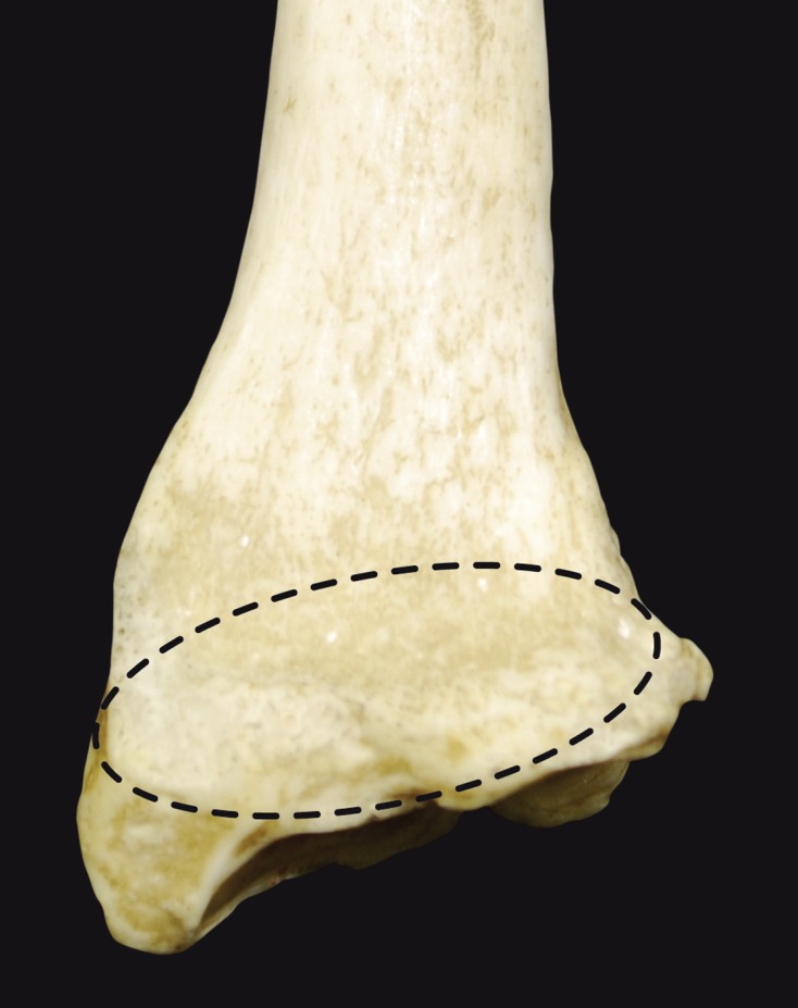
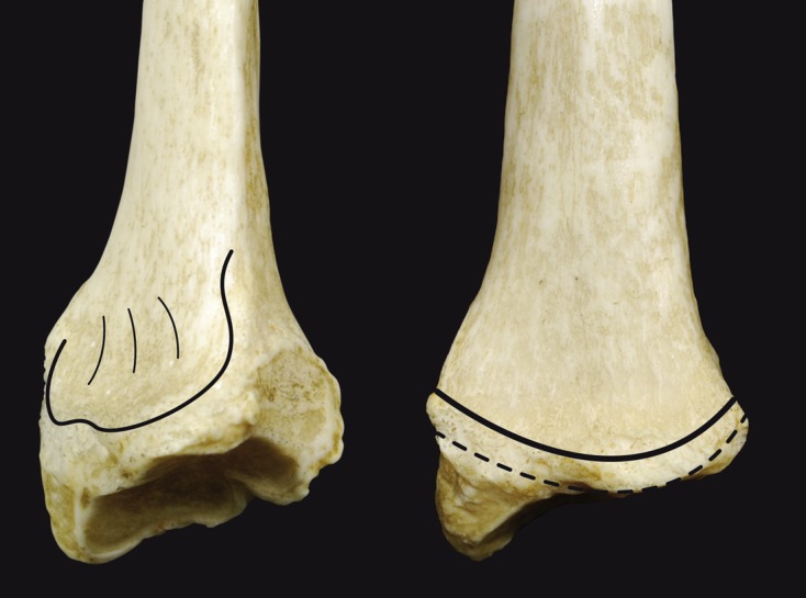
Few studies on the anatomy of the radial epiphysis have been published in the past 10 years. However, with the availability of new intra- and extra-medullary implants and the recent rash of avoidable iatrogenic injuries, now is the time for a more detailed description of the metaphyseal-epiphyseal regions in the distal radius. Published studies on distal radius anatomy in recent years have focused on three aspects: distal limit and watershed line, dorsal tubercle, and wrist columns. Furthermore, a fresh look at distal radius biomechanics shows that the loads experienced by the distal radius vary greatly. This information should be taken into account during volar plating of distal radius fractures.
Keywords: Anatomy; Distal radius; Volar plate.
Figures


References
-
- Obert L et al. (2012) Anatomy and biomechanics of distal radius fractures: a literature review. Chir Main 31(6), 287–297. - PubMed
-
- Herzberg G et al. (1998) Anatomie du radius distal. Cahiers d’enseignement de la SOFCOT. Paris, Expansion scientifique Publications, 14–27.
-
- Windisch G et al. (2001) Capsular attachment to the distal radius for extracapsular placement of pins. Surg Radiol Anat 23(5), 313–316. - PubMed
-
- Windisch G et al. (2007) Promontory of radius: a new anatomical description on the distal radius. Surg Radiol Anat 29(8), 629–633. - PubMed
-
- Nelson D (2013) Anatomy notes and their clinical significance for the volar approach By David L. Nelson, MD, http://www.davidlnelson.md/articles/Radius_Anatomy_Annotated.htm.
Publication types
LinkOut - more resources
Full Text Sources
Other Literature Sources
Miscellaneous