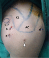
Osteoid osteoma (OO) of the coracoid: a case report of arthroscopic excision and review of literature
- PMID: 27163073
- PMCID: PMC4849236
- DOI: 10.1051/sicotj/2015016
Osteoid osteoma (OO) of the coracoid: a case report of arthroscopic excision and review of literature
Abstract
Osteoid osteoma (OO) of the coracoid is a rare entity that may present with variable symptoms from shoulder leading to delay in diagnosis and treatment. We present the clinical and radiological findings and management of one such case along with a review of similar cases reported in the literature. There was a delay of 2 years in diagnosis, which was later confirmed by computed tomography in addition to magnetic resonance imaging (MRI). The lesion was accessed arthroscopically and excised by unroofing and curettage. "OO" should be included in the differential diagnosis of shoulder pain in young patients not responding to long-term conservative treatment. Arthroscopic excision and curettage provide a good choice for management, with low morbidity and rapid recovery.
Keywords: Arthroscopic excision; Coracoid; Osteoid osteoma; Shoulder; Technique.
Figures


References
-
- Jaffe HL (1935) Osteoid osteoma of bone. Radiology 45, 319.
-
- Dorfman HD, Czerniak B (1998) Benign osteoblastic tumors, in Bone tumors. Gery L, Editor St. Louis, Mosby; pp. 85–104.
-
- Mosheiff R et al. (1991) Osteoid osteoma of the scapula: a case report and review of the literature. Clin Orthop 262, 129–131. - PubMed
-
- Kaempffe FA (1994) Osteoid osteoma of the coracoid process. Excision by posterior approach. A case report. Clin Orthop Relat Res 301, 260–262. - PubMed
-
- Ogose A, Sim FH, O’Connor MI, Unni KK (1999) Bone tumors of the coracoid process of the scapula. Clin orthop 358, 205–214. - PubMed
LinkOut - more resources
Full Text Sources
Other Literature Sources
