Review
. 2015 Oct 16:1:27.
doi: 10.1051/sicotj/2015027.

Hip arthroscopy and osteoarthritis: Where are the limits and indications?

Affiliations
Review

Hip arthroscopy and osteoarthritis: Where are the limits and indications?

Claudio Mella et al. SICOT J. .

Abstract

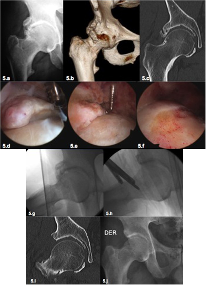
The use of hip arthroscopy, as a surgical technique, has increased significantly over the past ten years. The procedure has shown good and excellent results in symptom relief and function improvement for patients with femoro-acetabular impingement (FAI) and concurrent chondro-labral lesions. It is also a reliable method to correct the characteristic pathomorphologic alteration of FAI. However, surgical results are less successful among patients with advanced articular damage and secondary hip osteoarthritis. The aim of this article is to present some clinical and imagenological tools to discriminate the good candidates for arthroscopic FAI treatment from those who are not, due to extensive articular damage.

Keywords: Arthroscopy; Hip; Indication; Osteoarthritis.


Figures

Figure 1.
Figure 1.
(1a–1d) Overview of arthroscopic progression of chondral damage. In early stages of joint damage, a chondro-labral instability (chondromalacia, 1a) occurs in the anterolateral part of the acetabulum. The impact of the femoral bump leads to a disruption at the chondro-labral union (1b) or to degenerative lesions of the labrum. With the progression of the joint damage, chondral flaps were created toward the central area of the acetabulum (1c and 1d). Until these flaps do not reach the load-bearing surface, these damages can be considered early stages of osteoarthritis and satisfactory outcomes with hip arthroscopy can be expected. (1e and 1f) More advanced stages of osteoarthritis lead to thinning and ulceration of the cartilage in the acetabulum load-bearing surface (1e, white arrow), progression of degenerative cartilage damage of the femoral head (1e black arrow), and formation of osteophytes (1f). These advanced lesions should be considered beyond effective treatment with hip arthroscopy.
Figure 2.
Figure 2.
Male patient, 23 years old, medical student and amateur rugby player with sports-related pain in the right hip. The radiology demonstrated a FAI with a significant CAM deformity without significant joint space narrowing (2a, 2b). MRI showed a labral injury, a focal chondral lesion with subchondral cysts, and subchondral edema (2c). In spite of these ominous imaging signs given his young age and no other signs of osteoarthritis, hip arthroscopy was performed. At the arthroscopy an extensive full-thickness chondral lesion was found in the load-bearing surface of the acetabulum (2d); no femoral head cartilage lesions were present. The chondral lesion was treated by abrasive chondroplasty (2e) and microfractures (2f). In the peripheral compartment the extensive femoral bump (2g) was resected (2h). Intraoperative dynamic testing at the end of the femoroplasty demonstrated absence of impingement; the axial radiography demonstrated a satisfactory correction of the cam deformity (2i). Nevertheless after this arthroscopic repair, the future of this joint is uncertain with a high risk to progress to osteoarthritis in this young patient.
Figure 3.
Figure 3.
Male patient, age 32 years, intensive recreational athlete (triathlon, mountain climbing) with left hip pain in flexion and rotational movements. Clinical examination demonstrates a restriction of internal rotation in flexion above 90° associated with pain (anterior impingement). The radiograph demonstrates a FAI with a cam deformity and joint space narrowing in the peripheral joint area (3a–3c). The MRI showed a chondrolabral lesion and acetabular subchondral cyst (3d). Considering his young age and his rejection to a THR a hip arthroscopy was performed. Full-thickness chondral damage in acetabulum (3e) and diffuse fibrillation of cartilage in femoral head was found during the arthroscopy (3f). These should be considered an advanced osteoarthritis finding with a very unpredictable clinical outcome. The radiography one year later shows the progression of the chondral damage of the hip (3g). This case shows that with hip arthroscopy a temporary relief of pain can be achieved in cases of advanced chondral damage but it cannot change the course of the disease and progression to osteoarthritis of the hip.
Figure 4.
Figure 4.
Male patient, 32-year old physician, intensive recreational sports (Karate), 4 years after left hip arthroscopy for FAI with persistent pain and restriction of movement (ROM). Despite the evident radiological signs of osteoarthritis (4a, 4b) the patient insisted on a repeat arthroscopy due to the pain and limitations for sports. Considering the young age and the conserved joint space in the load-bearing area, a revision arthroscopy was performed. During the hip arthroscopy the chondrolabral damage was treated in the central compartment. In the peripheral compartment the osteophytes were identified and resected (4c) as well as the cam deformity. A satisfactory correction of the deformity was achieved (4d–4f). Even in this case with a satisfactory anatomic correction and a satisfactory clinical short-term result, the long-term outcome is still unknown with a high risk of progression to osteoarthritis.
Figure 5.
Figure 5.
(5a–5f) Male patient, 65 years with an advanced osteoarthritis of the hip with a significant ROM (5a). The patient and his wife (orthopedic surgeon) refused the proposed treatment with a total joint replacement insisting to perform a hip arthroscopy. The complemental imaging studies with CAT Scan demonstrate the extensive osteophytes (5b) with conservation of the joint space in the load-bearing area (5c). After a long discussion with the patient without creating false expectations, a hip arthroscopy was performed. The osteophytes in the peripheral compartment were resected and the acetabular labrum was debrided (5d, 5e). The acetabular rim was resected as well as the existing cam deformity (5f). (5g–5j) The same patient from Figures 5a–5f. The intraoperative radioscopy demonstrated the satisfactory resection of the osteophytes and the bone deformity (5g, 5h). The postoperative CAT scan (51) and the postoperative X-ray (5j) demonstrated also a satisfactory result. Five years after this surgery the patient has still an excellent clinical outcome (HHS 92) without progression of osteoarthritis. This case demonstrates that we still do not know where exactly are the limits for hip arthroscopy in cases with osteoarthritis. It demonstrated also that in advanced pincer cases with arthritis due to the resection of the deformity, good results can be achieved (5i, 5j). Independent of this satisfactory clinical result, in cases like these a total joint replacement (TJR) is the most effective and predictable option of treatment but it was refused by the patient.

References

    1. Kemp JL, MacDonald D, Collins NJ, Hatton AL, Crossley KM (2015) Hip arthroscopy in the setting of hip osteoarthritis: systematic review of outcomes and progression to hip arthroplasty. Clin Orthop Relat Res 473(3), 1055–1073. - PMC - PubMed
    1. Daivajna S, Bajwa A, Villar R (2015) Outcome of arthroscopy in patients with advanced osteoarthritis of the hip. PLoS One 10(1), e0113970. - PMC - PubMed
    1. Shearer DW, Kramer J, Bozic KJ, Feeley BT (2012) Is hip arthroscopy cost-effective for femoroacetabular impingement? Clin Orthop Relat Res 470(4), 1079–1089. - PMC - PubMed
    1. Mella C, del Rio J, Lara J, Parodi D, Moya L, Schmidt-Hebbel A et al. (2012) Arthroscopy after hip joint injury. Case studies and indications. Der Unfallchirurg 115(3), 273–278. - PubMed
    1. Ayeni OR, Wong I, Chien T, Musahl V, Kelly BT, Bhandari M (2012) Surgical indications for arthroscopic management of femoroacetabular impingement. Arthroscopy 28(8), 1170–1179. - PubMed

LinkOut - more resources