. 2016 May 10;14(5):e1002457.
doi: 10.1371/journal.pbio.1002457. eCollection 2016 May.

GPCRs Direct Germline Development and Somatic Gonad Function in Planarians

Affiliations

GPCRs Direct Germline Development and Somatic Gonad Function in Planarians

Amir Saberi et al. PLoS Biol. .

Abstract

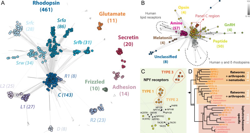
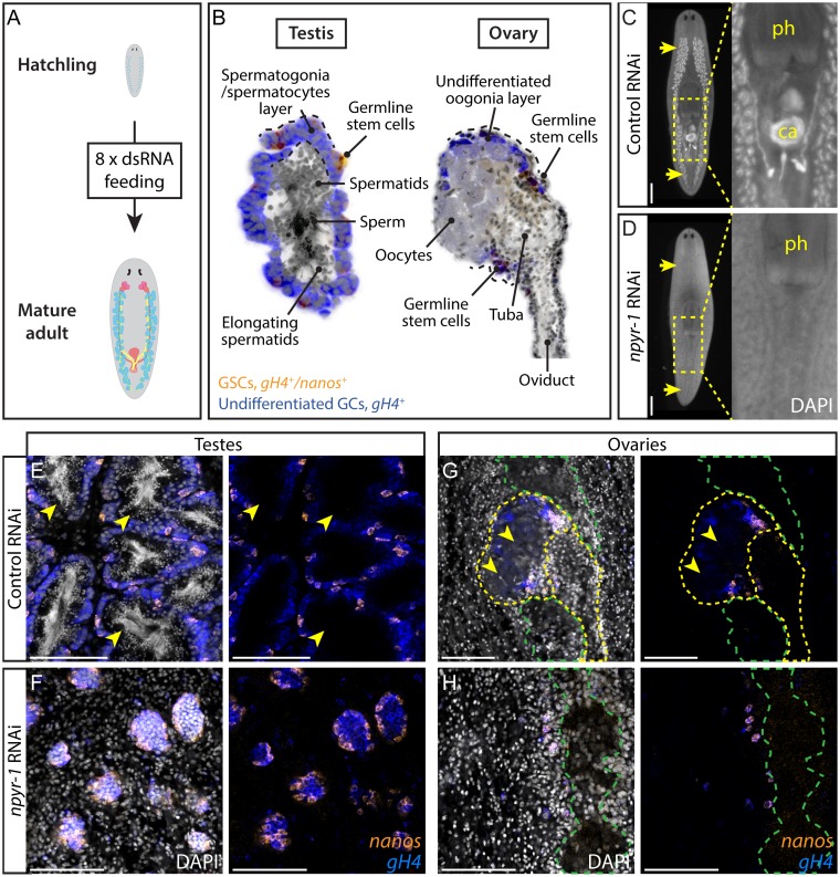
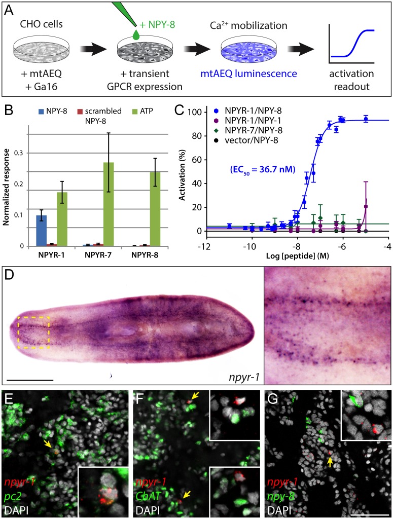
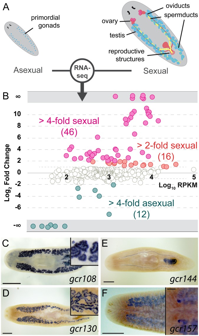
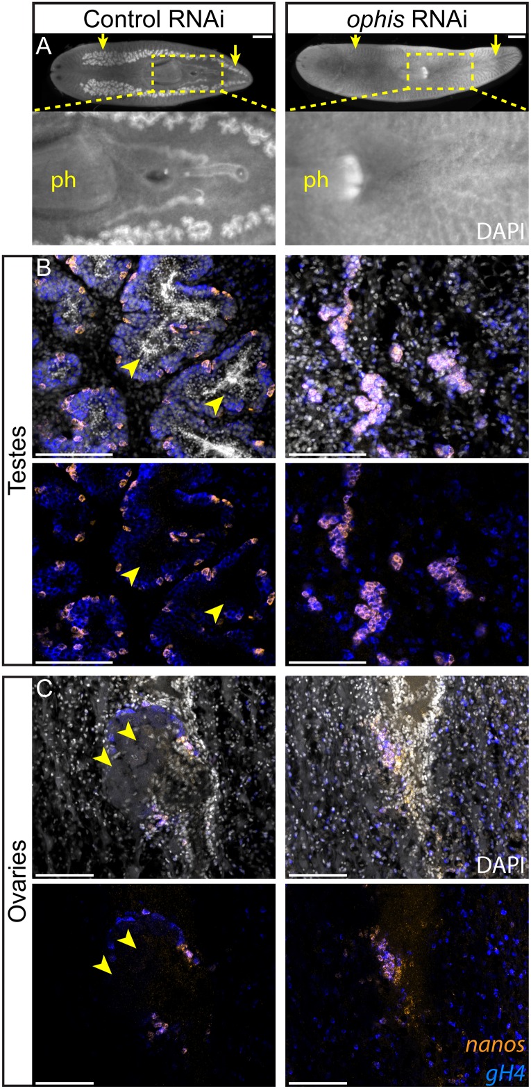
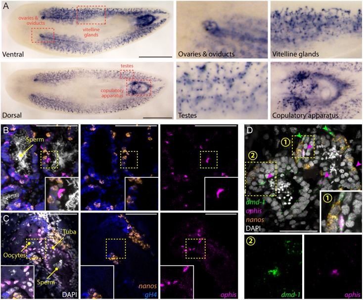
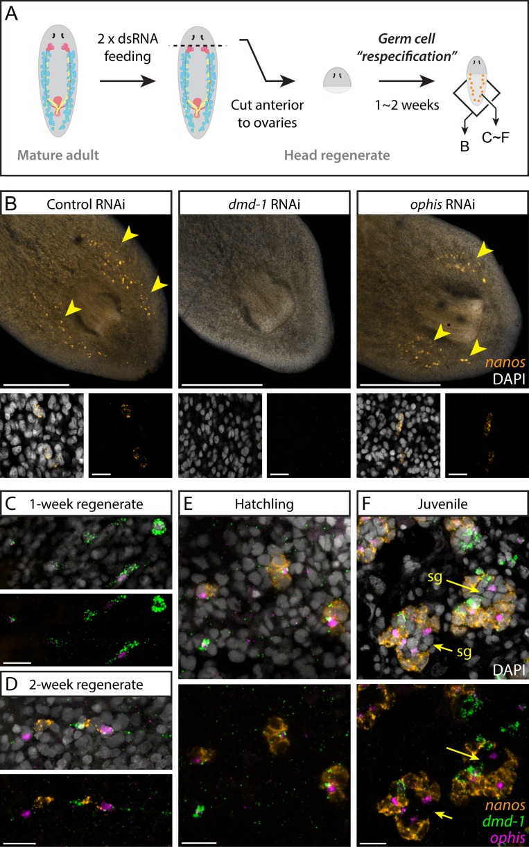
Planarians display remarkable plasticity in maintenance of their germline, with the ability to develop or dismantle reproductive tissues in response to systemic and environmental cues. Here, we investigated the role of G protein-coupled receptors (GPCRs) in this dynamic germline regulation. By genome-enabled receptor mining, we identified 566 putative planarian GPCRs and classified them into conserved and phylum-specific subfamilies. We performed a functional screen to identify NPYR-1 as the cognate receptor for NPY-8, a neuropeptide required for sexual maturation and germ cell differentiation. Similar to NPY-8, knockdown of this receptor results in loss of differentiated germ cells and sexual maturity. NPYR-1 is expressed in neuroendocrine cells of the central nervous system and can be activated specifically by NPY-8 in cell-based assays. Additionally, we screened the complement of GPCRs with expression enriched in sexually reproducing planarians, and identified an orphan chemoreceptor family member, ophis, that controls differentiation of germline stem cells (GSCs). ophis is expressed in somatic cells of male and female gonads, as well as in accessory reproductive tissues. We have previously shown that somatic gonadal cells are required for male GSC specification and maintenance in planarians. However, ophis is not essential for GSC specification or maintenance and, therefore, defines a secondary role for planarian gonadal niche cells in promoting GSC differentiation. Our studies uncover the complement of planarian GPCRs and reveal previously unappreciated roles for these receptors in systemic and local (i.e., niche) regulation of germ cell development.


Conflict of interest statement

The authors have declared that no competing interests exist.

Figures

Fig 1
Fig 1. Global view of planarian GPCRs and the NPY receptor family.
(A) Similarity clustering of 566 planarian GPCRs revealing members of all five metazoan GPCR families. Only GPCRs of the convex clusters are counted and highlighted by filled circles (see Materials and Methods). (B) Co-clustering of conserved planarian and human rhodopsin-like GPCRs reveals large groups of amine and peptide receptors, as well as smaller groups of receptors with other functionalities. Planarians appear to lack homologs of human γ and δ rhodopsins or lipid receptors. Planarian Rho-C and human receptors are shown by filled circles and grey crossmarks, respectively. Darker edges indicate higher similarity (lower p-value) between nodes. p-value scale shown at top-right corner. Area in dashed box is expanded in panel C. (C) Magnified view of the similarity network around NPY receptors (dashed box in B). Three groups of planarian receptors (16 total) are located adjacent to human NPY receptors. (D) Bayesian inference topology of candidate planarian NPY receptors (orange) and parasitic flatworm homologs (pink). Arrow indicates the root. Posterior probabilities are 1.00 at every node, except those with a value shown. Three monophyletic groups of NPY receptors are found that are parallel to groups identified by similarity clustering in panel C. Type 1 is conserved across flatworms, arthropods, and nematodes, type 2 receptors are found in flatworms and arthropods, and type 3 receptors are lophotrochozoan-specific. The complete phylogenetic analysis is shown in S1E Fig. See S1 and S2 Figs for more information on the planarian GPCR complement.
Fig 2
Fig 2. NPY receptor npyr-1 is required for germ cell maturation.
(A) Post-embryonic development RNAi paradigm used to determine the function of genes during normal planarian growth. Hatchlings (≤2 wk old) were fed dsRNA corresponding to each gene eight times to ensure that control worms achieve sexual maturity. (B) Schematic showing planarian testis and ovary structures. In both testes and ovaries, nanos+/gH4+ GSCs (orange) and nanos-/gH4+ spermatogonia/oogonia (blue) are located on the periphery, while more differentiated spermatids, sperm, or oocytes (grey) are in the middle of the gonads. (C, D) DAPI staining showing testes and stored sperm in whole-mount samples. Insets show the copulatory apparatus region. Pharynx and copulatory apparatus are marked by “ph” and “ca”, respectively. RNAi treatment followed the paradigm in A. Control worms (C) develop a complete reproductive system, while npyr-1(RNAi) worms (D) lack developed reproductive tissues and mature gametes. n = 5/5 for each of the three npyr-1 clones and control. RNAi quantification data can be found in S4 Data. (E–H) Double-FISH labeling GSCs (nanos+/gH4+, orange) and spermatogonia (nanos-/gH4+, blue) in whole-mount control and npyr-1(RNAi) samples. GSCs and spermatogonial cells are present in both conditions. Testis germ cells differentiate into sperm and spermatids (arrowheads) in control worms (E), but not in npyr-1(RNAi) worms (F). In ovaries, mature oocytes with large cytoplasm (arrowheads) are surrounded by gH4+ oogonia in control planarians (G), but are absent in npyr-1(RNAi) animals (H). DAPI (grey) labels nuclei. Yellow dashed lines indicate ovaries and oviducts in G and H. Green dashed lines indicate cephalic ganglia near the ovaries. Scale bars are 1 mm in C and D and 100 μm in E–H. See also S3 Fig.
Fig 3
Fig 3. NPY-8 targets npyr-1+ neuroendocrine cells in the CNS.
(A) Schematic of receptor-activation assay performed in CHO/mtAEQ/G16 cells. Candidate GPCRs were expressed transiently in a cell line that enables visualization of calcium mobilization upon receptor activation. (B) Normalized activation response of CHO cells expressing NPYR-1 or control receptors, challenged with NPY-8 or control peptides. NPY-8 specifically activates NPYR-1, while two other NPY receptors (NPYR-7 and NPYR-8) are not activated upon NPY-8 treatment. Scrambled NPY-8 was used as a negative control. Calcium responses were normalized to the total calcium response after addition of 0.1% Triton X-100. ATP, which activates an endogenous CHO receptor, was used to test the functionality of the assay. Peptides and ATP were tested at 10 and 1 μM, respectively. (C) Concentration-response curves for the activation of NPYR-1 by NPY-8 and control peptides. Data are shown as a percentage of the highest normalized response of the concentration series. NPY-8 activates NPYR-1 at EC50 = 36.7 nM (blue). Closely related NPY-1 fails to activate NPYR-1 (purple). Empty pcDNA3.1 vector and NPYR-7 were used as negative controls. Error bars in B and C represent standard error of the mean (SEM) (n ≥ 4). The underlying data for receptor assays can be found in S4 Data. (D) Colorimetric ISH showing expression of npyr-1 in a subset of cells in the brain and along the ventral nerve cords. Inset shows the brain region at higher magnification. (E–G) Double-FISH labeling npyr-1 (red) and other neural markers (green). npyr-1+ cells express neuroendocrine cell marker pc2 (E, 28/30 express pc2) but not the cholinergic neuronal marker ChAT (F, 0/30 express ChAT). npy-8 and npyr-1 are expressed in distinct populations of cells (G, 0/30 npyr-1 cells express npy-8 and 0/30 npy-8 cells express npyr-1). DAPI (grey) labels nuclei. Scale bars are 1 mm in D and 50 μm in E–G.
Fig 4
Fig 4. A subset of planarian GPCRs is enriched in reproductive tissues.
(A) Schematic of the reproductive system in the two biotypes of S. mediterranea. Sexual planarians (right) develop a complete reproductive system, including mature gonads and accessory reproductive organs. The asexuals (left) contain only presumptive gonads with PGCs. (B) Normalized RNA-seq RPKM ratios between sexual and asexual planarians plotted against relative abundance of each GPCR gene. Only data points with p-value < 0.05 are shown. (C–F) Representative colorimetric ISH experiments used to validate RNA-seq results (n = 24/27 genes tested expressed in sexual organs). Sexually enriched genes are expressed in various reproductive tissues, including spermatids (C), spermatogonia (D and F), oviducts and female copulatory apparatus (E), and ovaries (F). Scale bars are 1 mm. Insets in C and D show the area inside the dashed box. Inset in F shows the ventral side and the scale bar is 200 μm. See also S4 Fig and S3 Data.
Fig 5
Fig 5. ophis is required for male and female germ cell differentiation.
(A) Whole-mount DAPI staining of control and ophis(RNAi) worms shows testes (yellow arrows) and stored sperm (inset). Control animals (n = 5/5) possess a mature reproductive system, including all differentiated cell types of the testes and ovaries, ophis(RNAi) worms (n = 15/15, three independent experiments) lack gonads and mature gametes. RNAi treatment (8 dsRNA feedings) started in hatchlings (see Fig 2A for the dsRNA feeding paradigm). RNAi quantification data can be found in S4 Data. (B and C) Double-FISH labeling GSCs (nanos+, orange) and undifferentiated germ cells (nanos-/gH4+, blue) in control and ophis(RNAi) worms. In testes (B), ophis(RNAi) worms contain only nanos+ GSCs and are devoid of nanos-/gH4+ spermatogonial cells and DAPI-rich spermatids and sperm. Control worms have fully developed testes with spermatids and sperm in the middle of lobes (arrowheads). In ovaries (C), mature oocytes are observed in control animals (arrowheads) but not in ophis(RNAi) worms. See Fig 2B for a schematic representation of the spatial organization of the gonads. DAPI (grey) labels nuclei. Scale bars are 1 mm in A and 100 μm in B and C.
Fig 6
Fig 6. ophis is expressed in the somatic gonadal niche.
(A) Colorimetric ISH shows expression of ophis in somatic reproductive structures. Insets show magnified view of specific tissues indicated by red dashed boxes. (B and C) Triple-FISH labeling ophis (magenta), gH4 (blue), and nanos (orange). Within gonads, ophis expression is exclusive to somatic cells in the periphery of testis lobes (B) and in presumptive follicular cells of the ovaries (C). (D) Triple-FISH labeling ophis (magenta), nanos (orange), and dmd-1 (male somatic gonad cells, green) in the testes. ophis and dmd-1 are co-expressed inside testes (magenta arrowheads). dmd-1+/ophis- cells can be seen outside the testes (green arrowheads). Insets 1 and 2 show magnification of regions indicated by numbered yellow dashed boxes. DAPI (grey) labels nuclei in B–D. Scale bars are 1 mm in A, 100 μm in B and C, and 50 μm in D.
Fig 7
Fig 7. Somatic dmd-1+/ophis+ cells facilitate testis regeneration and development in planarians.
(A) Germ cell re-specification paradigm used to challenge worms to specify a germline de novo. Indicated genes were knocked down in worms prior to amputation. After 1 to 2 wk of posterior regeneration, regenerates were fixed and labeled to detect nanos expression. Schematic shows areas imaged in panel B and panels C–F. (B) FISH labels nanos+ GSCs in 2-wk head regenerates of control, dmd-1(RNAi), and ophis(RNAi) planarians. Unlike dmd-1, ophis is not required for de novo germ cell specification during regeneration of head fragments. Insets show early nanos+ GSCs. (C-F) FISH showing expression of dmd-1, ophis, and nanos during de novo gonad regeneration. At one week post-amputation (C), dmd-1+/ophis- and dmd1+/ophis+ cells are detected at the posterior half of head regenerates. Most worms are devoid of nanos+ germ cells (n = 8/10). At 2 wk (D), nanos+ cell clusters appear adjacent to dmd-1+/ophis+ cells (n = 10/10). Early hatchlings (<2 wk old, E) express clusters of nanos+ cells near dmd-1+/ophis+ somatic cells. No differentiated germ cells (nanos-) are observable within the clusters. In juveniles (F), in addition to all of the previous combinations, testis lobes with more differentiated spermatogonial cells (“sg” and arrows, nanos-) appear in the middle of the clusters. Quantification of the observations in C–F can be found in S4 Data. DAPI (grey) labels nuclei. Scale bars are 500 μm in B and 20 μm in insets and C–F. See also S5 Fig.
Fig 8
Fig 8. Schematic of the developmental mechanisms involved in planarian testis formation.
dmd-1+ cells in both sexual and asexual worms are required for specification of nanos+ GSCs. In sexual planarians, these dmd-1+ cells express ophis, which is required for further differentiation of GSCs into mature gametes. NPY-8 signaling, which occurs in the CNS, systemically promotes later stages of germ cell maturation.

References

    1. Noel SD, Kaiser UB. G protein-coupled receptors involved in GnRH regulation: molecular insights from human disease. Mol Cell Endocrinol. 2011;346: 91–101. 10.1016/j.mce.2011.06.022 - DOI - PMC - PubMed
    1. Tsutsumi M, Zhou W, Millar RP, Mellon PL, Roberts JL, Flanagan CA, et al. Cloning and functional expression of a mouse gonadotropin-releasing hormone receptor. Mol Endocrinol. 1992;6: 1163–1169. - PubMed
    1. Millar RP, Lu ZL, Pawson AJ, Flanagan CA, Morgan K, Maudsley SR. Gonadotropin-releasing hormone receptors. Endocr Rev. 2004;25: 235–275. - PubMed
    1. Cole LW, Sidis Y, Zhang C, Quinton R, Plummer L, Pignatelli D, et al. Mutations in prokineticin 2 and prokineticin receptor 2 genes in human gonadotrophin-releasing hormone deficiency: molecular genetics and clinical spectrum. J Clin Endocrinol Metab. 2008;93: 3551–3559. 10.1210/jc.2007-2654 - DOI - PMC - PubMed
    1. Topaloglu AK, Reimann F, Guclu M, Yalin AS, Kotan LD, Porter KM, et al. TAC3 and TACR3 mutations in familial hypogonadotropic hypogonadism reveal a key role for Neurokinin B in the central control of reproduction. Nat Genet. 2008;41: 354–358. 10.1038/ng.306 - DOI - PMC - PubMed

Publication types

MeSH terms