. 2016 May 10;12(5):e1004918.
doi: 10.1371/journal.pcbi.1004918. eCollection 2016 May.

Enzyme Sequestration as a Tuning Point in Controlling Response Dynamics of Signalling Networks

Affiliations

Enzyme Sequestration as a Tuning Point in Controlling Response Dynamics of Signalling Networks

Song Feng et al. PLoS Comput Biol. .

Abstract

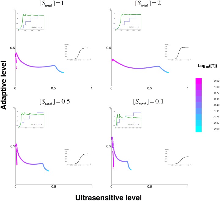
Signalling networks result from combinatorial interactions among many enzymes and scaffolding proteins. These complex systems generate response dynamics that are often essential for correct decision-making in cells. Uncovering biochemical design principles that underpin such response dynamics is a prerequisite to understand evolved signalling networks and to design synthetic ones. Here, we use in silico evolution to explore the possible biochemical design space for signalling networks displaying ultrasensitive and adaptive response dynamics. By running evolutionary simulations mimicking different biochemical scenarios, we find that enzyme sequestration emerges as a key mechanism for enabling such dynamics. Inspired by these findings, and to test the role of sequestration, we design a generic, minimalist model of a signalling cycle, featuring two enzymes and a single scaffolding protein. We show that this simple system is capable of displaying both ultrasensitive and adaptive response dynamics. Furthermore, we find that tuning the concentration or kinetics of the sequestering protein can shift system dynamics between these two response types. These empirical results suggest that enzyme sequestration through scaffolding proteins is exploited by evolution to generate diverse response dynamics in signalling networks and could provide an engineering point in synthetic biology applications.


Conflict of interest statement

The authors have declared that no competing interests exist.

Figures

Fig 1
Fig 1. Starting network topologies in evolutionary simulations.
Structure of three different seed networks labelled as Bipath (a signalling system featuring a branching point), Cascade (a simple, linear signalling system with phosphatases for each step), and Bifunctional (a signalling system featuring a bifunctional protein). The ligand and the output protein (e.g. a transcription factor) are shaped as oval, while all other signalling proteins (e.g. receptor/adaptor proteins, kinases, or phosphatases) are shaped as rectangle. Black line represents binding reaction between two sites. Red arrows represent phosphorylation reactions between a kinase site (red) and a phosphorylation site (purple). Blue arrows represent dephosphorylation reactions between a phosphatase site (blue) and a phosphorylation site.
Fig 2
Fig 2. Analysis of evolved ultrasensitive networks.
(A) Average saturation of enzymes in all of the evolved ultrasensitive networks. The average saturation of enzymes is calculated as the geometric mean of individual Michaelis-Menten constants of the different kinases and phosphatases and their allosteric states normalised by the substrate concentration (kinase, K1, or phosphatase, K2). The shape of each data point represents different starting structures to the evolutionary simulations (see S1 Fig). The colours of the data points represent two different evolutionary scenarios; blue: output protein [Stotal] = 1000, other signalling proteins (A*) concentrations [A*total] = 1; red: output protein [Stotal] = 10, other signalling proteins concentrations [A*total] = 10. The blue and red, star-shaped points indicate the average value of the enzyme saturation resulting from these initial concentrations at the start of the evolutionary simulations. Each data point is further labelled with the unique identification number used for each evolutionary simulation. (B) The fraction of different forms of the kinase (y-axis) against the ligand concentration (x-axis) for two different evolved networks (network 20 and 18 in S2 Fig). The fractions of the different forms of the kinase are the substrate-accessible (green), substrate-inaccessible (orange), and substrate-bound (blue) forms. This data is overlaid with the dose-response dynamics; the solid and dashed lines show the steady state concentration of phosphorylated (i.e. response) and unphosphorylated substrate respectively at a given input level. (C) Ratio between KM values of different conformational states (relaxed “R” state and tensioned “T” states) for kinase (x-axis) and phosphatase (y-axis). The colours, shapes and numbers on the dots are the same as in (A). For enzymes without allosteric regulation the ratio are set to one, so that there are no distinctive conformational differences. (D) The fraction of different forms of the phosphatase (top) and kinase (bottom) (y-axis) against the ligand concentration (x-axis) for two different evolved networks (network 18 and 23 in S2 Fig). The different forms of the enzymes are the different conformational states, relaxed “R” state (green) and tensioned “T” state (orange). These are overlaid with dose-response dynamics; the solid and dashed lines show the steady state concentration of phosphorylated (i.e. response) and unphosphorylated substrate respectively.
Fig 3
Fig 3. Analysis of evolved adaptive networks.
(A) Structure and dynamics of the evolved adaptive network number 1. The upper panel shows a cartoon of the network. The oval shapes represent ligand (top) and the output protein (bottom) (e.g. substrate with a phosphorylation site, S), while rectangles represent all other signalling proteins (e.g. receptors, scaffolds, kinases, or phosphatases). Black lines represent the binding reaction between two sites. Red arrows represent a kinase site (red) phosphorylating a substrate phosphorylation site (purple). Blue arrows represent a phosphatase site (blue) dephosphorylating a substrate site. The green rectangle with a self-pointing green arrow indicates a protein domain whose conformational switching is allosterically regulated. The lower panels show the temporal dynamics of the input ([L], top) and the output protein (black lines in lower panels). The dynamics of the output protein is overlaid with the distribution of the different kinase ([K], middle panels) and phosphatase ([P], bottom panels) complexes: blue for enzyme-substrate complexes, green for free form of the enzymes that are accessible by the substrate, and red for complexes of enzymes with other signalling proteins. (B) Structure and dynamics of the evolved adaptive network number 2. Panels are as in (A).
Fig 4
Fig 4. Designed signalling cycle motif and parameter space for adaptation and ultrasensitivity.
(A) Cartoon showing the designed signalling cycle motif with a sequestering protein. The sequestrating protein (T) binds both the kinase (K) and phosphatase (P), which catalyse the phosphorylation and dephosphorylation of unphosphorylated (S) and phosphorylated (Sp) substrate respectively. (B) The values of key parameters for achieving ultrasensitive (> 0.8) and adaptive (>0.3) responses, when assuming an enzyme-saturated regime ([Stotal] = 1, [Ptotal] = 0.1). Panels on the left show the distribution of Michaelis-Menten constants, for kinase: KM,K = (k2+k3)/k1 (x-axis) and phosphatase KM,P = (k5+k6)/k4 (y-axis). Panels on the right show the distribution of affinities of sequestrating protein T with kinase and phosphatase: KD,K = k8/k7 and KD,P = k10/k9 Note that all four panels are plotted on the same logarithmic range. Each black dot represents a parameter set and the colours shows density of parameters.
Fig 5
Fig 5. Modulation of response dynamics through tuning of scaffold protein concentration.
The four panels show sampling the total concentration of scaffolding protein, [Ttotal], when fixing all other parameters and with the total concentration of substrate [Stotal] as shown on the panels. The colour of each data point represents the concentration of the scaffolding protein concentration. In each panel, the best ultrasensitive or adaptive response dynamics that are achieved at a specific [Ttotal] level are shown as insets. For adaptive responses (top left of each panel), the inset shows the concentration of the input ([K]) as a blue dashed line and the concentration of the output ([Sp]) as a solid green line. For ultrasensitive dynamics (bottom right of each panel), the inset shows the steady state fraction of phosphorylated substrate ([Sp]/[Stot]) at a given input level ([K]) as a black solid line.

Similar articles

Cited by

References

    1. Tyson JJ, Chen KC, Novak B. Sniffers, buzzers, toggles and blinkers: dynamics of regulatory and signaling pathways in the cell. Curr Opin Cell Biol. 2003;15: 221–231. - PubMed
    1. Tyson JJ, Novak B. Functional motifs in biochemical reaction networks. Annu Rev Phys Chem. Annual Reviews; 2010;61: 219–240. 10.1146/annurev.physchem.012809.103457 - DOI - PMC - PubMed
    1. Slusarczyk AL, Lin A, Weiss R. Foundations for the design and implementation of synthetic genetic circuits. Nat Rev Genet. Nature Publishing Group; 2012;13: 406–420. 10.1038/nrg3227 - DOI - PubMed
    1. Purnick PEM, Weiss R. The second wave of synthetic biology: from modules to systems. Nat Rev Mol Cell Biol. Nature Publishing Group; 2009;10: 410–422. 10.1038/nrm2698 - DOI - PubMed
    1. Behar M, Hao N, Dohlman HG, Elston TC. Mathematical and computational analysis of adaptation via feedback inhibition in signal transduction pathways. Biophys J. Elsevier; 2007;93: 806–821. 10.1529/biophysj.107.107516 - DOI - PMC - PubMed

Publication types

LinkOut - more resources