. 2016 Nov 1;194(9):1127-1139.
doi: 10.1164/rccm.201601-0070OC.

Calcineurin Orchestrates Lateral Transfer of Aspergillus fumigatus during Macrophage Cell Death

Affiliations

Calcineurin Orchestrates Lateral Transfer of Aspergillus fumigatus during Macrophage Cell Death

Anand Shah et al. Am J Respir Crit Care Med. .

Abstract

Rationale: Pulmonary aspergillosis is a lethal mold infection in the immunocompromised host. Understanding initial control of infection and how this is altered in the immunocompromised host are key goals for comprehension of the pathogenesis of pulmonary aspergillosis.

Objectives: To characterize the outcome of human macrophage infection with Aspergillus fumigatus and how this is altered in transplant recipients on calcineurin inhibitor immunosuppressants.

Methods: We defined the outcome of human macrophage infection with A. fumigatus, as well as the impact of calcineurin inhibitors, through a combination of single-cell fluorescence imaging, transcriptomics, proteomics, and in vivo studies.

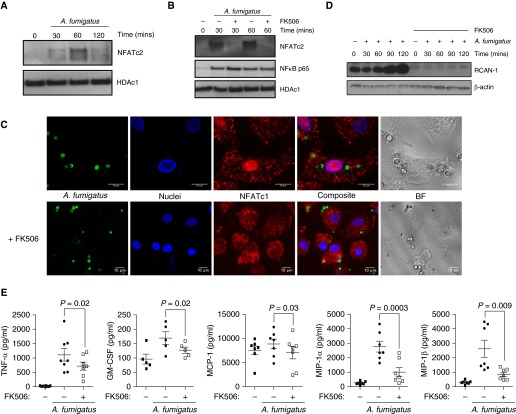
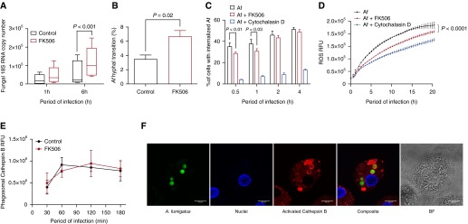
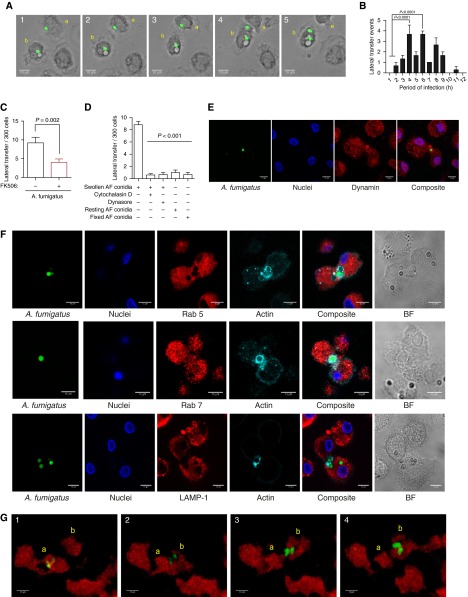
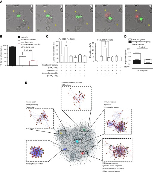
Measurements and main results: Macrophage phagocytosis of A. fumigatus enabled control of 90% of fungal germination. However, fungal germination in the late phagosome led to macrophage necrosis. During programmed necroptosis, we observed frequent cell-cell transfer of A. fumigatus between macrophages, which assists subsequent control of germination in recipient macrophages. Lateral transfer occurred through actin-dependent exocytosis of the late endosome in a vasodilator-stimulated phosphoprotein envelope. Its relevance to the control of fungal germination was also shown by direct visualization in our zebrafish aspergillosis model in vivo. The calcineurin inhibitor FK506 (tacrolimus) reduced cell death and lateral transfer in vitro by 50%. This resulted in uncontrolled fungal germination in macrophages and also resulted in hyphal escape.

Conclusions: These observations identify programmed, necrosis-dependent lateral transfer of A. fumigatus between macrophages as an important host strategy for controlling fungal germination. This process is critically dependent on calcineurin. Our studies provide fundamental insights into the pathogenesis of pulmonary aspergillosis in the immunocompromised host.

Keywords: Aspergillus; calcineurin; macrophage; necrosis; pulmonary fungal diseases.


Figures

Figure 1.
Figure 1.
Aspergillus fumigatus (Af) activates calcineurin–nuclear factor of activated T cells (NFAT)-dependent inflammatory responses in human macrophages. (A) Human monocyte-derived macrophages (hMDMs) were stimulated with live Af swollen conidia (SC) (multiplicity of infection [MOI], 1). hMDM nuclear extracts were collected at 30, 60, and 120 minutes postinfection and probed by Western blotting for NFAT, cytoplasmic, calcineurin-dependent 2 (NFATc2). The maximum translocation of NFATc2 to the nucleus was observed at 60 minutes postinfection. Western blots show representative data from four experiments. (B and C) hMDMs were pretreated with FK506 (10 ng/ml) and stimulated with live Af SC (MOI, 1). Nuclear extracts were probed by Western blotting for NFATc2 and whole-cell extracts for regulator of calcineurin 1 (RCAN-1). NFAT translocation was assessed by confocal microscopy. NFAT is shown in red, nuclei in blue, and Af conidia in green. Representative images of live conidia stimulating NFAT nuclear translocation are shown. Western blots show representative data from four experiments. (D) hMDMs were pretreated with FK506 (10 ng/ml) and stimulated with live Af SC (MOI, 1). FK506 completely inhibited RCAN-1 production. Western blot shows representative data from four experiments. (E) hMDMs were pretreated with FK506 (10 ng/ml) and stimulated with live Af SC (MOI, 1). Culture supernatant cytokines were measured at 6 hours postinfection by Luminex assay (n = 7 biological replicates). Scale bars = 10 μm. BF = brightfield; GM-CSF = granulocyte-macrophage colony–stimulating factor; HDAc1 = histone deacetylase 1; MCP-1 = monocyte chemoattractant protein 1; MIP-1 = macrophage inflammatory protein 1; NF-κB = nuclear factor-κB; TNF-α = tumor necrosis factor-α.
Figure 2.
Figure 2.
Human macrophage phagocytosis, reactive oxygen species (ROS) production, and control of Aspergillus fumigatus (Af) growth are calcineurin dependent. (A) Human monocyte-derived macrophages (hMDMs) pretreated with FK506 (10 ng/ml) or vehicle were stimulated with live Af swollen conidia (SC) (multiplicity of infection [MOI], 1), and total RNA was isolated at 1 and 6 hours postinfection. Fungal growth was assayed by reverse transcription–polymerase chain reaction of fungal RNA (n = 5 biological replicates). (B) hMDMs pretreated with FK506 (10 ng/ml) or vehicle were infected with live enhanced green fluorescent protein–expressing Af SC (MOI, 1), and hyphal transition was assessed using time-lapse video microscopy over a 10-hour period (n = 4 biological replicates). (C) hMDMs pretreated with FK506 (10 ng/ml) or cytochalasin D (5 nM) or vehicle were stimulated with live Af SC (MOI, 1) prestained with calcofluor white. Phagocytosis was quantified by using an ImageStream flow cytometer postinfection (n = 4 biological replicates). (D) hMDMs pretreated with FK506 (10 ng/ml) or vehicle were stimulated with live Af SC (MOI, 1), and ROS production was assayed by luminescence (n = 4 biological replicates). (E) hMDMs pretreated with FK506 (10 ng/ml) or vehicle were stimulated with live Af SC (MOI, 1), and cathepsin B activation was assayed by live confocal time-lapse microscopy. Phagosomal cathepsin B activation was quantified by using ImageJ software (National Institutes of Health, Bethesda, MD) (n = 4 biological replicates). (F) Representative confocal microscopic image showing cathepsin B activation in Af conidia–containing phagosomes in hMDMs. Activated cathepsin B is shown in red, nuclei in blue, and Af in green. Data are presented as mean ± SEM. Scale bars = 10 μm. BF = brightfield; RFU = relative fluorescence units.
Figure 3.
Figure 3.
Macrophages infected with swollen Aspergillus fumigatus (Af) undergo calcineurin-dependent lateral transfer. (A) Representative time-lapse widefield microscopic images of Af lateral transfer in human monocyte-derived macrophages (hMDMs) infected with live Af swollen conidia (SC) (multiplicity of infection [MOI], 1), shown in green. Transferring macrophage is labeled a, and receiving macrophage is labeled b. Images were taken at 10-minute intervals. (B) hMDMs were stimulated with live enhanced green fluorescent protein (eGFP)-Af SC (MOI, 1), and time-lapse widefield imaging was performed at 10-minute intervals for 12 hours postinfection. The timing of lateral transfer events per hour in macrophages infected with live Af SC is shown (n = 3 biological replicates). (C) hMDMs pretreated with FK506 (10 ng/ml) or vehicle were stimulated with live eGFP-Af SC (MOI, 1), and lateral transfer events were quantified by time-lapse widefield imaging at 10-minute intervals for 12 hours postinfection (n = 5 biological replicates). Total lateral transfer events over the 12-hour period were quantified for each biological replicate. (D) hMDMs were stimulated with live or fixed Af SC or resting conidia (MOI, 1), and lateral transfer events were quantified by time-lapse widefield microscopy. Cytochalasin D (5 nM) and dynasore (100 μM) were added at 90 minutes postinfection, and unbound conidia were washed away 45 minutes postinfection (n = 4 biological replicates). (E) Representative confocal microscopic image of dynamin-dependent lateral transfer in hMDMs stimulated with live Af SC. Dynamin is shown in red, nuclei in blue, and Af in green. (F) hMDMs were stimulated with live Af SC (MOI, 1), and nonphagocytosed conidia were washed away at 45 minutes postinfection. Lateral transfer events were captured by fixing cells every 10 minutes from 120 to 180 minutes postinfection. Characterization of Af lateral transfer was performed by staining for Ras-related protein 5 (Rab 5), Rab 7, and lysosomal-associated membrane protein 1 (LAMP-1) (shown in red) alongside actin (shown in cyan). Nuclei are shown in blue and live Af SC in green. Representative images show positive Rab 7 localization to Af-containing endosomes during lateral transfer, with little Rab 5 localization and no LAMP-1 localization. (G) mpeg:mCherry zebrafish larvae were infected with approximately 50 eGFP-expressing conidia of Af, and time-lapse confocal microscopy was performed. Representative images show Af lateral transfer between macrophages. The transferring macrophage is labeled a, and the receiving macrophage is labeled b. Images shown were taken at 7-minute intervals. Data are represented as mean ± SEM. Scale bars = 10 μm. BF = brightfield.
Figure 4.
Figure 4.
Macrophages undergoing programmed necrosis laterally transfer germinating Aspergillus fumigatus (Af) conidia in endosomes through an actin-dependent process. (A) Representative time-lapse confocal microscopic images of lateral transfer of live Af swollen conidia (SC) (shown in green) between human monocyte-derived macrophages (hMDMs). Propidium iodide (PI) staining (red) shows cell death of macrophage transferring Af conidia (labeled a) to a neighboring macrophage (labeled b). Images were taken at 10-minute intervals. (B) Time-lapse confocal microscopy was used to quantify the fate of transferred versus nontransferred conidia from dying hMDMs and to conidia within live macrophages. Germination was assessed over a 12-hour period. Control of fungal germination was significantly increased in dying hMDMs that transferred conidia to neighboring cells compared with those that did not (n = 4 biological replicates). (C) hMDMs pretreated with a pan-caspase inhibitor (50 μM Z-VAD-FMK), a receptor-interacting serine/threonine-protein kinase (RIPK-1) inhibitor (10 μM necrostatin-1), a caspase 1 inhibitor (50 μM Z-YVAD-FMK), 10 ng/ml FK506, or vehicle were stimulated with live Af SC (multiplicity of infection [MOI], 1), and lateral transfer events and cell death were quantified by PI fluorescence–based time-lapse confocal microscopy. Treatment with Z-VAD-FMK induced necroptotic cell death coupled to increased Af lateral transfer, which was inhibited by the addition of a RIPK-1 inhibitor (necrostatin-1) (n = 3 biological replicates). (D) hMDMs pretreated FK506 (10 ng/ml) or vehicle were stimulated with live Af SC (MOI, 1), and cell death was assayed by PI fluorescence and quantified by time-lapse confocal microscopy over a 6-hour period. The graph also shows the proportion of lateral transfer events in dying cells (approximately 30% of dying cells) (n = 4 biological replicates). (E) The network modules of the significantly expressed genes in FK506-pretreated macrophages stimulated with live Af SC (MOI, 1) for 6 hours. Significantly expressed genes were mapped onto a protein–protein interaction network, and network modules were identified using the MCODE plugin application of Cytoscape. Overrepresented pathways involving these network modules were identified using the Kyoto Encyclopedia of Genes and Genomes (“KEGG”) and WikiPathways databases (n = 6 biological replicates). Data are presented as mean ± SEM. Scale bars = 10 μm. AP-1 = activator protein 1; NOD = nucleotide-binding oligomerization domain.
Figure 5.
Figure 5.
Lateral transfer of Aspergillus fumigatus (Af) occurs through vasodilator-stimulated phosphoprotein (VASP) tunnel-like structures. (A) A phosphoprotein array analysis was performed with human monocyte-derived macrophages (hMDMs) pretreated with FK506 and stimulated with live Af swollen conidia (SC) (multiplicity of infection [MOI], 1) for 1 hour postinfection. Proteins with significant fold change differences in the phosphoprotein-to-nonphosphoprotein ratio between FK506 and control macrophages stimulated with live Af SC are shown. (B) hMDMs were pretreated with a cell-permeable cyclic guanosine monophosphate analog (25 mM 8-[4-chlorophenylthio]-guanosine 3′,5′-cyclic monophosphate [8-pCPT-cGMP]) to induce Ser238 VASP phosphorylation and with FK506 (10 ng/ml) or vehicle. They were then stimulated with a calcium ionophore (2 μg/ml ionomycin) to activate calcineurin. Macrophage whole-cell extracts were probed by Western blotting for Ser238 VASP and total VASP. (C) Representative confocal microscopic images of hMDM phagocytosis and lateral transfer of live Af SC (MOI, 1). VASP is shown in red, nuclei in blue, and Af in green. (1) Uninfected hMDMs. (2) Initial phagocytosis of Af by hMDMs. (3) Late phagocytosis of Af by hMDMs. (4) Lateral transfer of Af between hMDMs. (D) Three-dimensional reconstruction of confocal microscopic images of phagocytosis and lateral transfer of live Af SC (MOI, 1) between hMDMs. Af phagocytosis at 30 minutes postinfection with VASP is shown in red, Af in green, and nuclei in blue. (E) Three-dimensional reconstruction of confocal microscopic image of Af lateral transfer with VASP shown in yellow, actin in magenta, Af in green, and nuclei in blue. (F and G) Representative z-slice confocal microscopic image (F) and three-dimensional reconstruction (G) of Af lateral transfer. VASP is shown in red, and Af is shown in green. (H) THP-1 macrophages treated with either control or VASP small interfering RNA (siRNA) (50 µM) were infected with live Af SC and stained with Calcofluor White (Sigma-Aldrich, St. Louis, MO) (MOI, 1). Phagocytosis was quantified by flow cytometry (n = 3 biological replicates). Data are presented as mean ± SEM. Scale bars = 10 μm. AKT1 = RAC-α serine/threonine protein kinase; CDC25A = M-phase inducer phosphatase 1; HDAC1 = histone deacetylase 1; MAPK = mitogen-activated protein kinase; MEK1 = mitogen-activated protein kinase kinase 1; NFAT = nuclear factor of activated T cells; P-VASP = phospho-VASP; SMC1=structural maintenance of chromosomes protein 1; UT = untreated; VEGFR2 = vascular endothelial growth factor receptor 2.

Similar articles

Cited by

References

    1. Latgé JP. Aspergillus fumigatus and aspergillosis. Clin Microbiol Rev. 1999;12:310–350. - PMC - PubMed
    1. Bhatia S, Fei M, Yarlagadda M, Qi Z, Akira S, Saijo S, Iwakura Y, van Rooijen N, Gibson GA, St Croix CM, et al. Rapid host defense against Aspergillus fumigatus involves alveolar macrophages with a predominance of alternatively activated phenotype. PLoS One. 2011;6:e15943. - PMC - PubMed
    1. Chen GH, Teitz-Tennenbaum S, Neal LM, Murdock BJ, Malachowski AN, Dils AJ, Olszewski MA, Osterholzer JJ. Local GM-CSF-dependent differentiation and activation of pulmonary dendritic cells and macrophages protect against progressive cryptococcal lung infection in mice. J Immunol. 2016;196:1810–1821. - PMC - PubMed
    1. Herbst S, Shah A, Mazon Moya M, Marzola V, Jensen B, Reed A, Birrell MA, Saijo S, Mostowy S, Shaunak S, et al. Phagocytosis-dependent activation of a TLR9-BTK-calcineurin-NFAT pathway co-ordinates innate immunity to Aspergillus fumigatus. EMBO Mol Med. 2015;7:240–258. - PMC - PubMed
    1. Herbst S, Shah A, Carby M, Chusney G, Kikkeri N, Dorling A, Bignell E, Shaunak S, Armstrong-James D. A new and clinically relevant murine model of solid-organ transplant aspergillosis. Dis Model Mech. 2013;6:643–651. - PMC - PubMed

Publication types

MeSH terms