

Choroidal Caverns: A Novel Optical Coherence Tomography Finding in Geographic Atrophy
- PMID: 27163770
- PMCID: PMC4868100
- DOI: 10.1167/iovs.16-19083
Choroidal Caverns: A Novel Optical Coherence Tomography Finding in Geographic Atrophy
Abstract
Purpose: To describe and interpret "choroidal caverns," an unreported optical coherence tomography (OCT) finding in the choroid of patients with geographic atrophy (GA) secondary to atrophic AMD.
Methods: Retrospective analysis of patients with GA. Main outcomes measures included estimation of the prevalence of choroidal caverns, their localization and relation with retinal-choroidal structures by reviewing medical records and multimodal imaging.
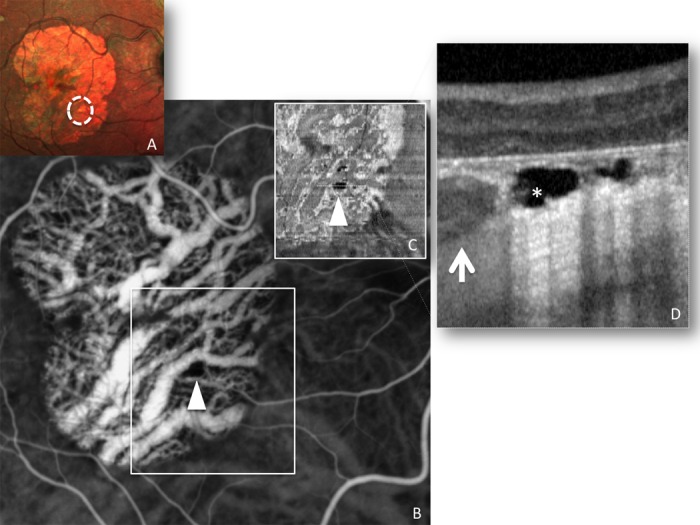
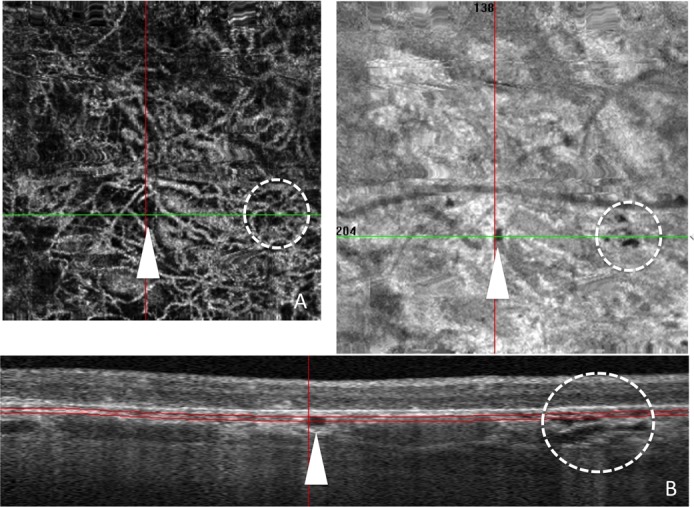
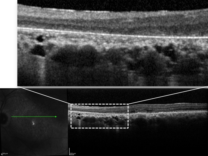
Results: One hundred twenty consecutive patients (mean age 80.5 ± 8.61 years) were included. Among the 201 eyes with GA, 17 eyes of 15 patients presented choroidal caverns on OCT B-scan in GA areas (a total of 43 choroidal caverns, mean 2.5/eye, variably localized in the Sattler and Haller layers, with relative preservation of the choriocapillaris). This accounts for 12.5% estimated prevalence (6.5-18.5, 95% confidence interval [CI]) of choroidal caverns in GA areas of atrophic AMD patients. Choroidal caverns appeared on OCT (both B-scan and en face) as gaping hyporeflective cavities in the choroid, typically empty, angular, without hyperreflective borders, often with punctate/linear hyperreflectivities internally. Indocyanine angiography and OCT-Angiography confirmed that the areas occupied by these cavities do not represent perfused choroidal blood vessels.
Conclusions: Choroidal caverns represent a relatively infrequent peculiar finding in GA areas of atrophic AMD eyes. They appear as gaping angular hyporeflective cavities in areas devoid of choroidal vessels, often with punctate/linear hyperreflectivities internally. Choroidal caverns may possibly arise from nonperfused ghost vessels and persistence of stromal pillars where the vessels were originally situated.
Figures
References
-
- Bressler NM,, Bressler SB,, Fine SL. Age-related macular degeneration. Surv Ophthalmol. 1988; 32: 375–413. - PubMed
-
- Klein R,, Klein BE,, Knudtson MD,, et al. Fifteen-year cumulative incidence of age-related macular degeneration: the Beaver Dam Eye Study. Ophthalmology. 2007; 114: 253–262. - PubMed
-
- Sunness JS. The natural history of geographic atrophy, the advanced atrophic form of age-related macular degeneration. Mol Vis. 1999; 5: 25. - PubMed
-
- Curcio CA,, Medeiros NE,, Millican CL. Photoreceptor loss in age-related macular degeneration. Invest Ophthalmol Vis Sci. 1996; 37: 1236–1249. - PubMed
-
- Dunaief JL,, Dentchev T,, Ying GS,, Milam AH. The role of apoptosis in age-related macular degeneration. Arch Ophthalmol. 2002; 120: 1435–1442. - PubMed
MeSH terms
LinkOut - more resources
Full Text Sources
Other Literature Sources
Research Materials
