. 2016 Jul;89(1063):20160072.
doi: 10.1259/bjr.20160072. Epub 2016 May 10.

MRI sequences for the detection of individual lymph nodes in regional breast radiotherapy planning

Affiliations

MRI sequences for the detection of individual lymph nodes in regional breast radiotherapy planning

Tristan C F van Heijst et al. Br J Radiol. 2016 Jul.

Abstract

Objective: In regional radiotherapy (RT) for patients with breast cancer, lymph node (LN) targets are delineated on CT, defined by anatomical boundaries. By identifying individual LNs, MRI-based delineations may reduce target volumes and thereby toxicity. We optimized MRI sequences for this purpose. Our aim was to evaluate the techniques for LN delineation in RT planning.

Methods: Supine MRI was explored at 1.5 T in RT position (arms in abduction). 5 MRI techniques were optimized in 10 and evaluated in 12 healthy female volunteers. The scans included one T1 weighted (T1w), three T2 weighted (T2w) and a diffusion-weighted imaging (DWI) technique. Quantitative evaluation was performed by scoring LN numbers per volunteer and per scan. Qualitatively, scans were assessed on seven aspects, including LN contrast, anatomical information and insensitivity to motion during acquisition.

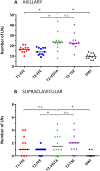
Results: Two T2w fast spin-echo (FSE) methods showed the highest LN numbers (median 24 axillary), high contrast, excellent fat suppression and relative insensitivity to motion during acquisition. A third T2w sequence and DWI showed significantly fewer LNs (14 and 10) and proved unsuitable due to motion sensitivity and geometrical uncertainties. T1w MRI showed an intermediate number of LNs (17), provided valuable anatomical information, but lacked LN contrast.

Conclusion: Explicit LN imaging was achieved, in supine RT position, using MRI. Two T2w FSE techniques had the highest detection rates and were motion insensitive. T1w MRI showed anatomical information. MRI enables direct delineation of individual LNs.

Advances in knowledge: Our optimized MRI scans enable accurate target definition in MRI-guided regional breast RT and development of personalized treatments.


Figures

Figure 1.
Figure 1.
Example of standard axillary delineations on CT in a patient with breast cancer in radiotherapy (RT) position. A transverse slice of the CT scan is shown (left), with the corresponding coronal view (right). Inner line is the clinical target volume (CTV) of the lower axilla for standard right-sided axillary RT; outer line is the planning target volume (not indicated in the figure), which is generated by an expansion of the CTV by 5 mm isotropic. Target delineations are performed based on guidelines with the use of anatomical boundaries. As a consequence, target volumes are relatively large. Other delineated structures are the ipsilateral lung, contralateral lung and heart.
Figure 2.
Figure 2.
MRI scanning setup. The volunteer is placed on a 10° Thorawedge board (CIVCO Medical Solutions, Reeuwijk, Netherlands) in supine RT position, i.e. with arms in abduction, resting on the board. Scanning is performed with a 1.5-T Ingenia (Philips, Best, Netherlands) wide-bore MRI scanner. An anterior FlexCoverage (Philips) receive coil is connected and placed on top of a custom-made polymethyl methacrylate support, which is adjustable, in order to prevent deformation of the outer body contour. A posterior receive coil (Philips) is located in the table.
Figure 3.
Figure 3.
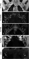
Coronal slices showing axillary lymph nodes (LNs) in five volunteers bilaterally, visualized with MRI. The MRI techniques represented are (a) T1 weighted fast field echo (T1-FFE; water-only image); (b) T2 weighted (T2w) volumetric isotropic T2w acquisition (T2-VISTA); (c) T2w turbo spin echo (T2-TSE; water-only image); (d) T2w fast field echo (T2-FFE); and (e) diffusion-weighted imaging (DWI; b = 1000 s mm−2). For the T1-FFE, T2-VISTA and T2-FFE examples, maximum intensity projections of three slices were used to increase signal-to-noise ratio and to cover the same volume as in the T2-TSE and DWI.
Figure 4.
Figure 4.
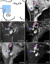
Coronal slices showing right-sided axillary lymph nodes (LNs) in one volunteer, visualized with MRI. The slices shown are cropped to approximately 80 × 80 mm2. Location with respect to anatomy is depicted schematically by the square in (a). MRI techniques represented are (b) T1 weighted fast field echo (T1-FFE; water-only image); (c) T2 weighted (T2w) volumetric isotropic T2w acquisition (T2-VISTA); (d) T2w turbo spin echo (T2-TSE; water-only image); (e) T2-FFE; and (f) diffusion-weighted imaging (DWI; b = 1000 s mm−2). For the T2-VISTA and T2-FFE examples, maximum intensity projections of three slices were used to increase signal-to-noise ratio and to cover the same volume as in the T2-TSE and DWI. The arrows indicate LNs, apparent in T1-FFE, T2-VISTA, DWI and T2-TSE but obscured in T2-FFE due to a pulsating artery nearby. The arrowheads indicate a LN that is clearly identifiable in the T2w and DWI sequences but lacks contrast in T1-FFE. ax. a. v., axillary arteria and vein; m. lat. d., musculus latissimus dorsi.
Figure 5.
Figure 5.
Numbers of lymph nodes (LNs) identified in images acquired with different MR sequences. For both (a) the axilla and (b) the supraclavicular area, the numbers of detected LNs are denoted, per MRI technique, for all 12 volunteers. Note that the y-axes in (a) and (b) differ. T1 weighted fast field echo (T1-FFE) is denoted by dots, T2 weighted (T2w) fast field echo (T2-FFE) by squares, T2w volumetric isotropic T2w acquisition (T2-VISTA) by triangles pointing upwards, T2w turbo spin echo (T2-TSE) by triangles pointing downwards and diffusion-weighted imaging (DWI) by black diamonds. Black horizontal lines indicate the median values. Statistical significances from paired Wilcoxon signed-rank tests are indicated with respect to T2-VISTA. *p < 0.05; n.s., non-significance or p > 0.05.

Similar articles

Cited by

References

    1. Ivens D, Hoe AL, Podd TJ, Hamilton CR, Taylor I, Royle GT. Assessment of morbidity from complete axillary dissection. Br J Cancer 1992; 66: 136–8. doi: 10.1038/bjc.1992.230 - DOI - PMC - PubMed
    1. Kakuda JT, Stuntz M, Trivedi V, Klein SR, Vargas HI. Objective assessment of axillary morbidity in breast cancer treatment. Am Surg 1999; 65: 995–8. - PubMed
    1. Giuliano AE, McCall L, Beitsch P, Whitworth PW, Blumencranz P, Leitch AM, et al. . Locoregional recurrence after sentinel lymph node dissection with or without axillary dissection in patients with sentinel lymph node metastases: the American College of Surgeons Oncology Group Z0011 randomized trial. Ann Surg 2010; 252: 426–32; discussion 432–3. doi: 10.1097/SLA.0b013e3181f08f32 - DOI - PMC - PubMed
    1. Galimberti V, Chifu C, Rodriguez Perez S, Veronesi P, Intra M, Botteri E, et al. . Positive axillary sentinel lymph node: is axillary dissection always necessary? Breast 2011; 20(Suppl. 3): S96–8. doi: 10.1016/S0960-9776(11)70303-4 - DOI - PubMed
    1. Jagsi R, Chadha M, Moni J, Ballman K, Laurie F, Buchholz TA, et al. . Radiation field design in the ACOSOG Z0011 (alliance) trial. J Clin Oncol 2014; 32: 3600–6. doi: 10.1200/JCO.2014.56.5838 - DOI - PMC - PubMed