

The Role of fadD19 and echA19 in Sterol Side Chain Degradation by Mycobacterium smegmatis
- PMID: 27164074
- PMCID: PMC6273163
- DOI: 10.3390/molecules21050598
The Role of fadD19 and echA19 in Sterol Side Chain Degradation by Mycobacterium smegmatis
Abstract
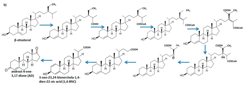
Mycobacteria are able to degrade natural sterols and use them as a source of carbon and energy. Several genes which play an important role in cholesterol ring degradation have been described in Mycobacterium smegmatis. However, there are limited data describing the molecular mechanism of the aliphatic side chain degradation by Mycobacterium spp. In this paper, we analyzed the role of the echA19 and fadD19 genes in the degradation process of the side chain of cholesterol and β-sitosterol. We demonstrated that the M. smegmatis fadD19 and echA19 genes are not essential for viability. FadD19 is required in the initial step of the biodegradation of C-24 branched sterol side chains in Mycobacterium smegmatis mc²155, but not those carrying a straight chain like cholesterol. Additionally, we have shown that echA19 is not essential in the degradation of either substrate. This is the first report, to our knowledge, on the molecular characterization of the genes playing an essential role in C-24 branched side chain sterol degradation in M. smegmatis mc²155.
Keywords: M. smegmatis; cholesterol; microbial sterol degradation; sterol side-chain degradation; β-sitosterol.
Conflict of interest statement
The authors declare no conflict of interest.
Figures


Similar articles
-
FadD19 of Rhodococcus rhodochrous DSM43269, a steroid-coenzyme A ligase essential for degradation of C-24 branched sterol side chains.Appl Environ Microbiol. 2011 Jul;77(13):4455-64. doi: 10.1128/AEM.00380-11. Epub 2011 May 20. Appl Environ Microbiol. 2011. PMID: 21602385 Free PMC article.
-
Post-translational Succinylation of Mycobacterium tuberculosis Enoyl-CoA Hydratase EchA19 Slows Catalytic Hydration of Cholesterol Catabolite 3-Oxo-chol-4,22-diene-24-oyl-CoA.ACS Infect Dis. 2020 Aug 14;6(8):2214-2224. doi: 10.1021/acsinfecdis.0c00329. Epub 2020 Jul 27. ACS Infect Dis. 2020. PMID: 32649175 Free PMC article.
-
Mycobacterium smegmatis is a suitable cell factory for the production of steroidic synthons.Microb Biotechnol. 2017 Jan;10(1):138-150. doi: 10.1111/1751-7915.12429. Epub 2016 Nov 2. Microb Biotechnol. 2017. PMID: 27804278 Free PMC article.
-
Microbial catabolism of sterols: focus on the enzymes that transform the sterol 3β-hydroxy-5-en into 3-keto-4-en.FEMS Microbiol Lett. 2017 Feb 1;364(3). doi: 10.1093/femsle/fnx007. FEMS Microbiol Lett. 2017. PMID: 28087615 Review.
-
Microbial cleavage of sterol side chains.Adv Appl Microbiol. 1977;22:29-58. doi: 10.1016/s0065-2164(08)70159-x. Adv Appl Microbiol. 1977. PMID: 337771 Review. No abstract available.
Cited by
-
Mycobacterium tuberculosis FadD18 Promotes Proinflammatory Cytokine Secretion to Inhibit the Intracellular Survival of Bacillus Calmette-Guérin.Cells. 2024 Jun 11;13(12):1019. doi: 10.3390/cells13121019. Cells. 2024. PMID: 38920649 Free PMC article.
-
Evolutionary insights into the selectivity of sterol oxidising cytochrome P450 enzymes based on ancestral sequence reconstruction.Chem Sci. 2025 May 13;16(24):11110-11122. doi: 10.1039/d5sc01863c. eCollection 2025 Jun 18. Chem Sci. 2025. PMID: 40417289 Free PMC article.
References
-
- Naghibi F., Tabatabai Yazdi M., Sahebgharani M., Noori Daloii M.R. Microbial transformation of cholesterol. J. Sci. Islam. Repub. Iran. 2002;13:103–106.
-
- Lad N. Optimalization of parameters for hydrocortisone succinate bioconversion. Malays. J. Microbiol. 2011;7:7–13.
-
- Field J.A., Bolesma F., Baten H., Rulkens W.H. Oxidation of anthracene in water solvent mixtures by the white-rot fungus Bjerkandera sp. strain BOS55. Appl. Microbiol. Biotechnol. 1995;44:234–240. doi: 10.1007/BF00164508. - DOI
-
- Cerniglia C.E. Biodegradation of polycyclic aromatic hydrocarbons. Biodegradation. 1992;3:351–368. doi: 10.1007/BF00129093. - DOI
MeSH terms
Substances
LinkOut - more resources
Full Text Sources
Other Literature Sources
Miscellaneous
