. 2016 Aug:68:142-52.
doi: 10.1016/j.archoralbio.2016.05.002. Epub 2016 May 3.

Expression of Evc2 in craniofacial tissues and craniofacial bone defects in Evc2 knockout mouse

Affiliations

Expression of Evc2 in craniofacial tissues and craniofacial bone defects in Evc2 knockout mouse

Mohammed K Badri et al. Arch Oral Biol. 2016 Aug.

Abstract

Objective: Our objectives were to determine the expression of EVC2 in craniofacial tissues and investigate the effect of Evc2 deficiency on craniofacial bones using Evc2 knockout (KO) mouse model.

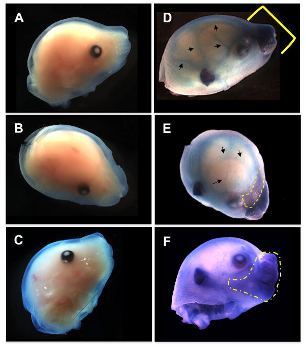
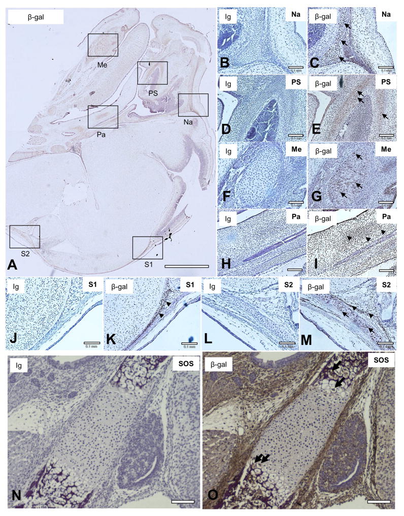
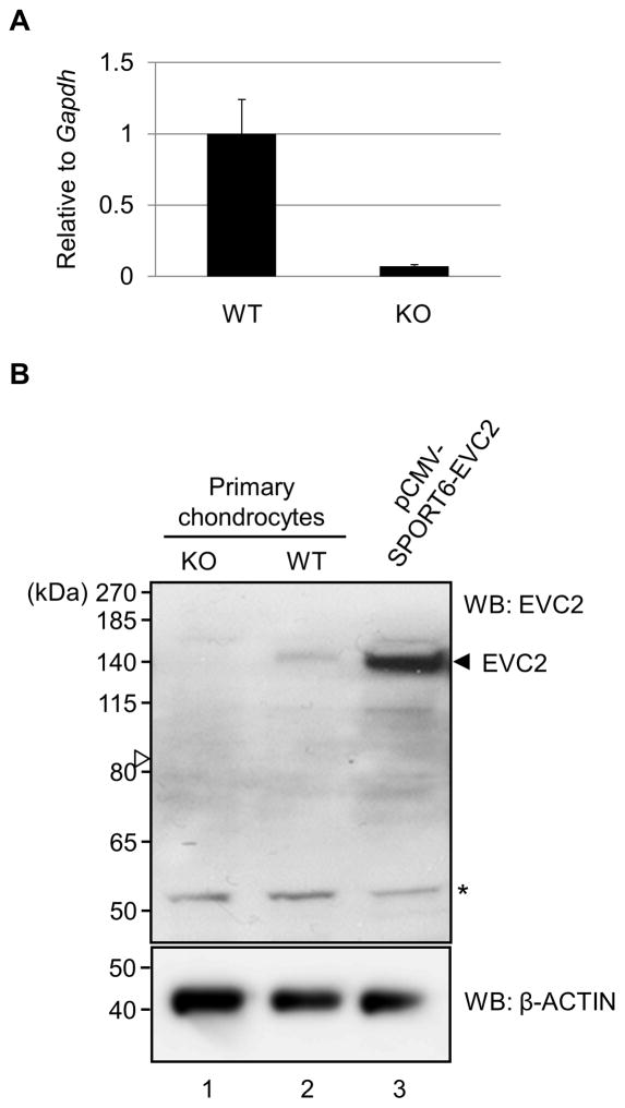
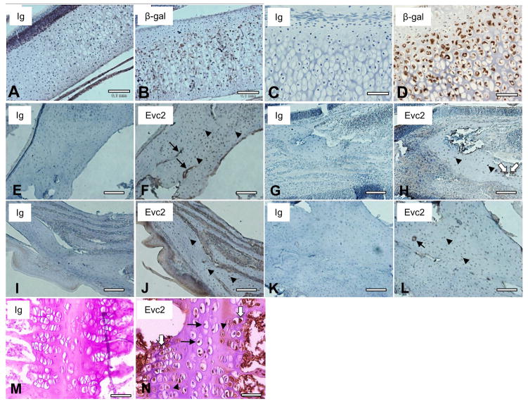
Design: Evc2 KO mice were generated by introducing a premature stop codon followed by the Internal Ribosomal Entry Site fused to β-galactosidase (LacZ). Samples from wild-type (WT), heterozygous (Het) and homozygous Evc2 KO mice were prepared. LacZ staining and immunohistochemistry (IHC) with anti-β-galactosidase, anti-EVC2 and anti-SOX9 antibodies were performed. The craniofacial bones were stained with alcian blue and alizarin red.

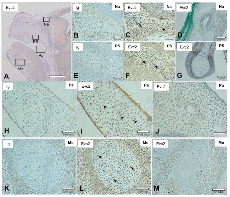
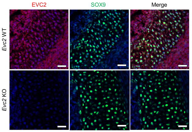
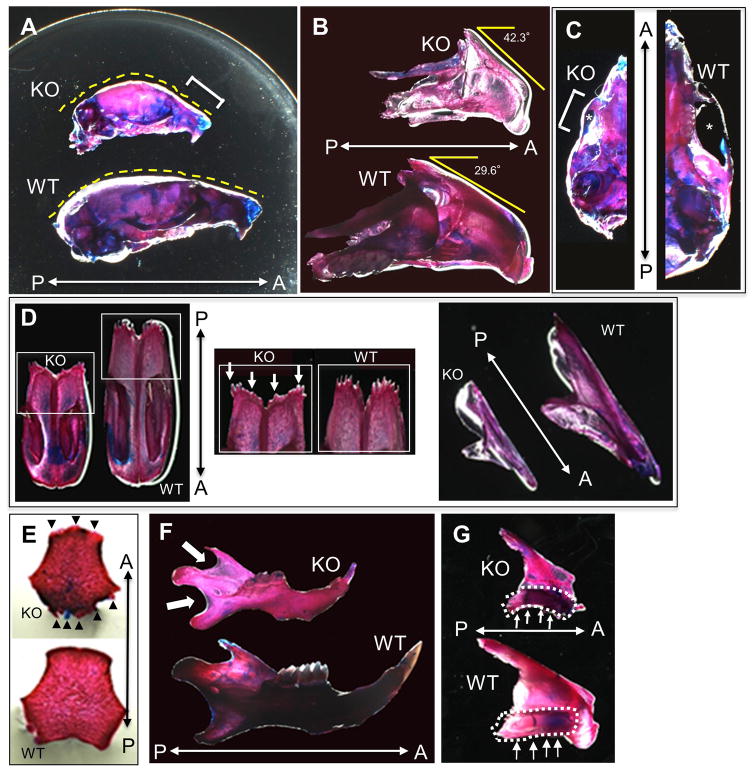
Results: The LacZ activity in KO was mainly observed in the anterior parts of viscerocranium. The Evc2-expressing cells were identified in many cartilageous regions by IHC with anti-β-galactosidase antibody in KO and Het embryos. The endogenous EVC2 protein was observed in these areas in WT embryos. Double labeling with anti-SOX9 antibody showed that these cells were mainly chondrocytes. At adult stages, the expression of EVC2 was found in chondrocytes of nasal bones and spheno-occipital synchondrosis, and osteocytes and endothelial-like cells of the premaxilla and mandible. The skeletal double staining demonstrated that craniofacial bones, where the expression of EVC2 was observed, in KO had the morphological defects as compared to WT.

Conclusion: To our knowledge, our study was the first to identify the types of Evc2-expressing cells in craniofacial tissues. Consistent with the expression pattern, abnormal craniofacial bone morphology was found in the Evc2 KO mice, suggesting that EVC2 may be important during craniofacial growth and development.

Keywords: Craniofacial bone; Development; EVC2; Ellis-van Creveld syndrome; Expression; Knockout (KO) mouse.


Conflict of interest statement

Competing interests: The authors declare no potential conflicts of interest with respect to the authorship and/or publication of this article.

Figures

Figure 1
Figure 1. Expression pattern of Evc2 in the craniofacial tissues at the embryonic stage
(A–F) Visualization of Evc2-expressing cell distribution by X-gal staining at E15.5. Evc2 WT (AC) and KO (D–F) embryos were stained with X-gal. The KO embryo exhibited a higher LacZ activity in the anterior portion of the viscerocranium in the nasal, premaxilla, maxilla, and mandibular (D and F; indicated by a bracket and a dotted line) areas. Expression was also detected in the anterior part of frontal bones (E; indicated by a dotted line) and in the suture area of the neurocranium (D and E; indicated by arrows). WT embryos used as a negative control showed no LacZ activity (A–C). At least 3 samples per each genotype have been investigated and the representative head images were acquired and presented.
Figure 2
Figure 2. Detection of Evc2-expressing cells in the homozygous Evc2 KO embryo
IHC staining using anti-β-gal antibody was performed to visualize the Evc2-expressing cells in the craniofacial structures of homozygous Evc2 KO embryos at E15.5 (A–M) and E18.5 (N, O) (brown). Sagittal sections of KO head were prepared and the area of nasal capsule (Na), paranasal sinus (PS), Meckel’s cartilage (Me), posterior palate (Pa), or cranial suture (S1, S2) was indicated by an open box (A), respectively. Expression of β-gal was detected in chondrocytes (indicated by arrows) of Na (C), PS (E), Me (G), S2 (M), and spheno-occipital synchondrosis (SOS) (O) as well as in mesenchymal cells (indicated by arrowheads) of Pa (I) and S1 (K). Serial sections of the KO head were also stained with non-immune immunoglobulin (Ig) as a negative control and no immunoreactivity in the corresponding craniofacial areas was observed (Na in (B), PS in (D), Me in (F), Pa in (H), S1 in (J), S2 in (L), SOS in (N), respectively). Sections were counterstained with hematoxylin (blue). At least 3 head samples have been investigated using at least 6 sections per sample and the representative images were acquired and presented. Scale bars; A = 1 mm, B–O = 0.1 mm.
Figure 3
Figure 3. Expression of EVC2 in Evc2 WT and KO primary chondrocytes
(A) Gene expression of Evc2 in primary chondrocytes. Quantitative real-time PCR analysis was performed using Evc2 WT and KO primary chondrocytes. The mean fold change in the expression of Evc2 was calculated based on the normalization to that of glyceraldehyde-3-phosphate dehydrogenase (Gapdh) using the value of Evc2 in WT chondrocytes as a calibrator. The values are shown as the mean + S.D. based on triplicate assays. (B) Analysis of EVC2 protein levels in Evc2 WT and KO chondrocytes by rabbit anti-EVC2 antibody. Western blot (WB) analysis showed the detection of EVC2 in WT chondrocytes (upper panel, lane 2, at ~140kDa) and a lack of detection of EVC2 in Evc2 KO chondrocytes (upper panel, lane 1). Immunodetection of a band at ~140kDa in a positive control (i.e. pCMV-SPORT6-Evc2 plasmid-transfected ATDC5 chondrogenic cells, indicated by an arrowhead) indicated successful detection and validation of rabbit anti-EVC2 antibody. An open arrowhead indicated the expected molecular weight of the truncated EVC2 protein, which was not identified. β-ACTIN was used as loading control. *; non-specific band.
Figure 4
Figure 4. Expression of endogenous EVC2 protein in the craniofacial tissues
(A, C, F, I, L) The endogenous EVC2 expression pattern was visualized by IHC analysis using anti-EVC2 antibody with WT embryo at E15.5. Evc2 KO embryo at E15.5 was also used as a negative control for anti-EVC2 antibody (D, G, J, M). The immunoreactivity was detected in the areas of the nasal capsule (Na), paranasal sinus (PS), palate (Pa) and Meckel’s cartilage (Me). The EVC2 protein expression was clearly observed in chondrocytes in these areas (Na in (C), PS in (F), Me in (L)) as well as in mesenchymal cells of Pa in (I). Serial section of the WT head was also stained with non-immune Ig as a negative control and no immunoreactivity in the corresponding craniofacial areas was observed (Na in (B), PS in (E), Pa in (H), Me in (K), respectively). No immunoreactivity was detected using Evc2 KO embryo with anti-EVC2 antibody in areas of Na (D), PS (G), Pa (J) and Me (M). Sections were counterstained with hematoxylin. At least 3 samples per each genotype have been investigated using at least 6 sections per sample and the representative images were acquired and presented. Scale bars; A= 1 mm, B, C, E, F, H, I, K, L = 0.05 mm, D, G, J, M = 0.1 mm.
Figure 5
Figure 5. The cell type that expresses EVC2 is chondrocyte
Fluorescence IHC with double labeling was performed to identify the cell type that expresses EVC2. Sagittal sections of WT and KO heads at E16.5 were prepared. The endogenous EVC2 expression was visualized by fluorescence IHC analysis with anti-EVC2 (left panels, red) and anti-SOX9 (middle panels, green) antibodies using WT (upper panels) and KO (lower panels) embryos. The immunoreactivity was detected in the area of paranasal sinus (PS) and is shown. No immunoreactivity was detected using Evc2 KO embryo with anti-EVC2 antibody (lower left panel). Sections were counterstained with DAPI. Scale bars; 0.04 mm.
Figure 6
Figure 6. EVC2 expression at postnatal stages
Sagittal sections of Het (A, B) and KO (C, D) heads at 3 weeks were used to visualize Evc2-expressing cells. IHC staining with anti-β-gal antibody showed Evc2-expressing cells (brown) in hypertrophic chondrocytes in the nasal septum area in Het (B) and KO (D). Corresponding areas to (B, D), respectively, were stained with non-immune Ig as negative controls (A, C). (E–N) Sagittal sections of WT heads at 6 weeks were used to perform IHC staining by anti-EVC2 antibody. Expression of EVC2 was detected in osteocytes of the premaxilla (F, arrowheads), paranasal (ethmoid) sinus (H, arrowheads), palate (J, arrowheads) and mandible (L, arrowheads). Expression was also detected in osteoblasts of paranasal sinuses (H; indicated by open arrows), endothelial-like cells (F, L, filled arrows) and hematopoietic cells (N). In addition, EVC2 expression was observed in chondrocytes in the resting (arrows), proliferative (arrowheads) and hypertrophic (open arrows) zones at spheno-occipital synchondrosis (N). No immunoreactivity was observed with non-immune Ig as a negative control in the corresponding areas (E, G, I, K, M). Sections were counterstained with hematoxylin. At least 3 head samples have been investigated using at least 6 sections per sample and the representative images were acquired and presented. Scale bars; A, B, E–L = 0.1mm, C, D = 0.05 mm, M, N = 0.04 mm.
Figure 7
Figure 7
Skeletal double staining of Evc2 WT and KO craniofacial tissues. The heads of Evc2 WT and KO mice at postnatal day (P) 0, P18, and P24 were stained with alizarin red and alcian blue, and craniofacial bones were further dissected for comparison. (A) Lateral view of WT and KO mouse skulls at P18. The size of the KO surface area was smaller than that of WT. The nasal-premaxilla area in KO (bracket) is more depressed and retruded with an overall convex skull outline (yellow dotted line) as compared to WT. A; anterior, P; posterior. (B) Lateral view of the premaxilla-maxilla-palatine bone complex at P18, showing that KO exhibited the retruded premaxilla with a larger degree than WT. A; anterior, P; posterior. (C) Ventral view of WT and KO mouse skulls at P18 showing depressed zygomatic arch in KO (bracket). Note that the infra-temporal space (asterisk) below the arch in KO was smaller than that in WT. A; anterior, P; posterior. (D) Ventral (left and middle panels) and lateral (right panel) view of nasal bones at P24. Irregular and wide border of the frontal process of the nasal bone (middle panel, arrows) was found in KO as compared to WT. A; anterior, P; posterior. (E) Ventral view of basioccipital bones. Irregular borders and abnormal shape of the basioccipital bone in KO (arrowheads) at P0 as compared to WT. A; anterior, P; posterior. (F) Lateral view of mandibles at P18. KO showed transparent areas at the mandibular notch and the posterior mandibular bone border (arrows) compared to WT. A; anterior, P; posterior. (G) Lateral view of the palatine bones at P24. The palatine bone in KO was deformed in a more curved shape as compared to WT (indicated by dotted circle areas and arrows). A; anterior, P; posterior.

Similar articles

Cited by

References

    1. Ellis RW, van Creveld S. A Syndrome Characterized by Ectodermal Dysplasia, Polydactyly, Chondro-Dysplasia and Congenital Morbus Cordis: Report of Three Cases. Arch Dis Child. 1940;15(82):65–84. - PMC - PubMed
    1. Baujat G, Le Merrer M. Ellis-van Creveld syndrome. Orphanet J Rare Dis. 2007;2:27. - PMC - PubMed
    1. Metrakos JD, Fraser FC. Evidence for a hereditary factor in chondroectodermal dysplasia (Ellis-van Creveld syndrome) American journal of human genetics. 1954;6(2):260–269. - PMC - PubMed
    1. Polymeropoulos MH, Ide SE, Wright M, Goodship J, Weissenbach J, Pyeritz RE, et al. The gene for the Ellis-van Creveld syndrome is located on chromosome 4p16. Genomics. 1996;35(1):1–5. - PubMed
    1. Ruiz-Perez VL, Ide SE, Strom TM, Lorenz B, Wilson D, Woods K, et al. Mutations in a new gene in Ellis-van Creveld syndrome and Weyers acrodental dysostosis. Nat Genet. 2000;24(3):283–286. - PubMed

MeSH terms