

Exclusion of dysfunctional mitochondria from Balbiani body during early oogenesis of Thermobia
- PMID: 27164893
- PMCID: PMC5031756
- DOI: 10.1007/s00441-016-2414-x
Exclusion of dysfunctional mitochondria from Balbiani body during early oogenesis of Thermobia
Abstract
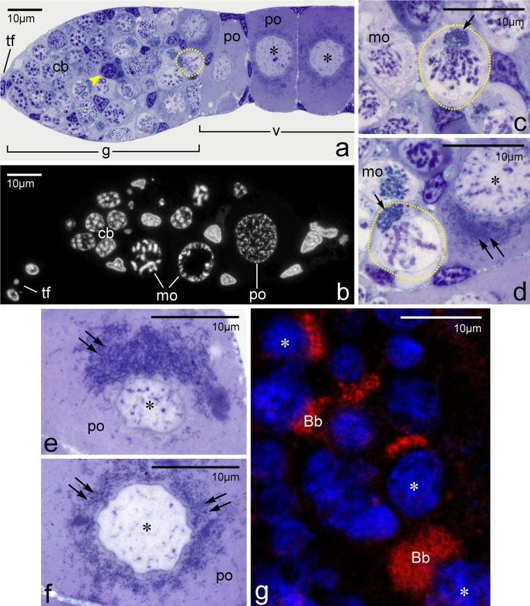
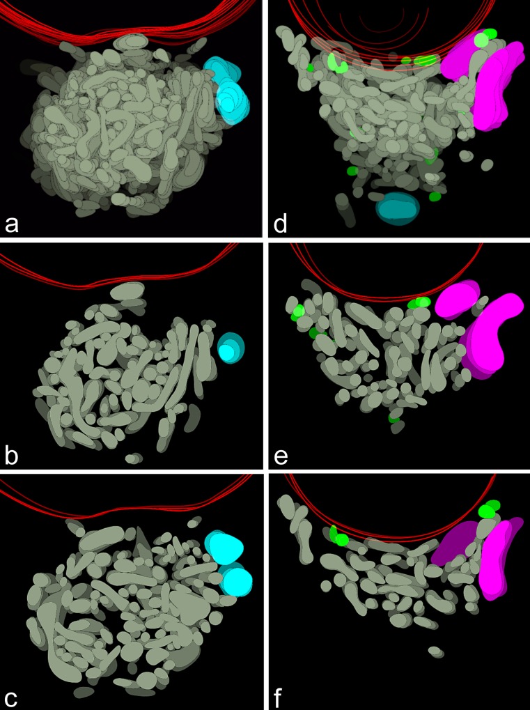
Oocytes of many invertebrate and vertebrate species contain a characteristic organelle complex known as the Balbiani body (Bb). Until now, three principal functions have been ascribed to this complex: delivery of germ cell determinants and localized RNAs to the vegetal cortex/posterior pole of the oocyte, transport of the mitochondria towards the germ plasm, and participation in the formation of lipid droplets. Here, we present the results of a computer-aided 3D reconstruction of the Bb in the growing oocytes of an insect, Thermobia domestica. Our analyses have shown that, in Thermobia, the central part of each fully developed Bb comprises a single intricate mitochondrial network. This "core" network is surrounded by several isolated bean-shaped mitochondrial units that display lowered membrane potential and clear signs of degeneration. In light of the above results and recent theoretical models of mitochondrial quality control, the role of the Bb is discussed. We suggest that, in addition to the aforementioned functions, the Bb is implicated in the selective elimination of dysfunctional mitochondria during oogenesis.
Keywords: Balbiani body; Mitochondrial dynamics; Mitochondrial network; Oogenesis.
Figures


Similar articles
-
Meiosis, Balbiani body and early asymmetry of Thermobia oocyte.Protoplasma. 2017 Mar;254(2):649-655. doi: 10.1007/s00709-016-0978-7. Epub 2016 May 14. Protoplasma. 2017. PMID: 27180195 Free PMC article.
-
Morphogenesis of the Balbiani body in developing oocytes of an orthopteran, Metrioptera brachyptera, and multiplication of female germline mitochondria.J Morphol. 2020 Sep;281(9):1142-1151. doi: 10.1002/jmor.21242. Epub 2020 Aug 7. J Morphol. 2020. PMID: 32767591
-
Balbiani body of basal insects is potentially involved in multiplication and selective elimination of mitochondria.Sci Rep. 2024 Apr 9;14(1):8263. doi: 10.1038/s41598-024-58997-6. Sci Rep. 2024. PMID: 38594333 Free PMC article.
-
The vertebrate Balbiani body, germ plasm, and oocyte polarity.Curr Top Dev Biol. 2019;135:1-34. doi: 10.1016/bs.ctdb.2019.04.003. Epub 2019 May 3. Curr Top Dev Biol. 2019. PMID: 31155356 Review.
-
Selection of mitochondria in female germline cells: is Balbiani body implicated in this process?J Assist Reprod Genet. 2017 Nov;34(11):1405-1412. doi: 10.1007/s10815-017-1006-3. Epub 2017 Jul 28. J Assist Reprod Genet. 2017. PMID: 28755153 Free PMC article. Review.
Cited by
-
Morphology of Mitochondria in Syncytial Annelid Female Germ-Line Cyst Visualized by Serial Block-Face SEM.Int J Cell Biol. 2020 Jan 7;2020:7483467. doi: 10.1155/2020/7483467. eCollection 2020. Int J Cell Biol. 2020. PMID: 32395131 Free PMC article.
-
Meiosis, Balbiani body and early asymmetry of Thermobia oocyte.Protoplasma. 2017 Mar;254(2):649-655. doi: 10.1007/s00709-016-0978-7. Epub 2016 May 14. Protoplasma. 2017. PMID: 27180195 Free PMC article.
-
The constructive and destructive impact of autophagy on both genders' reproducibility, a comprehensive review.Autophagy. 2023 Dec;19(12):3033-3061. doi: 10.1080/15548627.2023.2238577. Epub 2023 Jul 28. Autophagy. 2023. PMID: 37505071 Free PMC article. Review.
-
Female Germline Cysts in Animals: Evolution and Function.Results Probl Cell Differ. 2024;71:23-46. doi: 10.1007/978-3-031-37936-9_2. Results Probl Cell Differ. 2024. PMID: 37996671 Review.
-
All for one: changes in mitochondrial morphology and activity during syncytial oogenesis†.Biol Reprod. 2022 Jun 13;106(6):1232-1253. doi: 10.1093/biolre/ioac035. Biol Reprod. 2022. PMID: 35156116 Free PMC article.
References
-
- Bilinski SM. Introductory remarks. Folia Cytoch Cytobiol. 1998;36:143–145. - PubMed
-
- Buning J. The insect ovary. Ultrastructure, previtellogenic growth and evolution. London: Chapman & Hall; 1994.
Publication types
MeSH terms
LinkOut - more resources
Full Text Sources
Other Literature Sources
