Review
. 2016 Aug;7(4):523-9.
doi: 10.1007/s13244-016-0497-2. Epub 2016 May 10.

Idiopathic granulomatous mastitis: a diagnostic dilemma for the breast radiologist

Affiliations
Review

Idiopathic granulomatous mastitis: a diagnostic dilemma for the breast radiologist

Smiti Sripathi et al. Insights Imaging. 2016 Aug.

Abstract

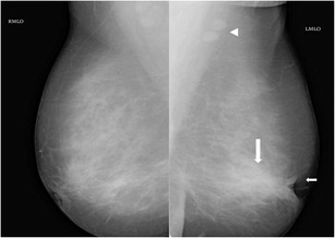
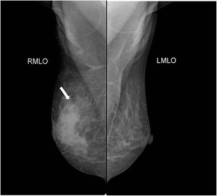
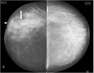
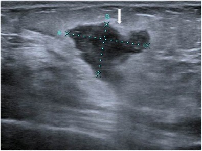
Background: Idiopathic granulomatous mastitis is a chronic inflammatory disease of the breast, which is often difficult to differentiate both clinically and radiologically from infectious aetiologies such as tuberculosis, fungal infections, and also from malignancy, thus posing a diagnostic dilemma. We present a pictorial review of the commonly encountered imaging findings in idiopathic granulomatous mastitis on mammography and ultrasound.

Materials and methods: Mammographic and ultrasound findings of histopathologically proven cases of granulomatous mastitis are discussed.

Conclusion: Idiopathic granulomatous mastitis has varied and non-specific appearances on ultrasound and mammography. Histopathology is essential to establish diagnosis.

Teaching points: • Idiopathic granulomatous mastitis often poses a diagnostic dilemma for the radiologist by mimicking malignancy. • It has varied and non-specific appearances on mammography and ultrasound. • Histopathology is mandatory to establish the diagnosis and decide management.

Keywords: Granulomatous; Histopathology; Mammography; Mastitis; Ultrasound.


Figures

Fig. 1
Fig. 1
A 38-year-old woman presented with right breast lump of 3-week duration. Mammogram (cranio-caudal view) of the right breast shows an asymmetric opacity (arrow). Histopathology was s/o idiopathic granulomatous mastitis
Fig. 2
Fig. 2
Mammogram (MLO view) of left breast of a 35-year-old woman presenting with a tender lump in left breast of 1-week duration revealed retraction of the left nipple (small arrow) with increased density in the retroareolar region (large arrow). A few benign axillary lymph nodes appearing enlarged were also seen (arrowhead). Histopathology from breast was suggestive of idiopathic granulomatous mastitis
Fig. 3
Fig. 3
Mammogram (CC view) of a 30-year-old woman who presented with painless lump in the right breast of 20-day duration showed an ill-defined dense irregular opacity (arrow) with architectural distortion involving outer quadrant of right breast. A possibility of BIRADS category IV lesion was considered and biopsy of lump was done, which showed features of idiopathic granulomatous mastitis
Fig. 4
Fig. 4
Mammogram (MLO view) of right breast in a 42-year-old woman presenting with progressively increasing lump in the right breast showed an ill-defined asymmetric opacity in the retroareolar region of the right breast (arrow) and right areolar skin thickening, compared to the opposite side
Fig. 5
Fig. 5
A 48-year-old woman presented with a tender lump of 2-week duration. There was no history of fever. Mammogram (cranio-caudal view) of right breast revealed increased density in retroareolar region (arrow) with overlying skin thickening (arrowhead). Histopathology was suggestive of granulomatous mastitis
Fig. 6
Fig. 6
A middle-aged woman presented with swelling in the left breast associated with nipple discharge. (a, b) Mammogram (CC view) shows asymmetric breast density with skin thickening in nipple areolar region of the left breast. Histopathology was s/o. Granulomatous mastitis and patient was managed conservatively. The patient presented with swelling and pain in the right breast after 2 years with nipple discharge. Mammogram (c, d) revealed skin thickening and increased density in the right breast and was once again managed conservatively
Fig. 7
Fig. 7
Ultrasound of the left breast in a 28-year-old woman who presented with breast lump and pain showed a few ill-defined hypoechoic lesions containing internal echoes (arrows) communicating with each other by tubular hypoechoic extensions (arrowhead). Biopsy was done under ultrasound guidance, and histopathology was suggestive of granulomatous mastitis
Fig. 8
Fig. 8
Ultrasound of right breast in a 33-year-old woman presenting with hard painless lump of 3 months duration shows a well-defined hypoechoic lesion with irregular margins. Ultrasound guided biopsy was done and histopathology showed granulomatous mastitis
Fig. 9
Fig. 9
Ultrasound of left breast in a 35-year-old woman presenting with a left breast lump of 1-month duration shows a small ill-defined heterogeneously hypoechoic lesion with central echogenic areas, which was confirmed to be granulomatous mastitis on histopathology
Fig. 10
Fig. 10
Ultrasound of the right breast from the same patient mentioned in Fig. 1 shows an ill-defined heterogenous lesion with increased vascularity (arrow) and tubular extension (arrowhead). Biopsy was done and histopathology was suggestive of granulomatous mastitis
Fig. 11
Fig. 11
A 32-year-old woman presented with a lump in the left breast of 6-week duration with no history of associated fever. Ultrasound of the left breast showed an ill-defined heterogeneously hypoechoic collection/lesion (arrow) with overlying skin thickening (arrowhead), which was proven to be granulomatous mastitis on histopathology
Fig. 12
Fig. 12
Mammogram (MLO view) of the right breast of a 48-year-old woman presenting with a lump in right breast of 1-month duration shows diffusely increased density in central glandular component (thick arrow) extending to the retromammary space with trabecular thickening (thin arrow). Associated areolar skin thickening (open arrow) and multiple enlarged axillary lymphnodes (arrowhead) are seen. The lesion was proven to be infiltrating ductal carcinoma on histopathology
Fig. 13
Fig. 13
A 42-year-old female presented with swelling and lump in the left breast of 1-month duration. Mammogram (a, b) showed focal asymmetric density in the lower inner quadrant of the left breast. Ultrasound (c) showed a well-defined heterogeneously hypoechoic lesion in retroareolar region with surrounding inflammation extending along the inner quadrant measuring 5 x 3.5 cm in size. The axilla showed enlarged hypoechoic lymph node (d) with loss of fatty hilum. Histopathology was s/o idiopathic granulomatous mastitis, and patient was managed conservatively
Fig. 14
Fig. 14
Granulomatous mastitis: 4x magnification showing a forming granuloma (thin arrow) in the background of diffuse lympho-plasmacytic infiltration (thick arrow) of breast parenchyma with scattered giant cells (inset, 40x)

References

    1. Kessler E, Wolloch Y. Granulomatous mastitis: a lesion clinically simulating carcinoma. Am J Clin Pathol. 1972;58:642–646. doi: 10.1093/ajcp/58.6.642. - DOI - PubMed
    1. Baslaim MM, Khayat HA, Al-Amoudi SA. Idiopathic granulomatous mastitis: a heterogeneous disease with variable clinical presentation. World J Surg. 2007;31:1677–1681. doi: 10.1007/s00268-007-9116-1. - DOI - PubMed
    1. Going JJ, Anderson TJ, Wilkinson S, Chetty U. Granulomatous lobular mastitis. J Clin Pathol. 1987;40:535–540. doi: 10.1136/jcp.40.5.535. - DOI - PMC - PubMed
    1. Akcan A, Akyildiz H, Deneme MA, Akgun H, Aritas Y. Granulomatous lobular mastitis: a complex diagnostic and therapeutic problem. World J Surg. 2006;30:1403–1409. doi: 10.1007/s00268-005-0476-0. - DOI - PubMed
    1. Imoto S, Kitaya T, Kodama T, Hasebe T, Mukai K. Idiopathic granulomatous mastitis: case report and review of the literature. Jpn J Clin Oncol. 1997;27:274–277. doi: 10.1093/jjco/27.4.274. - DOI - PubMed

LinkOut - more resources