. 2016 Oct;29(10):1383-91.
doi: 10.5713/ajas.16.0032. Epub 2016 Apr 22.

Characterization of the Nanog 5'-flanking Region in Bovine

Affiliations

Characterization of the Nanog 5'-flanking Region in Bovine

Don-Ho Choi et al. Asian-Australas J Anim Sci. 2016 Oct.

Abstract

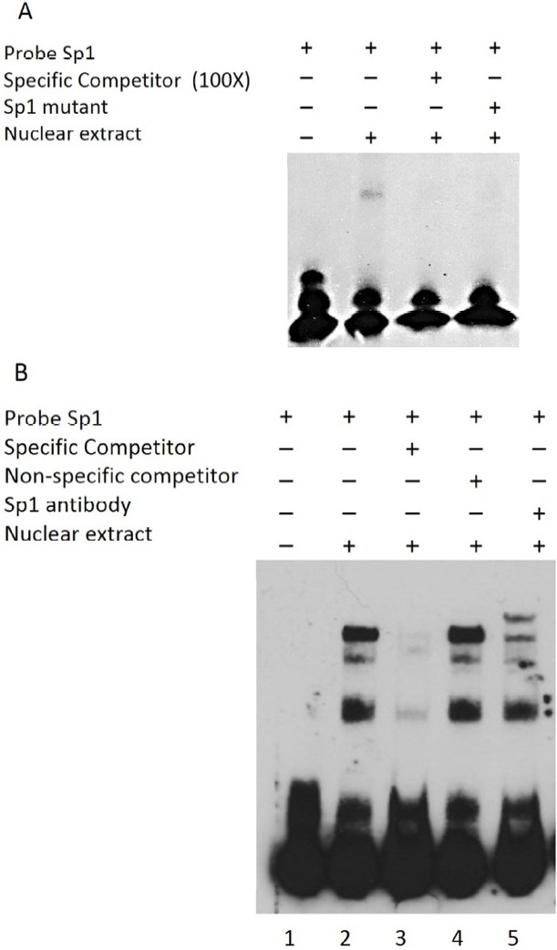
Bovine embryonic stem cells have potential for use in research, such as transgenic cattle generation and the study of developmental gene regulation. The Nanog may play a critical role in maintenance of the undifferentiated state of embryonic stem cells in the bovine, as in murine and human. Nevertheless, efforts to study the bovine Nanog for pluripotency-maintaining factors have been insufficient. In this study, in order to understand the mechanisms of transcriptional regulation of the bovine Nanog, the 5'-flanking region of the Nanog was isolated from ear cells of Hanwoo. Results of transient transfection using a luciferase reporter gene under the control of serially deleted 5'-flanking sequences revealed that the -134 to -19 region contained the positive regulatory sequences for the transcription of the bovine Nanog. Results from mutagenesis studies demonstrated that the Sp1-binding site that is located in the proximal promoter region plays an important role in transcriptional activity of the bovine Nanog promoter. The electrophoretic mobility shift assay with the Sp1 specific antibody confirmed the specific binding of Sp1 transcription factor to this site. In addition, significant inhibition of Nanog promoter activity by the Sp1 mutant was observed in murine embryonic stem cells. Furthermore, chromatin-immunoprecipitation assay with the Sp1 specific antibody confirmed the specific binding of Sp1 transcription factor to this site. These results suggest that Sp1 is an essential regulatory factor for bovine Nanog transcriptional activity.

Keywords: Bovine; Embryonic Stem Cells; Nanog; Sp1; Transcription Factors.


Figures

Figure 1
Figure 1
Regulation of the bovine Nanog 5′-flanking region and promoter activities in HEK293T, EBTr, and MBDK cell lines. (A) Schematics of the series of the bovine Nanog promoter deletion construct. (B) Bovine Nanog 5′-deletion constructs and pRL-TK were transfected into HEK293T cells (human embryonic kidney cell line, top), EBTr cells (bovine trachea cell line, middle), and MDBK cells (bovine kidney cell line, bottom). Cells were harvested after 48 h, followed by measurement of luciferase activity. Firefly luciferase activity was normalized to Renilla luciferase activity. Data shown represent the mean±standard deviation of three independent experiments. HEK293T cells, human embryonic kidney cell line; EBTr cells, bovine trachea cell line; MDBK cells, bovine kidney cell line.
Figure 2
Figure 2
Effects of site-directed mutations within the NF-κB- and Sp1-binding region of the proximal promoter region of bovine Nanog. (A) Sequence alignment of bovine, human, and mouse Nanog 5′-proximal promoter sequences. Oct4/Sox2–binding site was marked in italic, NF-κB–bindsing site is in bold and Sp1A-binding site is in bold/italic. Sp1A was identified in this study, and was also identified by Wu and Yao (2006); Sp1B was identified by Wu and Yao (2006). (B) Role of NF-κB- and Sp1-binding sites in bovine Nanog promoter activity in HEK293T cells. Wild-type or mutants of the bovine Nanog 5′-deletion construct (pbNanog-Luc [−134/+84], pbNanog-Luc [−134/+84]-mutSp1, and pbNanog-Luc [−134/+84]-mutNF-κB) (left) were co-transfected with pRL-Tk into HEK293T cells and cultured for two days. Each activity value represents the average of at least three independent experiments and is normalized according to the activity of co-transfected Renilla (* p<0.05). (C) Comparison of Nanog 5′-flanking activity between and wild- and Sp1 mutant type of GFP reporter vector. HEK293T cells, human embryonic kidney cell line.
Figure 3
Figure 3
Gel shift analyses of nuclear protein binding to Sp1 probes. Results from incubation of Biotin 3′-end labeled Sp1 probes and mutated probes (Sp1-mt) with nuclear extract proteins from HEK293T cells. (A) Lane 1, Biotin labeled Sp1 probes incubated without HEK293T nuclear extract proteins; Lane 2, Sp1 probes incubated with nuclear extract; Lane 3, Sp1-mt probes incubated with nuclear extract; Lanes 4, specific competition with unlabeled Sp1-mt probes. (B) Supershift assay using Sp1 Ab. Lane 1, without nuclear extract; Lane 2, Sp1 probes with nuclear extract; Lanes 3 and 4, specific and nonspecific competition; Lane 5, Supershift assays using Sp1 Ab. Sp-1mut: Sp1 mutant probe; NS: nonspecific competitor; HEK293T NE, HEK293T cell nuclear extracts.
Figure 4
Figure 4
Chromatin immunoprecipitation assay demonstrating the in vivo potential of Sp1 protein to bind to Nanog 5′-flanking region. Lane 1, input only; Lane 2, input with Sp1 antibody; Lane 3, input with isotype antibody; Lane 4, no template.

References

    1. Black AR, Black JD, Azizkhan-Clifford J. Sp1 and kruppel-like factor family of transcription factors in cell growth regulation and cancer. J Cell Physiol. 2001;188:143–160. - PubMed
    1. Bouwman P, Philipsen S. Regulation of the activity of Sp1-related transcription factors. Mol Cell Endocrinol. 2002;195:27–38. - PubMed
    1. Chambers I, Colby D, Robertson M, Nichols J, Lee S, Tweedie S, Smith A. Functional expression cloning of Nanog, a pluripotency sustaining factor in embryonic stem cells. Cell. 2003;113:643–655. - PubMed
    1. Chan KK, Zhang J, Chia NY, Chan YS, Sim HS, Tan KS, Oh SK, Ng HH, Choo AB. KLF4 and PBX1 directly regulate NANOG expression in human embryonic stem cells. Stem Cells. 2009;27:2114–2125. - PubMed
    1. Ginis I, Luo Y, Miura T, Thies S, Brandenberger R, Gerecht-Nir S, Amit M, Hoke A, Carpenter MK, Itskovitz-Eldor J, Rao MS. Differences between human and mouse embryonic stem cells. Dev Biol. 2004;269:360–380. - PubMed

LinkOut - more resources