. 2016;25(5):408-16.
doi: 10.1159/000446480. Epub 2016 Jun 4.

Proliferation and Migration of Peripheral Retinal Pigment Epithelial Cells Are Associated with the Upregulation of Wingless-Related Integration and Bone Morphogenetic Protein Signaling in Dark Agouti Rats

Affiliations

Proliferation and Migration of Peripheral Retinal Pigment Epithelial Cells Are Associated with the Upregulation of Wingless-Related Integration and Bone Morphogenetic Protein Signaling in Dark Agouti Rats

Heba Al-Hussaini et al. Med Princ Pract. 2016.

Abstract

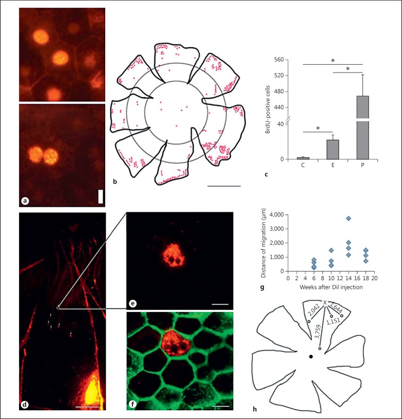
Objective: The aim of this study was to investigate the possible migration of proliferating peripheral retinal pigment epithelial (RPE) cells and their association with differential gene expressions.

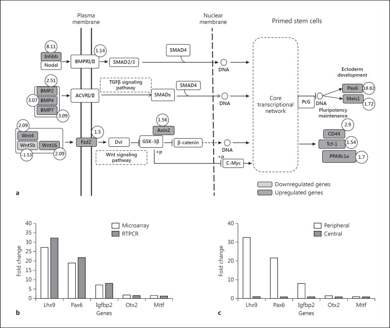
Materials and methods: The RPE layer was obtained from the inner aspect of the eyeball of dark agouti rats (12-13 weeks old) and was mounted on glass slides. The peripheral RPE cell proliferation was evaluated using bromodeoxyuridine immunohistochemistry (n = 10). The cell migration was examined using the Dil tracer technique (n = 40) at the end of weeks 6, 10, 14 and 18. Affymetrix microarray analysis was used to investigate differential gene expressions in peripheral and central RPE cells, which was authenticated by RT-PCR using 4 RPE-specific genes (n = 10).

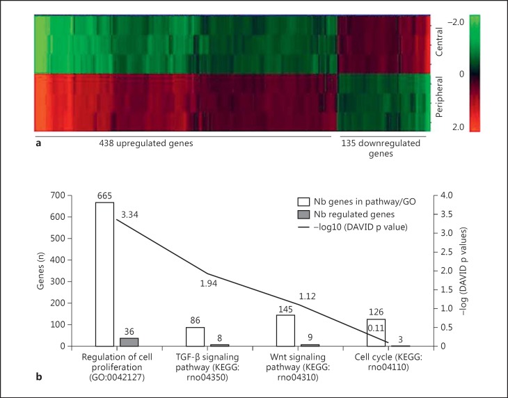
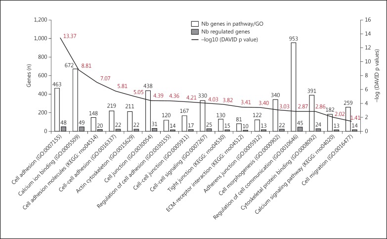
Results: In this study, peripheral RPE cells divided and appeared in clusters, but equatorial and central RPE cells rarely divided. The peripheral RPE cells migrated to the central RPE region in a time-dependent manner up to the end of week 14, but not later. The microarray analysis showed the expression of 9,645 out of a total of 35,220 genes studied. Among the 9,645 genes, 573 were differentially expressed (438 were upregulated and 135 were downregulated) in peripheral RPE cells as compared to central RPE cells. Of these 573 genes, 56 were associated with signaling pathways related to the regulation of cell proliferation, including Pax6, TGFβ, BMP and Wnt, and 404 were associated with pathways of cell migration.

Conclusions: In this study, peripheral RPE cells divided and migrated to the central region. This process was associated with differential gene expressions in these cells.


Figures

Fig. 1
Fig. 1
Quantification of RPE cell proliferation and migration in adult DA rats. a Representative photomicrograph of binucleated and mononucleated RPE cells indicating BrdU-labeled cells. Scale bar = 10 µm. b Distribution of BrdU-labeled cells mostly in the peripheral RPE cells. Scale bar = 2 mm. c The number of BrdU-labeled cells at the three RPE regions: central (C), equatorial (E) and peripheral (P). d A low-magnification photomicrograph showing the DiI crystal injection site on the lower right; a few Dil-labeled RPE cells are indicated by arrows. e, f A fluorescent DiI crystal-incorporated cell from a is shown at a higher magnification. In f, the same cell shown in e is presented with phalloidin counterstain to mark the cell membrane. Scale bars = 500 µm (d) and 10 µm (e, f). g Distances (in µm) travelled by migrating RPE cells at the end of weeks 6, 10, 14 and 18, measured from the sites of Dil crystal injections at the peripheral retina. h The floral representation of distances travelled by migrating RPE cells by week 14. The letter X indicates the site of the injection and the numbers indicate the distances (in µm) travelled by the migrating cells.
Fig. 2
Fig. 2
a Hierarchical clustering of differentially expressed genes in the two regions of the RPE layer. The color intensities show the extent of gene expressions in peripheral RPE cells either above (red) or below (green) the average gene expression (indicated by 0.0). b KEGG pathways and GO terms relating to proliferation. White bars correspond to the number of genes involved in KEGG pathways/GO terms and the gray bars correspond to the number of regulated genes involved in the 4 signaling pathways associated with cell proliferation. The dark curve corresponds to -log (DAVID p value) for each KEGG pathway/GO term (uncorrected p values are indicated above and below the curve, calculated using the EASE score (modified Fisher's exact p value) from DAVID.
Fig. 3
Fig. 3
KEGG pathways/GO terms relating to migration. White bars correspond to the number of genes involved in the KEGG pathways/GO terms and the gray bars correspond to the number of regulated genes for the 17 signaling pathways associated with cell migration. The black line corresponds to -log (DAVID p value) for each KEGG pathway/GO terms (uncorrected p values are indicated above and below the curve, calculated using the EASE score (modified Fisher's exact p value) from DAVID.
Fig. 4
Fig. 4
a BMP and Wnt signaling pathways in peripheral RPE cells. Fold changes for upregulated (dark gray) and downregulated (light gray) genes in peripheral RPE cells as compared to those in central RPE cells are indicated. The fold changes are indicated by numbers inside circles. b Comparison of RT-PCR results for the 5 genes with those of the microarray in peripheral RPE cells indicating the reproducibility of fold changes by the two methods. c RT-PCR results for the 5 genes indicating fold changes in the peripheral RPE as compared to those in the central RPE.

References

    1. Strauss O. The retinal pigment epithelium in visual function. Physiol Rev. 2005;85:845–881. - PubMed
    1. Al-Hussaini H, Kam JH, Vugler A, et al. Mature retinal pigment epithelium cells are retained in the cell cycle and proliferate in vivo. Mol Vis. 2008;14:1784–1791. - PMC - PubMed
    1. Kokkinopoulos I, Shahabi G, Colman A, et al. Mature peripheral RPE cells have an intrinsic capacity to proliferate; a potential regulatory mechanism for age-related cell loss. PLoS One. 2011;6:e18921. - PMC - PubMed
    1. Del Priore LV, Kuo YH, Tezel TH. Age-related changes in human RPE cell density and apoptosis proportion in situ. Invest Ophthalmol Vis Sci. 2002;43:3312–3318. - PubMed
    1. Alexander P, Thomson HA, Luff AJ, et al. Retinal pigment epithelium transplantation: concepts, challenges, and future prospects. Eye (Lond) 2015;29:992–1002. - PMC - PubMed