. 2016 May 10;15(1):264.
doi: 10.1186/s12936-016-1318-2.

Delayed acquisition of Plasmodium falciparum antigen-specific CD4(+) T cell responses in HIV-exposed uninfected Malawian children receiving daily cotrimoxazole prophylaxis

Affiliations

Delayed acquisition of Plasmodium falciparum antigen-specific CD4(+) T cell responses in HIV-exposed uninfected Malawian children receiving daily cotrimoxazole prophylaxis

Herbert Longwe et al. Malar J. .

Abstract

Background: Cotrimoxazole (CTX) prophylaxis, recommended in HIV-exposed uninfected (HEU) children primarily against HIV-related opportunistic infections, has been shown to have some efficacy against Plasmodium falciparum malaria. The effects of CTX prophylaxis on the acquisition of P. falciparum antigen specific CD4(+) T cells-mediated immunity in HEU children is still not fully understood.

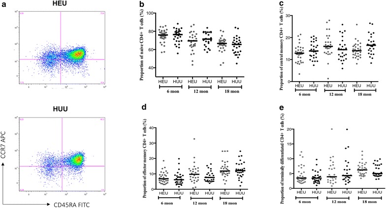
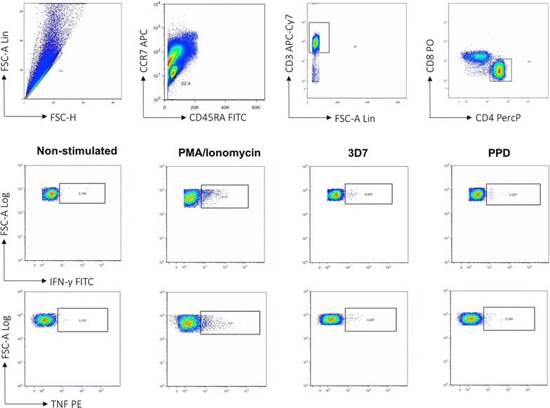
Methods: Peripheral blood was collected from HEU and HIV-unexposed uninfected (HUU) children at 6, 12 and 18 months of age. Proportion of CD4(+) T cells subsets were determined by immunophenotyping. P. falciparum antigen-specific CD4(+) T cells responses were measured by intracellular cytokine staining assay.

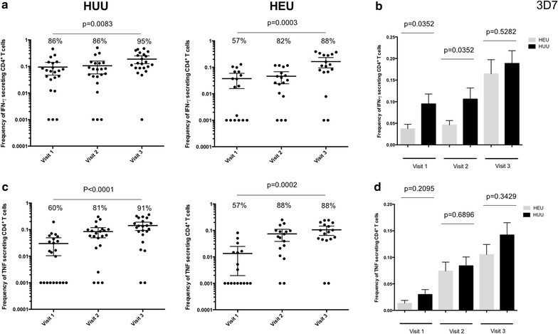
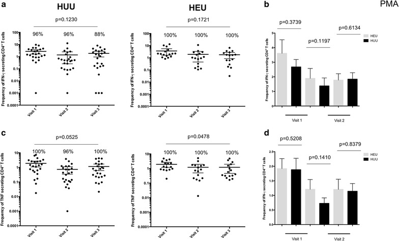
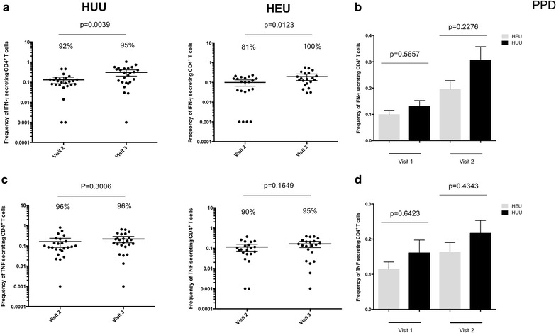
Results: There were no differences in the proportions of naïve, effector and memory CD4(+) T cell subsets between HEU and HUU children at all ages. There was a trend showing acquisition of P. falciparum-specific IFN-γ and TNF-producing CD4(+) T cells with age in both HUU and HEU children. There was, however, lower frequency of P. falciparum-specific IFN-γ-producing CD4(+) T cells in HEU compared to HUU at 6 and 12 months, which normalized 6 months after stopping CTX prophylaxis.

Conclusion: The results demonstrate that there is delayed acquisition of P. falciparum-specific IFN-γ-producing CD4(+) T cells in HEU children on daily cotrimoxazole prophylaxis, which is evident at 6 and 12 months of age in comparison to HUU age-matched controls. However, whether this delayed acquisition of P. falciparum-specific IFN-γ-producing CD4(+) T cells leads to higher risk to malaria disease remains unknown and warrants further investigation.

Keywords: CD4+ T cells; Cotrimoxazole; HIV-exposed children; Plasmodium falciparum.


Figures

Fig. 1
Fig. 1
The proportions of naive and memory CD4+ T cell subsets in HEU and HUU children at different ages. Cells were stained anti CD3 PE, anti CD4 PerCP, anti CD8 APC-H7, anti CD45RA FITC and anti CCR7 APC monoclonal antibodies. a A gate was drawn on CD4 CD8(non T cells) and CD4+ CD8 (CD4+ T cells) populations and plotted on CD45RA FITC against CCR7 APC. Using quadrant statistics, distinct populations of naïve (CD45RA+ CCR7+), central memory (CD45RACCR7+), effector memory (CD45RA CCR7), and terminally differentiated (CD45RA+ CCR7) CD4+ T cells were identified. Median proportions of Naïve CD4+ (b), central memory CD4+ (c), effector memory CD4+ (d), and terminally differentiated CD4+T lymphocytes (e) were determined in peripheral blood of HEU and HUU children at 6, 12 and 18 months. X-axis represents age of children in months. Horizontal bars represent medians
Fig. 2
Fig. 2
Representative FACS plots showing gating strategy for identifying antigen specific CD4+ T cell by intracellular cytokine staining assay. Heparinized whole blood collected at 6, 12 and 18 months was stimulated with PMA, P. falciparum 3D7 strain, purified protein derivative (PPD) and no antigen for 24 h. Lymphocytes were gated using FSC vs. SSC plots, FSC vs. CD3+ T cells and then CD4+T cells vs. CD8+T cells to isolate CD4+ T cells. Non-stimulated control samples were used to establish the threshold quadrants of background responses that were applied to quantify positive cytokine-producing cells in paired antigen-stimulated samples
Fig. 3
Fig. 3
Frequency of PMA/Ionomycin stimulated CD4+ T cells in HEU and HUU children at different ages. a Total percentage of all IFN-γ cytokine producing CD4+ T cells was determined in HEU children and compared to HUU children at 6, 12 and 18 months of age. b Histogram comparing total percentage of all IFN-γ cytokine producing CD4+ T cells between HEU and HUU children overtime. c Total percentage of all TNF cytokine producing CD4+ T cells was determined in HEU children and compared to HUU children at 6, 12 and 18 months of age. d Histogram comparing total percentage of all TNF cytokine producing CD4+ T cells between HEU and HUU children overtime. X-axis represents time of visit. Black horizontal bars represent (a, c) mean with confidence intervals or (b, d) mean with SEM. Statistical significance was determined if p value was equal to or less than 0.05
Fig. 4
Fig. 4
Frequency of Plasmodium falciparum antigen specific CD4+ T cells in HEU and HUU children at different ages. a Total percentage of all IFN-γ cytokine producing CD4+ T cells was determined in HEU children and compared to HUU children at 6, 12 and 18 months of age. b Histogram comparing total percentage of all IFN-γ cytokine producing CD4+ T cells between HEU and HUU children overtime. c Total percentage of all TNF cytokine producing CD4+ T cells was determined in HEU children and compared to HUU children at 6, 12 and 18 months of age. d Histogram comparing total percentage of all TNF cytokine producing CD4+ T cells between HEU and HUU children overtime. X-axis represents time of visit. Black horizontal bars represent (a, c) mean with confidence intervals or (b, d) mean with SEM. Statistical significance was determined if p value was equal to or less than 0.05
Fig. 5
Fig. 5
Frequency of mycobacterium antigen specific CD4+ T cells in HEU and HUU children at different ages. a Total percentage of all IFN-γ cytokine producing CD4+ T cells was determined in HEU children and compared to HUU children at 6, 12 and 18 months of age. b Histogram comparing total percentage of all IFN-γ cytokine producing CD4+ T cells between HEU and HUU children overtime. c Total percentage of all TNF cytokine producing CD4+ T cells was determined in HEU children and compared to HUU children at 6, 12 and 18 months of age. d Histogram comparing total percentage of all TNF cytokine producing CD4+ T cells between HEU and HUU children overtime. X-axis represents time of visit. Black horizontal bars represent (a, c) mean with confidence intervals or (b, d) mean with SEM. Statistical significance was determined if p value was equal to or less than 0.05

References

    1. Marsh K, Kinyanjui S. Immune effector mechanisms in malaria. Parasite Immunol. 2006;28:51–60. doi: 10.1111/j.1365-3024.2006.00808.x. - DOI - PubMed
    1. Schumacher RF, Spinelli E. Malaria in children. Mediterr J Hematol Infect Dis. 2012;4:e2012073. doi: 10.4084/mjhid.2012.073. - DOI - PMC - PubMed
    1. Kimura D, Miyakoda M, Honma K, Shibata Y, Yuda M, Chinzei Y, et al. Production of IFN-gamma by CD4(+) T cells in response to malaria antigens is IL-2 dependent. Int Immunol. 2010;22:941–952. doi: 10.1093/intimm/dxq448. - DOI - PubMed
    1. Overstreet MG, Cockburn IA, Chen YC, Zavala F. Protective CD8 T cells against Plasmodium liver stages: immunobiology of an ‘unnatural’ immune response. Immunol Rev. 2008;225:272–283. doi: 10.1111/j.1600-065X.2008.00671.x. - DOI - PMC - PubMed
    1. Roestenberg M, Teirlinck AC, McCall MB, Teelen K, Makamdop KN, Wiersma J, et al. Long-term protection against malaria after experimental sporozoite inoculation: an open-label follow-up study. Lancet. 2011;377:1770–1776. doi: 10.1016/S0140-6736(11)60360-7. - DOI - PubMed

Publication types

Substances