

The intracellular trafficking mechanism of Lipofectamine-based transfection reagents and its implication for gene delivery
- PMID: 27165510
- PMCID: PMC4863168
- DOI: 10.1038/srep25879
The intracellular trafficking mechanism of Lipofectamine-based transfection reagents and its implication for gene delivery
Abstract
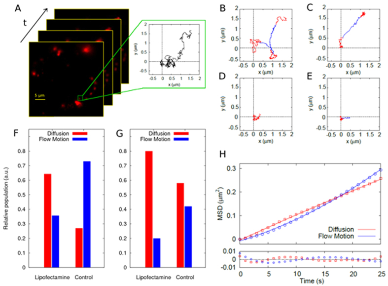
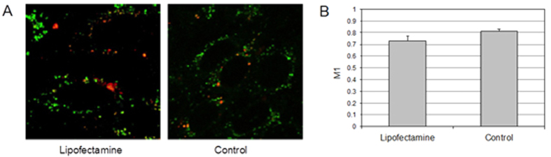
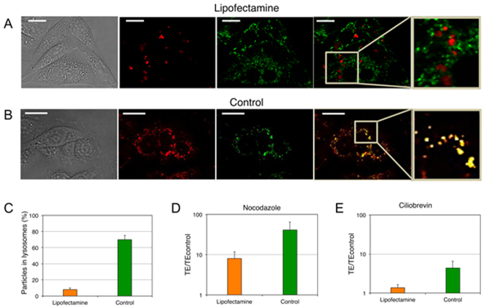
Lipofectamine reagents are widely accepted as "gold-standard" for the safe delivery of exogenous DNA or RNA into cells. Despite this, a satisfactory mechanism-based explanation of their superior efficacy has remained mostly elusive thus far. Here we apply a straightforward combination of live cell imaging, single-particle tracking microscopy, and quantitative transfection-efficiency assays on live cells to unveil the intracellular trafficking mechanism of Lipofectamine/DNA complexes. We find that Lipofectamine, contrary to alternative formulations, is able to efficiently avoid active intracellular transport along microtubules, and the subsequent entrapment and degradation of the payload within acidic/digestive lysosomal compartments. This result is achieved by random Brownian motion of Lipofectamine-containing vesicles within the cytoplasm. We demonstrate here that Brownian diffusion is an efficient route for Lipofectamine/DNA complexes to avoid metabolic degradation, thus leading to optimal transfection. By contrast, active transport along microtubules results in DNA degradation and subsequent poor transfection. Intracellular trafficking, endosomal escape and lysosomal degradation appear therefore as highly interdependent phenomena, in such a way that they should be viewed as a single barrier on the route for efficient transfection. As a matter of fact, they should be evaluated in their entirety for the development of optimized non-viral gene delivery vectors.
Figures
Similar articles
-
Polyethyleneimine-Coated Gold Nanoparticles: Straightforward Preparation of Efficient DNA Delivery Nanocarriers.Chem Asian J. 2016 Dec 6;11(23):3365-3375. doi: 10.1002/asia.201600951. Epub 2016 Nov 10. Chem Asian J. 2016. PMID: 27685032
-
The role of cytoskeleton networks on lipid-mediated delivery of DNA.Ther Deliv. 2013 Feb;4(2):191-202. doi: 10.4155/tde.12.151. Ther Deliv. 2013. PMID: 23343159 Free PMC article.
-
[Development of non-viral vector based on the quantitative comparison of intracellular trafficking with viral vector].Yakugaku Zasshi. 2006 Nov;126(11):1047-57. doi: 10.1248/yakushi.126.1047. Yakugaku Zasshi. 2006. PMID: 17077611 Review. Japanese.
-
Quantitative and mechanism-based investigation of post-nuclear delivery events between adenovirus and lipoplex.Nucleic Acids Res. 2007;35(5):1533-43. doi: 10.1093/nar/gkl1165. Epub 2007 Feb 7. Nucleic Acids Res. 2007. PMID: 17287293 Free PMC article.
-
Cationic lipids, lipoplexes and intracellular delivery of genes.J Control Release. 2006 Nov 28;116(2):255-64. doi: 10.1016/j.jconrel.2006.06.024. Epub 2006 Jun 28. J Control Release. 2006. PMID: 16914222 Review.
Cited by
-
Synthesis and bioactivity of a novel surfactin-based lipopeptide for mRNA delivery.Nanoscale Adv. 2024 Sep 4;6(20):5193-206. doi: 10.1039/d4na00404c. Online ahead of print. Nanoscale Adv. 2024. PMID: 39247856 Free PMC article.
-
Actin cytoskeleton remodeling primes RIG-I-like receptor activation.Cell. 2022 Sep 15;185(19):3588-3602.e21. doi: 10.1016/j.cell.2022.08.011. Cell. 2022. PMID: 36113429 Free PMC article.
-
Looking Back, Moving Forward: Lipid Nanoparticles as a Promising Frontier in Gene Delivery.ACS Pharmacol Transl Sci. 2023 Oct 24;6(11):1561-1573. doi: 10.1021/acsptsci.3c00185. eCollection 2023 Nov 10. ACS Pharmacol Transl Sci. 2023. PMID: 37974625 Free PMC article. Review.
-
Optimized workflow to modify microRNA expression in primary human intravascular cells.BMC Immunol. 2023 Feb 15;24(1):5. doi: 10.1186/s12865-023-00540-9. BMC Immunol. 2023. PMID: 36792999 Free PMC article.
-
Technological Approaches in the Analysis of Extracellular Vesicle Nucleotide Sequences.Front Bioeng Biotechnol. 2021 Dec 23;9:787551. doi: 10.3389/fbioe.2021.787551. eCollection 2021. Front Bioeng Biotechnol. 2021. PMID: 35004647 Free PMC article. Review.
References
-
- Akita H., Ito R., Khalil I. A., Futaki S. & Harashima H. Quantitative three-dimensional analysis of the intracellular trafficking of plasmid DNA transfected by a nonviral gene delivery system using confocal laser scanning microscopy. Mol. Ther. 9, 443–451 (2004). - PubMed
-
- Suh J., Wirtz D. & Hanes J. Real-time intracellular transport of gene nanocarriers studied by multiple particle tracking. Biotechnol. Prog. 20, 598–602 (2004). - PubMed
-
- Elouahabi A. & Ruysschaert J. M. Formation and intracellular trafficking of lipoplexes and polyplexes. Mol. Ther. 11, 336–347 (2005). - PubMed
-
- Lucas B. et al.. Towards a better understanding of the dissociation behavior of liposome-oligonucleotide complexes in the cytosol of cells. J. Control. Release 103, 435–450 (2005). - PubMed
MeSH terms
Substances
LinkOut - more resources
Full Text Sources
Other Literature Sources
Research Materials
