Comparative Study
. 2016 Jul;90(1):109-22.
doi: 10.1016/j.kint.2016.02.036. Epub 2016 May 7.

Distinct roles for the complement regulators factor H and Crry in protection of the kidney from injury

Affiliations
Comparative Study

Distinct roles for the complement regulators factor H and Crry in protection of the kidney from injury

Jennifer Laskowski et al. Kidney Int. 2016 Jul.

Abstract

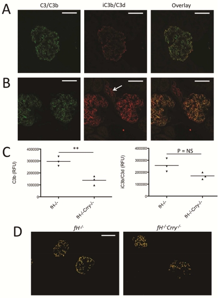
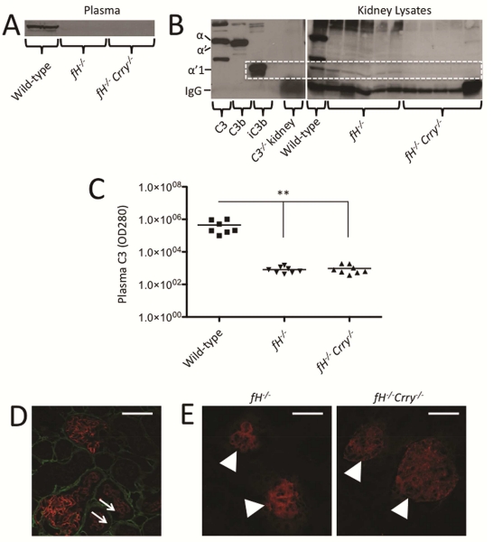
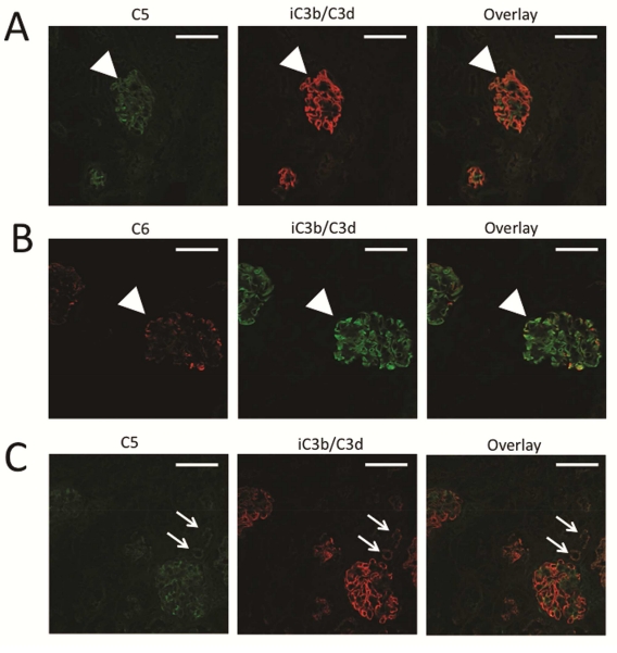
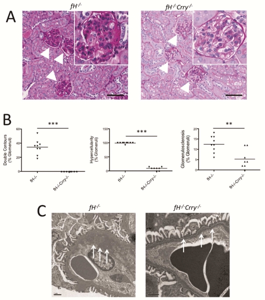
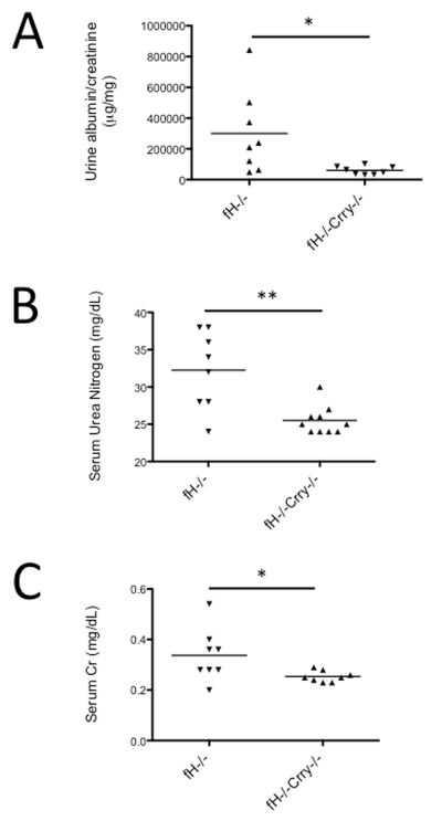
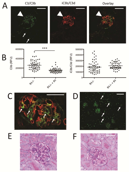
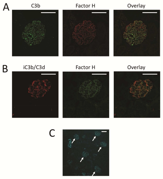
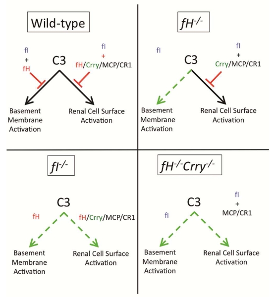
Mutations in the complement regulatory proteins are associated with several different diseases. Although these mutations cause dysregulated alternative pathway activation throughout the body, the kidneys are the most common site of injury. The susceptibility of the kidney to alternative pathway-mediated injury may be due to limited expression of complement regulatory proteins on several tissue surfaces within the kidney. To examine the roles of the complement regulatory proteins factor H and Crry in protecting distinct renal surfaces from alternative pathway mediated injury, we generated mice with targeted deletions of the genes for both proteins. Surprisingly, mice with combined genetic deletions of factor H and Crry developed significantly milder renal injury than mice deficient in only factor H. Deficiency of both factor H and Crry was associated with C3 deposition at multiple locations within the kidney, but glomerular C3 deposition was lower than that in factor H alone deficient mice. Thus, factor H and Crry are critical for regulating complement activation at distinct anatomic sites within the kidney. However, widespread activation of the alternative pathway reduces injury by depleting the pool of C3 available at any 1 location.

Keywords: complement; glomerulonephritis; inflammation.


Figures

Figure 1
Figure 1. C3 metabolism in the glomeruli of mice with targeted deletion of the genes for factor H and Crry
We examined the deposition and metabolism of C3 in the glomeruli of fH−/− and fH−/−Crry−/− mice using confocal microscopy. A) Kidney sections of fH−/− mice were stained using antibodies to detect C3b (green) and iC3b/C3d (red). Both forms of C3 were detected along the capillary loops. Original magnification ×600. Bar = 50 μm. B) In fH−/−Crry−/− mice, C3b and iC3b/C3d co-localized within the mesangium. iC3b/C3d was also detected within the proximal tubules (arrow). Original magnification ×600. C) The intensities of C3b and iC3b/C3d were measured in the glomeruli of fH−/− and fH−/−Crry−/− mice. Less glomerular C3b was detected in the fH−/−Crry−/− mice. There was a trend towards lower levels of iC3b/C3d in the fH−/−Crry−/− mice, but some iC3b/C3d was seen in all of the glomeruli. D) To examine the sites of iC3b/C3d generation, the co-localization function of Olympus FV10-ASW software was used to isolate areas of C3b and iC3b/C3d co-localization in the kidneys of fH−/− and fH−/−Crry−/− mice. Co-localized C3b and iC3b/C3d deposits were seen in the capillary loops of fH−/− mice and in the mesangium of fH−/−Crry−/− mice. Original magnification ×200. Bar = 50 μm.
Figure 2
Figure 2. Complement activation in mice with targeted deletions of the genes for both factor H and Crry
A) Western blot analysis of C3 fragments in the plasma of fH−/− and fH−/−Crry−/− mice demonstrates that intact C3 levels are lower in both strains of mice than in wild-type mice. B) A western blot of kidney lysates demonstrates that the C3α’1 fragment of iC3b is more abundant in fH−/− mice than in fH−/−Crry−/− mice. Purified proteins were used to identify the different C3α chain fragments, and the C3α’1 band is indicated with the dashed line. C) Plasma C3 levels were measured by ELISA. C3 levels were significantly lower in the plasma of fH−/− and fH−/−Crry−/− mice than in that of wild-type mice. **P < 0.01. D) Kidney sections of fH−/−Crry−/− mice were stained using antibodies to detect collagen IV (green) and iC3b/C3d (red). Collagen IV was seen in the tubular basement membrane at the basolateral side of the tubules, demonstrating that iC3b/C3d was deposited on the apical surface of the proximal tubules (arrows). E) DAF was detected in the glomeruli (arrowheads) of both fH−/− and fH−/−Crry−/− mice. Original magnification ×600. Bar = 50 μm.
Figure 3
Figure 3. Terminal complement complex generation in factor H deficient mice
To assess the degree of complement activation in the glomeruli of fH−/− and fH−/−Crry−/− mice, we examined the deposition of C5 and C6 using confocal microscopy. A) C5 (green) was detected in the glomeruli of fH−/− mice in a pattern similar to that of iC3b/C3d (red). B) C6 (red) and iC3b/C3d (green) co-localized in the glomeruli and in the proximal tubules of fH−/− mice, indicating that complement activation generated the terminal complement complex at this location. C) Segmental deposits of C5 (green) were also detected in the glomeruli of fH−/−Crry−/− mice in a pattern similar to iC3b/C3d (red). Original magnification ×600. Bar = 50 μm.
Figure 4
Figure 4. Mice with targeted deletions of the genes for both factor H and Crry are protected from glomerular injury
A) Kidney sections from 14 month-old fH−/− and fH−/−Crry−/− mice were stained with PAS and examined for histologic evidence of injury. Glomeruli in fH−/− mice demonstrated double contours of the GBM, hypercellularity, mesangial expansion, and glomerulosclerosis. Most of the glomeruli in the fH−/−Crry−/− mice were normal appearing. Expanded view of a representative glomerulus is shown in the inset. Original magnification ×200 (×400 for inset). Bar = 100 μm. B) Kidney sections from 14 month-old fH−/− and fH−/−Crry−/− mice were examined by a renal pathologist in a blinded fashion. Significantly fewer glomeruli in fH−/−Crry−/− mice demonstrated double contours of the GBM, hypercellularity, or glomerulosclerosis. All of the fH−/− mice had both endocapillary proliferation and mesangial expansion, whereas hypercellularity in the fH−/− mice was only seen in the mesangium. **P < 0.01, ***P < 0.001. C) Kidney sections from 14 month-old fH−/− and fH−/−Crry−/− mice were examined by electron microscopy. The GBMs of fH−/− mice were markedly thickened and contained electron dense material (arrows). The GBMs of fH−/−Crry−/− mice were normal appearing (arrows). The size bar represents 1 μm.
Figure 5
Figure 5. Mice with targeted deletions of the genes for both factor H and Crry are protected from development of albuminuria and renal failure
A) Albumin and creatinine levels were measured in the urine from 14 month-old fH−/− and fH−/−Crry−/− mice. The albumin/creatinine content of fH−/−Crry−/− mice was significantly lower than that of fH−/− mice. B) Serum urea nitrogen and C) serum creatinine levels were measured in 14 month-old fH−/− and fH−/−Crry−/− mice. Serum urea nitrogen and creatinine were both significantly lower in fH−/−Crry−/− mice compared to fH−/− mice. *P < 0.05, **P < 0.01.
Figure 6
Figure 6. Factor H regulates complement activation on the GBM
Factor H deficient mice were reconstituted with factor H purified from the plasma of wild-type mice, and complement proteins in the glomeruli were examined using confocal microscopy. A) Kidney sections of reconstituted fH−/− mice were stained using antibodies to detect C3b (green) and iC3b/C3d (red). The pattern of C3b deposits suggested deposition in the mesangium (arrowhead) and along the tubular basement membranes (small arrows). iC3b/C3d was heavily deposited along the GBM. Original magnification ×600. Bar = 50 μm. B) The intensities of C3b and iC3b/C3d were measured in the glomeruli of a fH−/− mouse injected with a vehicle control or with purified factor H. Injection with factor H reduced the intensity of glomerular C3b. C) A high-power view of a glomerulus from a fH−/− mouse injected with purified factor H demonstrates that areas of C3b without iC3b/C3d are seen in the mesangium, and iC3b/C3d is seen in the capillary loops. Original magnification ×600. Bar = 50 μm. D) A low power view of a kidney sections from a fH−/− mouse reconstituted with factor H shows that injection of factor H restored C3b deposition on the tubular basement membranes (arrows). Original magnification ×200. Bar = 100 μm. PAS stained kidneys from a representative fH−/− mouse injected with E) vehicle and F) factor H did are shown. Original magnification ×600. Bar = 50 μm.
Figure 7
Figure 7. Factor H binds to the glomerular basement membrane of factor H deficient mice
fH−/− mice were injected with DyLight 650-labeled recombinant factor H, and kidneys from these mice were examined by confocal microscopy. A) C3b was detected in the capillaries and the mesangium of injected mice, and factor H (shown as red in this panel) was deposited along the capillary walls. B) iC3b/C3d was detected along the capillary walls in a pattern similar to the pattern of the injected factor H deposition (shown as green in this panel). Original magnification ×600. Bar = 50 μm. C) A low power view of a kidney section demonstrates that factor H primarily localized to the glomeruli of fH−/− mice. Original magnification ×200. Bar = 50 μm.
Figure 8
Figure 8. Model of glomerular complement regulation
Factor I and factor H together control AP activation on the GBM. Factor I also controls AP activation on renal cell surfaces. Cofactor function on cell surfaces is provided by factor H, MCP, CR1, and Crry (in rodents). Deficiency of factor H in fH−/− mice permits uncontrolled AP activation on the GBM. Factor I deficiency permits uncontrolled activation on the GBM and on cell surfaces. Combined factor H and Crry deficiency also permits activation on the GBM and on cell surfaces.

Similar articles

Cited by

References

    1. Ricklin D, Hajishengallis G, Yang K, et al. Complement: a key system for immune surveillance and homeostasis. Nat Immunol. 2010;11:785–797. - PMC - PubMed
    1. Zipfel PF, Skerka C. Complement regulators and inhibitory proteins. Nat Rev Immunol. 2009;9:729–740. - PubMed
    1. de Cordoba SR, de Jorge EG. Translational mini-review series on complement factor H: genetics and disease associations of human complement factor H. Clin Exp Immunol. 2008;151:1–13. - PMC - PubMed
    1. Pickering MC, D’Agati VD, Nester CM, et al. C3 glomerulopathy: consensus report. Kidney Int. 2013;84:1079–1089. - PMC - PubMed
    1. Servais A, Fremeaux-Bacchi V, Lequintrec M, et al. Primary glomerulonephritis with isolated C3 deposits: a new entity which shares common genetic risk factors with haemolytic uraemic syndrome. Journal of medical genetics. 2007;44:193–199. - PMC - PubMed

Publication types