. 2016 Aug;90(2):300-310.
doi: 10.1016/j.kint.2016.02.018. Epub 2016 Apr 27.

Endothelin A receptor activation on mesangial cells initiates Alport glomerular disease

Affiliations

Endothelin A receptor activation on mesangial cells initiates Alport glomerular disease

Brianna Dufek et al. Kidney Int. 2016 Aug.

Abstract

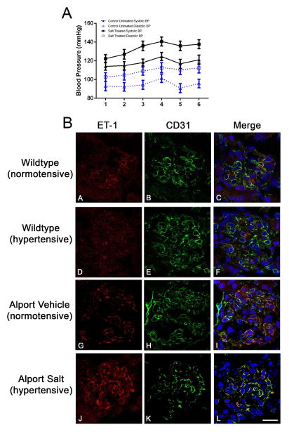
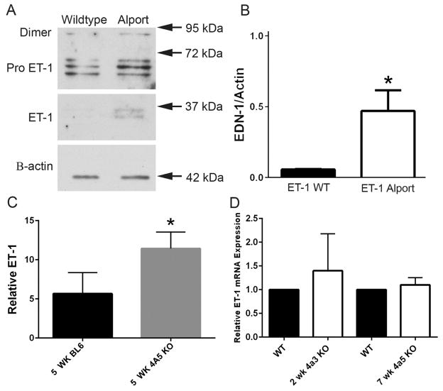
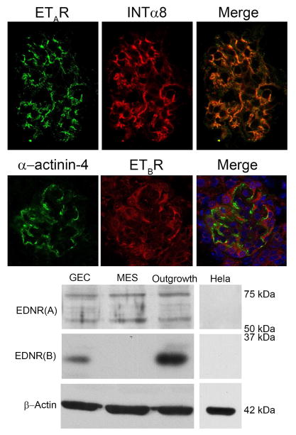
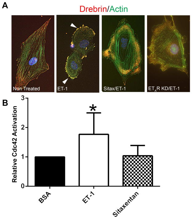
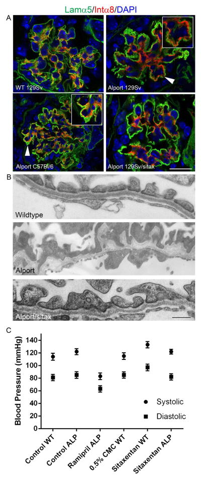
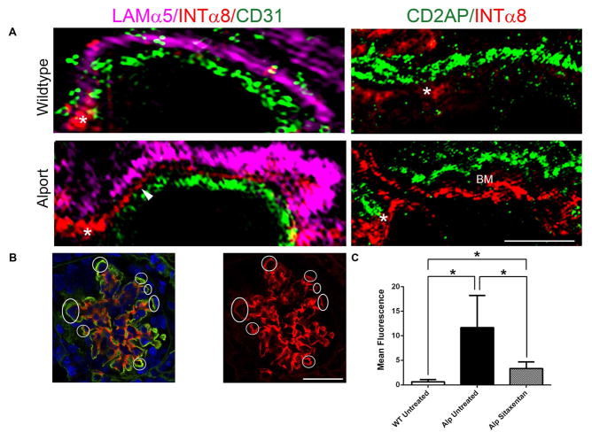
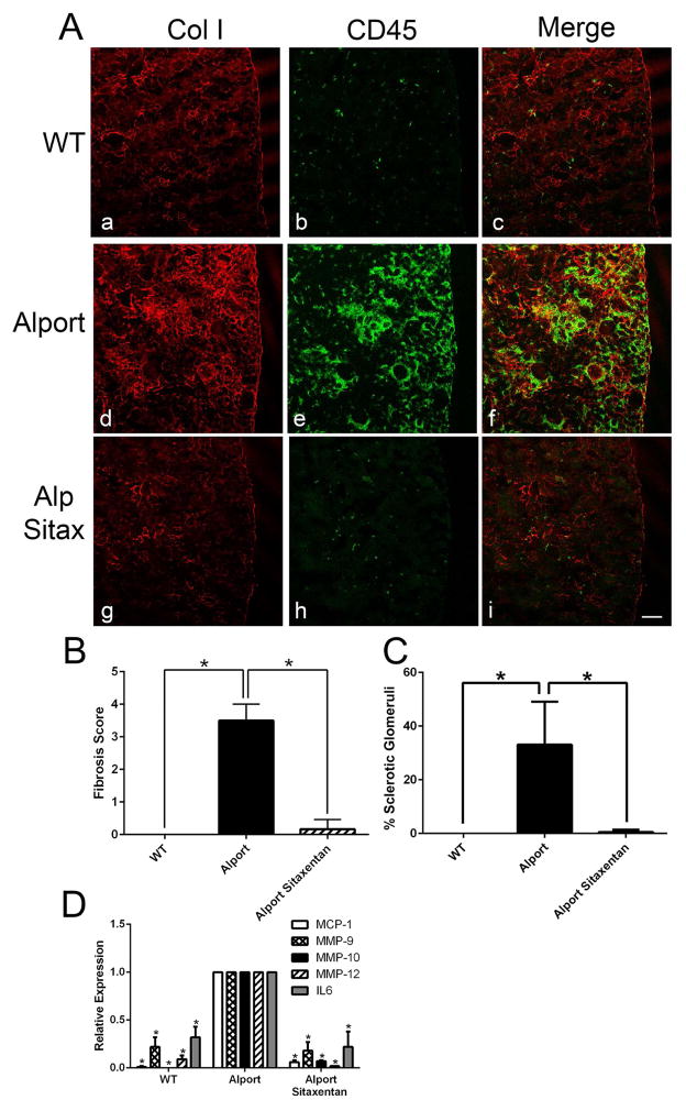
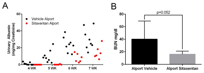
Recent work demonstrates that Alport glomerular disease is mediated through a biomechanical strain-sensitive activation of mesangial actin dynamics. This occurs through a Rac1/CDC42 cross-talk mechanism that results in the invasion of the subcapillary spaces by mesangial filopodia. The filopodia deposit mesangial matrix proteins in the glomerular basement membrane, including laminin 211, which activates focal adhesion kinase in podocytes culminating in the up-regulation of proinflammatory cytokines and metalloproteinases. These events drive the progression of glomerulonephritis. Here we test whether endothelial cell-derived endothelin-1 is up-regulated in Alport glomeruli and further elevated by hypertension. Treatment of cultured mesangial cells with endothelin-1 activates the formation of drebrin-positive actin microspikes. These microspikes do not form when cells are treated with the endothelin A receptor antagonist sitaxentan or under conditions of small, interfering RNA knockdown of endothelin A receptor mRNA. Treatment of Alport mice with sitaxentan results in delayed onset of proteinuria, normalized glomerular basement membrane morphology, inhibition of mesangial filopodial invasion of the glomerular capillaries, normalization of glomerular expression of metalloproteinases and proinflammatory cytokines, increased life span, and prevention of glomerulosclerosis and interstitial fibrosis. Thus endothelin A receptor activation on mesangial cells is a key event in initiation of Alport glomerular disease in this model.

Keywords: Alport syndrome; actin dynamics; endothelin; glomerulonephritis.


Figures

Figure 1
Figure 1
1A: Induction of hypertension in Alport mice. C57Bl/6 X-linked Alport mice were made hypertensive by giving them the L-NAME salts in their drinking water from 4 to 7 weeks of age. Normotensive mice were given plain drinking water. Blood pressures were measured longitudinally using the CODA-2 tail cuff system. Numbers on the X-axis represent individual measurements which were performed twice weekly from 4 to 7 weeks of age. Both systolic and diastolic changes comparing normotensive and hypertensive mice were stastically significant (P<0.05). 1B: Hypertension induces endothelin-1 in Alport glomerular endothelial cells. Cryosections were dual immunostained with antibodies specific for either endothelin-1 or CD31 (a marker for endothelial cells). ET-1, endothelin-1. Elevated expression of endothelin-1 is clearly evident in glomeruli from hypertensive mice relative to normotensive mice (Compare G and J). This was not observed in wild type mice (compare A and D). Co-localization of endothelin-1 with CD31 demonstrates this induction is coming from the endothelial cell compartment. Bar=30μm.
Figure 2
Figure 2
ET-1 protein expression is elevated in glomeruli and urine from Alport mice relative to age/ strain-matched wild type mice. Panel A. Glomeruli were isolated from 2 week old (pre-proteinurc) 129 Sv Alport mice and wild type mice. Lysates were analyzed by western blots and probed with anti-ET-1 antibodies. Panel B. Active ET-1 was quantified on quadruplicate blots by densitometry, demonstrating significantly elevated ET-1 in Alport versus wild type glomeruli. Panel C. Urine from 5 week C57 Bl/6 wild type and X-linked Alport mice was analyzed for active ET-1 using a microplate ELISA assay. Panel D. Glomerular RNA analysis by real time RT-PCR shows that ET-1 mRNA levels are not significantly elevated in either the pre-proteinuric 129 Sv autosomal Alport model or the C57 Bl/6 X-linked Alport model. *P< 0.05
Figure 3
Figure 3
Endothelin A receptor is the primary endothelin receptor on mouse mesangial cells. Upper panel: Kidney cryosections from wild type mice were dual stained with antibodies specific for the endothelin A receptor and the mesangial cell marker integrin α8. Staining in a single glomerulus is shown. The merged image shows that the endothelin A receptor staining is primarily in the mesangial cell compartment. Bar=30μm. The lower panel shows that Endothelin B receptors are principally expressed on podocytes, consistent with earlier reports (10). Alpha-actinin-4 is used as a podocyte marker. Panel B. Western blots from cultured mesangial cells (MES) and podocytes (GEC) confirm that ETAR is robustly expressed on mesangial cells, while ETBR is not detected. Cultured podocytes and glomerular outgrowths (cultured for 24 hours after glomerular isolation, used as a positive control for both endothelin receptors) express both ETAR and ETBR. Hela cell extracts were used as a negative control, and expressed neither ETARs or ETBRs.
Figure 4
Figure 4
Treatment of mesangial cells with endothelin-1 activates CDC42 and induces the formation of drebrin-positive actin microspikes; microspikes and CDC42 activation are inhibited by pre-treatment of cells with Sitaxentan. Panel A: Cultured mesangial cells were serum starved, pretreated for 1 hour with or without Sitaxentan, treated for 30 minutes with endothelin-1, fixed with acetone, and dual stained with anti-drebrin antibodies (red) and phalloidin (green). Drebrin-positive microspikes (filopodia, denoted by arrowheads) are highly abundant on the endothelin-treated cells, but not detected when the cells are pre-treated with Sitaxentan or in ETAR knockdown cells (control). Bar=12μm. Panel B: Cells were treated the same way as in panel A, then lysates prepared and assayed by ELISA for GTP-CDC42. Endothelin treatment significantly activates CDC42 in the cultured mesangial cells, and its activation is inhibited by pre-treatment of cells with Sitaxentan.
Figure 5
Figure 5
Endothelin A receptor blockade prevents mesangial process invasion of glomerular capillaries and ameliorates GBM damage in Alport mice. 129 Sv Alport animals were treated with the endothelin A receptor specific blocking agent Sitaxentan from 2 weeks to 7 weeks of age. Dual staining demonstrates absence of integrin α8 immunostaining in the glomerular capillaries, which are dual stained with anti-laminin α5 antibodies (Panel A). Arrows denote integrin α8 immunopositivity in the capillary loops of the glomeruli from untreated 129 Sv Autosomal Alport mice and C57 Bl/6 X-linked Alport mice, and the relative absence of integrin α8 immunopositivity in the sitaxentan-treated mice. Sitaxentan ameliorates GBM dysmorphology and (Panel B), largely normalizing the irregular thickening and thinning observed for the GBM of 7 week old Alport mice. Bar=500nm. Sitaxentan treatment does not affect blood pressure (Panel C). Wild type and Alport mice were treated with nothing (control), vehicle (CMC), ramipril (positive control for an agent known to effect blood pressure), or sitaxentan for 1 week and blood pressures measured (5 measures per condition) using a tail cuff method (CODA 2). While blood pressure was lower in the ramipril-treated mice, sitaxentan treatment had no effect on blood pressure.
Figure 6
Figure 6
Panel A (left panels) Ultrahigh resolution Structured Illumination Microscopy (SIM) of Alport mouse glomeruli reveals that mesangial filopodia (arrowhead) emanate from the mesangial angles (asterisk), and flow between the glomerular endothelial cells, labeled using anti-CD31 antibodies, and the GBM, labeled using anti-laminin α5 antibodies. These processes are not observed in wild type glomeruli. Panel A (right panels) SIM analysis of dual staining with the podocyte pedicle marker CD2AP and integrin α8 demonstrate non-overlapping staining in the alport capillary loops with a space in between the labeled area where the basemtn membrane (BM) is located. Bar=2.5μm. Panel B. Integrin α8 immunolabeling in the GBM demonstrates extensive filopodial invasion (circled) in 7 week Alport glomeruli that is prevented by treatment of animals with Sitaxentan. Bar=15μm. Panel C. Sitaxentan treatment significantly reduces integrin α8 immunopositivity in the glomerular capillary loops. At least 6 glomeruli from at least 3 independent animals per group were analyzed by quantifying red fluorescence in circumferential capillary loops (denoted in Panel B as circled regions) using NIH image J software. Mesangial angles were excluded.
Figure 7
Figure 7
Panel A Sitaxentan treatment ameliorates interstitial fibrosis and monocytic infiltration in Alport mouse kidneys. Cryosections from Sitaxentan-treated and vehicle treated wild type and Alport mice were immunostained with antibodies specific for collagen I (a marker for fibrosis) and CD45 (a pan-leukocyte marker). Note the complete absence of fibrosis and interstitial leukocytes in the treated mice. Bar=50μM. Panel B. Sitaxentan treatment ameliorates Alport interstitial fibrosis. Fibrosis scores were determined by blinded quantification of the relative cortical area with dense collagen I immunostaining (five anials per group). A score of 1 is 10%; a score of 4 is 40%. Panel C. Sitaxentan treatment ameliorates glomerulosclerosis in Alport mice. Sclerotic glomeruli were counted (blindly) and plotted as percent relatve to the total number of glomeruli (five animals per group). Panel D. Sitaxentan treatment reduces mRNA expression of MMP-9, MMP-10, MMP-12, MCP-1, and IL-6 in glomeruli from Alport mice. Glomerular RNA from Sitaxentan-treated and vehicle-treated mice was analyzed by real time RT-PCR for the indicated transcripts. MMP, matrix metalloproteinase; MCP-1, monocyte chemoattractant protein-1; IL-6, interleukin 6. *P<0.001
Figure 8
Figure 8
Sitaxentan treatment of Alport mice significantly delays the onset and progression of proteinuria, and reduces serum BUN levels. Urine was collected at the indicated timepoints and analyzed for albumin using an ELISA kit. Albumin measures were normalized to urinary creatinine. Note that measurable albumin in the Sitaxentan-treated mice was not detected until 6 weeks of age. Consistent with a delayed onset of glomerular disease. Bun measures were performed on serum from 7 week old vehicle or sitaxentan-treated Alport mice. *P<0.05

Comment in

References

    1. Kleppel MM, Fan W, Cheong HI, et al. Evidence for separate networks of classical and novel basement membrane collagen. Characterization of alpha 3(IV)-Alport antigen heterodimer. J Biol Chem. 1992;267:4137–4142. - PubMed
    1. Gunwar S, Ballester F, Noelken ME, et al. Glomerular basement membrane. J Biol Chem. 1998;273:8767–8775. - PubMed
    1. LeBleu V, Sund M, Sugimoto H, et al. Identification of the NC1 domain of α3 chain as critical for α3α4α5 type IV collagen network assembly. J Biol Chem. 2010 Dec 31;285(53):41874–41885. - PMC - PubMed
    1. Meehan DT, Delimont D, Cheung L, et al. Biomechanical strain causes maladaptive gene regulation, contributing to Alport glomerular disease. Kidney Int. 2009;76:968–976. - PMC - PubMed
    1. Zallocchi M, Johnson B, Meehan DT, et al. α1β1 integrin/Rac1-dependent mesangial invasion of glomerular capillaries in Alport syndrome. Am J Pathol. 2013;183:1269–1280. - PMC - PubMed

Publication types

MeSH terms