. 2016 Jun;89(6):1231-43.
doi: 10.1016/j.kint.2016.02.002. Epub 2016 Mar 11.

Ligand trap for the activin type IIA receptor protects against vascular disease and renal fibrosis in mice with chronic kidney disease

Affiliations

Ligand trap for the activin type IIA receptor protects against vascular disease and renal fibrosis in mice with chronic kidney disease

Olga A Agapova et al. Kidney Int. 2016 Jun.

Abstract

The causes of cardiovascular mortality associated with chronic kidney disease (CKD) are partly attributed to the CKD-mineral bone disorder (CKD-MBD). The causes of the early CKD-MBD are not well known. Our discovery of Wnt (portmanteau of wingless and int) inhibitors, especially Dickkopf 1, produced during renal repair as participating in the pathogenesis of the vascular and skeletal components of the CKD-MBD implied that additional pathogenic factors are critical. In the search for such factors, we studied the effects of activin receptor type IIA (ActRIIA) signaling by using a ligand trap for the receptor, RAP-011 (a soluble extracellular domain of ActRIIA fused to a murine IgG-Fc fragment). In a mouse model of CKD that stimulated atherosclerotic calcification, RAP-011 significantly increased aortic ActRIIA signaling assessed by the levels of phosphorylated Smad2/3. Furthermore, RAP-011 treatment significantly reversed CKD-induced vascular smooth muscle dedifferentiation as assessed by smooth muscle 22α levels, osteoblastic transition, and neointimal plaque calcification. In the diseased kidneys, RAP-011 significantly stimulated αklotho levels and it inhibited ActRIIA signaling and decreased renal fibrosis and proteinuria. RAP-011 treatment significantly decreased both renal and circulating Dickkopf 1 levels, showing that Wnt activation was downstream of ActRIIA. Thus, ActRIIA signaling in CKD contributes to the CKD-MBD and renal fibrosis. ActRIIA signaling may be a potential therapeutic target in CKD.

Keywords: chronic kidney disease; fibrosis; signaling; vascular calcification.


Figures

Figure One
Figure One
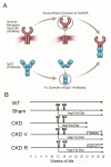
Schematic of ActRIIA ligand trap and experimental design of its use in the ldlr−/− high fat fed CKD vascular calcification model. A, schematic diagram of the mouse fusion protein of the extracellular domain of ActRIIA and the Fc domain of IgG1 (RAP-011). B, experimental design of RAP-011 effects on the CKD-MBD in ldlr−/− high fat fed mice. Mice in four groups were fed the high fat diet beginning at 12 weeks (wks) of age. At 12 wks either sham operation (SO) or electrocautery cortical injury (EC) was performed. At 14 wks either SO or contralateral nephrectomy (NX) was performed. At 22 wks of life, vehicle treatment or RAP-011, 10mg/kg subcutaneous twice weekly, was instituted. WT, wild type mice on chow diet for normal reference levels. Sham, sham operated ldlr−/− high fat fed mice; CKD, CKD ldlr−/− high fat fed mice studied at 22 wks to establish levels of vascular calcium at the start of therapy; CKD V, CKD ldlr−/− high fat fed mice vehicle treated; CKD R, CKD ldlr−/− high fat fed mice RAP-011 treated; WT, Sham, CKD V and CKD R mice were euthanized at 28 weeks of age.
Figure Two
Figure Two
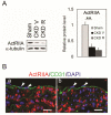
Expression of ActRIIA in mouse aortas. A, Westerns for ActRIIA in aortic homogenates and immunoblot quantitation to the right. For the quantitation, n=4, **p<0.0l. B, Immunofluorescent detection of ActRIIA (a,b) in the aortas of sham (a), and CKD (b) mice. a,b, ActRIIA (red) was expressed in aortic VSMC, but was not detected in endothelial cells. VSMC ActRIIA levels remained detectable in CKD compared to sham. CD31 (green) (arrowheads) was used as an endothelial cell marker. Nuclei were stained by DAPI. Scale bar 20 µm.
Figure Three
Figure Three
Effects of CKD and the ActRIIA ligand trap on aortic gene expression and protein levels in ldlr−/− high fat fed mice with CKD. A, CKD causes increased mRNA expression of osteoblastic proteins (Runx2 and alkaline phosphatase (Alpl)), and decreased levels of aortic smooth muscle cell 22α (Tagln) which were all reversed by treatment with the ActRIIA ligand trap, RAP-011. Aortic myocardin (Myocd) levels were decreased by CKD, but not affected by RAP-011. ***p<0.00l,. **p<0.0l. *p<0.05. .B, Westerns for proteins in aortic homogenates and immunoblot quantitation to the right. CKD causes decreased levels of aortic α-smooth muscle cell actin protein and increased levels of osteoblastic Runx2, which were reversed by treatment with RAP-011, but myocardin levels were not changed. For the quantitation, n=4, **p<0.0l.
Figure Four
Figure Four
Effects of CKD and the ActRIIA ligand trap on aortic calcification in ldlr−/− high fat fed mice with CKD. A, Alizarin Red stained sections of proximal aortic atherosclerotic plaques from vehicle and RAP-011 treated CKD mice. Arrow head indicates calcium deposition in intima (i); m – media. Scale bar 100 µm. B, Aortic Calcium levels in the groups of mice: wild type (WT); sham operated ldlr−/− high fat fed (Sham); CKD euthanasia at 22 weeks, the time of institution of treatment (CKD); CKD treated with vehicle from 22 to 28 weeks (CKD V); CKD treated with RAP-011, 10mg/kg subcutaneous twice weekly from 22 to 28 weeks (CKD R). The boxes represent median (line in box) and interquartile ranges from 25th to 75th percentile. The error bars represent 1.5 fold of the interquartile range. Groups were compared using ANOVA Holm-Sidak method for multiple comparisons with p<0.05 as level for significant difference. *p<0.02; n for each goup 8-12.
Figure Five
Figure Five
ActRIIA signaling in aorta. A, analysis of ActRIIA signaling by westerns of aortic homogenates from sham, CKD vehicle and CKD RAP-011 treated mice. Immunoblots of homogenates from two aortas of animals in each group. ActRIIA and Activin (inhibin β-A) levels were decreased in aortic homogenates from CKD mice. The Alk4 (AcvR1B) and Alk1 (AcvRL1) type 1 receptors were present in aortic homogenates but not affected by CKD.. RAP-011 decreased Smad 2/3 levels, but CKD decreased smad2/3 phosphorylation in aortas which was increased by RAP-011 treatment. CKD decreased phospho-Erk 1/2 levels. Runx2 levels were increased by CKD and normalized by RAP-011 treatment. B, p-Smad2/3 immunoblot quantitation, n=4, **p<0.0l. C, When RAP-011 was administered to WT mice, the decrease in Smad 2/3 levels observed in CKD mice was reproduced, but ActRIIA levels and p-Smad 2/3 levels were very low and not affected by RAP-011 treatment.
Figure Six
Figure Six
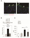
Wnt signaling in aorta, and circulating Dkk1 levels. A, immunofluorescence microscopy of beta-catenin expression in the aortas of: a, wild-type mouse ; b, CKD mouse. Red – beta catenin, Bright green - CD31, an endothelial cell marker, yellow – co-localization. There was no immunofluorescence for beta-catenin in the vascular smooth muscle cells. There was beta-catenin expression in the endothelium of aortas from CKD mice. Arrow heads, beta-catenin and CD31 colocalization in endothelial cells. Scale bar 20 µm. B, analysis of Wnt signaling as marked by Dkk1 protein expression in westerns of aortic homogenates from sham, CKD V and CKD R treated mice, immunoblot quantitation below, **p<0.01, n=4. C, effect of CKD V and RAP-011 treatment on plasma Dkk1 levels. *p<0.05, **p<0.01
Figure Seven
Figure Seven
Renal αklotho levels and ActRIIA signaling in kidney. A, effects of RAP-011 treatment on renal αklotho mRNA levels . CKD decreased αklotho gene expression levels in kidney homogenates, and RAP-011 treatment significantly increased them compared to CKD-3V. *p<0.05, **p<0.01, ***p<0.005. B, analysis of ActRIIA signaling by westerns of kidney homogenates from sham, CKD vehicle, and RAP-011 treated mice. The immunoblots are representative of homogenates from 4 kidneys. ActRIIA levels were not affected by CKD or RAP-011. Activin A (inhibin β-A) levels were increased in kidney homogenates of CKD mice (quantitation in Figure 9) and decreased by RAP-011. The Alk4 (AcvR1B) and Alk2 (AcvR1) type 1 receptors were present in kidney homogenates, but CKD V or CKD R did not significantly affect Alk4 phosphorylation. CKD increased renal smad2/3 phosphorylation (p-Smad2/3), and RAP-011 treatment decreased kidney p-Smad2/3 levels. (immunoblot quantitationin 5C),. CKD increased Col1A1 and fibronectin levels, while RAP-011 treatment decreased them. C, Quantitation of the p-Smad 2/3, Col1A1 and fibronectin immunoblots, *p<0.01, **p<0.01 .
Figure Eight
Figure Eight
Effects of RAP-011 treatment on renal fibrosis. A, Trichrome staining of kidney sections from two CKDV mice (a and b) and two CKD RAP-011 (c and d) treated mice. Areas of interstitial fibrosis marked by arrowheads. Kidneys of CKD RAP-011 treated mice had decreased interstitial fibrosis. Scale bar 50 µm. See supplemental figure 3 for marking of whole kidney coronal sections as to where the photomicrograph sections were taken. B, Quantitation of fibrosis by measuring interstitial volume. Wild type mice were used in this experiment to establish the normal reference. **p<0.01 C, Effects of RAP-011 on urinary protein. There was significant proteinuria in the CKDV mice, which was decreased by RAP-011 treatment, *p<0.05.
Figure Nine
Figure Nine
CKD increases Activin in the circulation and the kidney. A, induction of circulating activin-A by CKD in two disease models, atherosclerotic ldlr−/− high fat fed CKD-3 mice and Alport’s syndrome mice as described in methods; B, inhibin betaA (Inhba) mRNA expression in mouse kidney (activin-A is formed of homodimers of inhibin betaA); C, Westerns for inhibin β-A in kidney homogenates and immunoblot quantitation to the right, n=6 for the immunoblot quantitation; *p<0.05, **p<0.01, ***p<0.005.
Figure Ten
Figure Ten
Kidney InhbA immunostaining in: a, Sham and b, CKD Vehicle mice, In Sham mice occasional peritubular interstitial cells express activin-A, but in CKD mice many more peritubular interstitial cells are positive for activin-A at varying levels of intensity. Scale bar 50 µm.

Comment in

References

    1. Sarnak MJ, Levey AS, Schoolwerth AC, et al. Kidney Disease as a Risk Factor for Development of Cardiovascular Disease. Circulation. 2003;108:2154–2169. - PubMed
    1. Papademetriou V, Lovato L, Doumas M, et al. Chronic kidney disease and intensive glycemic control increase cardiovascular risk in patients with type 2 diabetes. Kidney Int. 2015;87:649–659. - PubMed
    1. Pletcher MJ, Tice JA, Pignone M, et al. Using the Coronary Artery Calcium Score to Predict Coronary Heart Disease Events: A Systematic Review and Meta-analysis. Arch Intern Med. 2004;164:1285–1292. - PubMed
    1. Wexler L, Brundage B, Crouse J, et al. Coronary artery calcification: Pathophysiology, epidemiology, imaging methods, and clinical implications. Circulation. 1996;94:1175–1192. - PubMed
    1. Barraclough KA, Stevens LA, Er L, et al. Coronary artery calcification scores in patients with chronic kidney disease prior to dialysis: reliability as a trial outcome measure. Nephrol Dial Transplant. 2008;23:3199–3205. - PMC - PubMed

MeSH terms