

Case of eastern equine encephalitis presenting in winter
- PMID: 27165999
- PMCID: PMC4885365
- DOI: 10.1136/bcr-2016-215270
Case of eastern equine encephalitis presenting in winter
Abstract
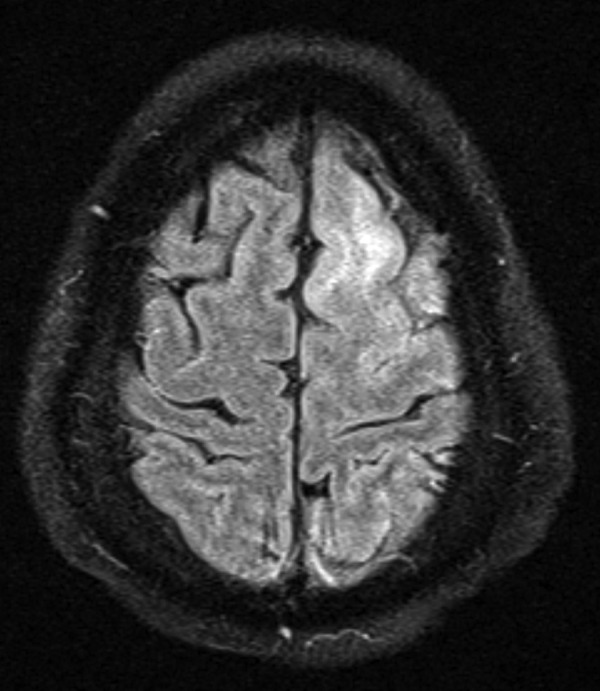
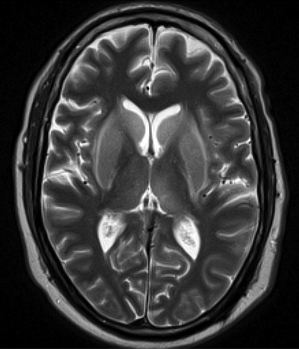
A 50-year-old man was admitted in midwinter with fever, altered mental status and new onset generalised tonic-clonic seizure with urinary incontinence. Cerebrospinal fluid (CSF) analysis revealed an opening pressure of 14.5 cm of water, normal glucose and protein 82 mg/dL (reference range: 15-45 mg/dL). Cell count showed: red cells 11 (reference range: <5 mm(3)), white cell count 1 (reference range: <5 mm(3)). The patient's blood and CSF cultures had no growth. MRI of the brain with and without gadolinium contrast showed abnormal T2 and fluid-attenuated inversion recovery signals within bilateral ventricular nuclei, hippocampi, left frontal and parietal regions. Eastern equine encephalitis (EEE) antibody, IgG titre was 1:64 and IgM titre was <1:16. Three weeks later, his repeat/convalescent titres increased to 1:1024 and 1:32, respectively. Hence, a diagnosis of EEE was established. The patient was treated with supportive care. He recovered well with mildly impaired memory but no other cognitive deficits.
2016 BMJ Publishing Group Ltd.
Figures
References
-
- CDC. Epidemiology and geographic distribution—Eastern equine encephalitis: arbonet, arboviral diseases branch, centers for disease control and prevention. http://www.cdc.gov/EasternEquineEncephalitis/tech/epi.html
-
- Morris C. Eastern equine encephalomyelitis. In: Monath TP, ed. The arboviruses: epidemiology and ecology. Vol 3 Boca Raton, FL: CRC Press, 1988:1–20.
-
- Deresiewicz RL, Thaler SJ, Hsu L et al. . Clinical and neuroradiographic manifestations of eastern equine encephalitis. N Engl J Med 1997;336:1867–74. doi:10.1056/NEJM199706263362604 - DOI - PubMed
-
- [No authors listed] Case definitions for infectious conditions under public health surveillance. Centers for disease control and prevention. MMWR Recomm Rep 1997;46:1–55. - PubMed
-
- Wendell LC, Potter NS, Roth JL et al. . Successful management of severe neuroinvasive eastern equine encephalitis. Neurocrit Care 2013;19:111–15. doi:10.1007/s12028-013-9822-5 - DOI - PubMed
Publication types
MeSH terms
LinkOut - more resources
Full Text Sources
Other Literature Sources
Medical