. 2016 Aug;30(8):2926-44.
doi: 10.1096/fj.201600330RR. Epub 2016 May 10.

Interaction of human biliverdin reductase with Akt/protein kinase B and phosphatidylinositol-dependent kinase 1 regulates glycogen synthase kinase 3 activity: a novel mechanism of Akt activation

Affiliations

Interaction of human biliverdin reductase with Akt/protein kinase B and phosphatidylinositol-dependent kinase 1 regulates glycogen synthase kinase 3 activity: a novel mechanism of Akt activation

Tihomir Miralem et al. FASEB J. 2016 Aug.

Abstract

Biliverdin reductase A (BVR) and Akt isozymes have overlapping pleiotropic functions in the insulin/PI3K/MAPK pathway. Human BVR (hBVR) also reduces the hemeoxygenase activity product biliverdin to bilirubin and is directly activated by insulin receptor kinase (IRK). Akt isoenzymes (Akt1-3) are downstream of IRK and are activated by phosphatidylinositol-dependent kinase 1 (PDK1) phosphorylating T(308) before S(473) autophosphorylation. Akt (RxRxxSF) and PDK1 (RFxFPxFS) binding motifs are present in hBVR. Phosphorylation of glycogen synthase kinase 3 (GSK3) isoforms α/β by Akts inhibits their activity; nonphosphorylated GSK3β inhibits activation of various genes. We examined the role of hBVR in PDK1/Akt1/GSK3 signaling and Akt1 in hBVR phosphorylation. hBVR activates phosphorylation of Akt1 at S(473) independent of hBVR's kinase competency. hBVR and Akt1 coimmunoprecipitated, and in-cell Förster resonance energy transfer (FRET) and glutathione S-transferase pulldown analyses identified Akt1 pleckstrin homology domain as the interactive domain. hBVR activates phosphorylation of Akt1 at S(473) independent of hBVR's kinase competency. Site-directed mutagenesis, mass spectrometry, and kinetic analyses identified S(230) in hBVR (225)RNRYLSF sequence as the Akt1 target. Underlined amino acids are the essential residues of the signaling motifs. In cells, hBVR-activated Akt1 increased both GSK3α/β and forkhead box of the O class transcription class 3 (FoxO3) phosphorylation and inhibited total GSK3 activity; depletion of hBVR released inhibition and stimulated glucose uptake. Immunoprecipitation analysis showed that PDK1 and hBVR interact through hBVR's PDK1 binding (161)RFGFPAFS motif and formation of the PDK1/hBVR/Akt1 complex. sihBVR blocked complex formation. Findings identify hBVR as a previously unknown coactivator of Akt1 and as a key mediator of Akt1/GSK3 pathway, as well as define a key role for hBVR in Akt1 activation by PDK1.-Miralem, T., Lerner-Marmarosh, N., Gibbs, P. E. M., Jenkins, J. L., Heimiller, C., Maines, M. D. Interaction of human biliverdin reductase with Akt/protein kinase B and phosphatidylinositol-dependent kinase 1 regulates glycogen synthase kinase 3 activity: a novel mechanism of Akt activation.

Keywords: Akt/hBVR/GSK3 axis; activation of hBVR; bilirubin; heme oxygenase 1.


Figures

Figure 1.
Figure 1.
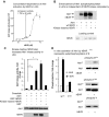
A) hBVR binds and stimulates Akt1 kinase activity in cell. Cells were transfected with pcDNA-Akt1 with or without pcDNA-HA-hBVR or pcDNA3 vector. Some cells, as indicated, were treated with 40 ng/ml IGF-1. Immunoprecipitates were prepared using anti-Akt1 antibody, and Akt1 kinase activity was measured using Crosstide as substrate. Western blot of immunoprecipitated protein was probed with anti-Akt1. Expression of hBVR and Akt1 was verified by Western blot test. ***P < 0.001. B) hBVR and Akt1 coimmunoprecipitate. HEK cells overexpressing HA-hBVR either alone or cotransfected with pcDNA-Akt1 were treated with 40 ng/ml IGF-1 for 15 min; cell lysates were subjected to immunoprecipitation using anti-HA antibody. Immunoprecipitates were analyzed by Western blot analysis with anti-Akt1 antibody followed by anti-hBVR. Expression of hBVR and Akt1 was verified by Western blot test. C) hBVR and Akt association is mediated by Akt PH domain. GST-BVR bound to GSH agarose was added to lysates of cells that had been transfected with indicated EGFP fusion plasmids. Proteins bound by immobilized BVR were assayed by Western blot test, which was probed with anti-GFP antibodies. GST was included as additional control.
Figure 2.
Figure 2.
A) In-cell FRET analysis of hBVR and Akt1 interaction. Cells cotransfected with pDsRed2-hBVR and indicated pEGFP-Akt1 constructs, wt or truncation mutants, were examined by confocal microscopy after stimulation with 40 ng/ml IGF-1. Empty pEGFP vector was used as control. FRET was detected from multiple images as described in Materials and Methods; FRET images shown depict same cells as shown for confocal microscopy. B) FRET efficiency was calculated for at least 3 microscope fields for each condition. Values for no donor were calculated for cells that had been transfected with pDsRed2-hBVR only. **P < 0.01.
Figure 3.
Figure 3.
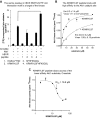
A) In-cell FRET analysis of hBVR and Akt2 PH domain interaction. Cells cotransfected with pEGFP-Akt2 constructs and pDsRed2-hBVR were examined by FRET as in Fig. 2A. B) FRET efficiency was calculated as in Fig. 2B; efficiency for Akt21–250 construct was measured before and after IGF-1 treatment. C) hBVR does not display preferential affinity for Akt enzymes. His6-tagged Akts, expressed in E. coli and bound to Ni-NTA agarose, were incubated with GST-BVR. After washing, proteins bound on beads were analyzed by gel electrophoresis and Western blot test using anti-GST antibodies. A series of loading controls for GST-BVR was also included on blot to allow approximate quantification of GST-hBVR bound to Akt. The blot was subsequently stripped and probed with anti-Akt1 antibodies to verify input Akt1 levels. D) E. coli–expressed Akt1 is kinase active. his6-tagged Akt1 bound to Ni-NTA agarose was equilibrated in PDK1 kinase buffer and treated with PDK1. PDK1 was then washed from agarose beads, immobilized protein was equilibrated in Akt kinase buffer, and kinase activity was measured with Aktide substrate. E) Akt1 expressed in E. coli is phosphorylated at T308 by PDK1. PDK1 kinase reaction products from panel D were examined for T308 phosphorylation by Western blot test.
Figure 4.
Figure 4.
hBVR activates Akt1 in vitro. A) Concentration dependence of Akt1 activation by hBVR in vitro. Phosphorylation of Crosstide substrate by his6-tagged recombinant Akt1 was measured in presence of increasing concentrations of GST-tagged hBVR. Data were fitted to sigmoid curve using nonlinear regression. B) Enhancement of Akt1 autophosphorylation in vitro is independent of hBVR kinase competency. Autophosphorylation of Akt1 was measured in presence or absence of 2 µg wt or kinase dead (G17 > A) hBVR. Kinase reaction products were separated by SDS-PAGE and detected by autoradiography; loading of hBVR was detected by Ponceau S staining. C) Kinase-inactive hBVR also increases Akt1 kinase activity in vitro. Akt1 activity was measured as in (A) using Crosstide as substrate and wt GST-hBVR, G17 > A kinase inactive mutant, or GST alone. Akt1 and hBVR input is indicated by Western blots. *P < 0.05. D) In vitro activation of Akt1 by hBVR represents autophosphorylation of S473. Akt1 was incubated under conditions favoring Akt kinase activity in presence or absence of hBVR, as described in (C). T308, S473, and tyrosine phosphorylated products were detected by Western blot test by sequentially probing membranes with antibody against specific phosphorylation products, followed by anti-Akt1. Densitometry of T308, S473, and Akt1 blots was used to correct antiphosphopeptide signals for differences in loading.
Figure 5.
Figure 5.
Sequence coverage of phosphorylated hBVR by proteolysis and mass spectrometry. Products of kinase reaction that included Akt1 and his6-hBVR (purified from E. coli) were resolved by gel electrophoresis. Three identical samples, each containing 2 µg of protein, were digested with trypsin, chymotrypsin, or Arg-C protease. Products were identified by mass spectrometry. Extent of sequence coverage by each digest is indicated by underlining; serine residues identified as phosphorylation sites are indicated by dots above sequence. Red shading indicates Akt binding/phosphorylation site as predicted by computational analysis, and blue shading indicates RFxFPxFS PDK1 binding motif.
Figure 6.
Figure 6.
A) Serine residue in hBVR RNRYLS230F Akt interaction motif is target of kinase. Akt1 was assayed in vitro in presence of Crosstide as substrate or peptides (each at 20 µM) derived from hBVR, as described in Materials and Methods. Effect of adding 1 µg hBVR to reaction mixture with Crosstide was also examined. B) KRNRYLSF peptide binds with high affinity to Akt1 catalytic site. Akt was assayed in presence or absence of 20 µM KRNRYLSF while varying Crosstide concentration. Data were fitted to Michaelis-Menten equation by nonlinear regression. C) KRNRYLSF peptide blocks access of lower-affinity Akt1 substrate Crosstide. Akt1 was assayed in vitro in presence of 30 µM Crosstide as substrate and increasing amounts of KRNRYLSF.
Figure 7.
Figure 7.
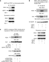
A) Coimmunoprecipitation of PDK1 and hBVR. Cells were cotransfected with PDK1 expression plasmid and pcDNA-HA-hBVR. Lysates were prepared from cells with or without stimulation with 40 ng/ml IGF-1 and immunoprecipitated with anti-HA antibody. Immunoprecipitates were analyzed by Western blot test; blot was sequentially probed with antibodies to PDK1 and hBVR. Expression of PDK1 and hBVR in lysates was verified by Western blot test. B) PDK1, Akt1, and hBVR coimmunoprecipitate in IGF-1 stimulated cells. Lysates prepared from cells cotransfected with pcDNA-HA-hBVR and pcDNA-Akt1 and stimulated with 40 ng/ml IGF-1 or 50 ng/ml TNF-α were immunoprecipitated with anti-HA antibodies. Immunoprecipitates were analyzed by Western blot test, which was sequentially probed with antibodies to PDK1, Akt1, and hBVR. Overexpression of Akt1 and HA-hBVR, and presence of PDK1 were verified by Western blot test of cell lysates. C) hBVR consensus PDK1 binding site is important in PDK1 and Akt1 interaction. Cells were cotransfected with PDK1 expression plasmid with or without pcDNA-HA-hBVR or pcDNA-HA-hBVR C-Box (RFGFPAFS to RVGAPAVS) mutant. Immunoprecipitates obtained with Akt1 antibody were analyzed for PDK1 as in (B); equality of loading was confirmed by probing blot with anti-Akt1 antibody. Overexpression of proteins was verified by Western blot test of cell lysates.
Figure 8.
Figure 8.
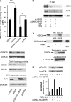
A) Presence of hBVR decreases GSK3α/β activity in cell. Cells were transfected with either pcDNA-HA-hBVR or with siBVR. Lysates prepared from cells were assayed for GSK3α/β activity using peptide substrate from glycogen synthase, as described in text. **P < 0.01, ***P < 0.001. Western blots of lysates were probed with antibodies to phospho-GSK3β, GSK3α, GSK3β, hBVR, and anti–tubulin. B) sihBVR treatment prevents formation of hBVR/PDK1/Akt1 complex. Immunoprecipitates were analyzed by Western blot test, which was sequentially probed with PDK1 and Akt1 antibodies. Observed pattern of PDK1 is consistent with presence of splice variants that are recognized by antibody. C) GST pulldown shows association of GSK3β from insulin-treated cells with hBVR. Cells were transfected with pcDNA-GSK3β and subsequently treated with insulin or left untreated. Lysates were analyzed by GST pulldown using GST-hBVR, followed by Western blot analysis. Blots were probed with antibodies to GSK3β followed by anti-hBVR. Overexpression of GSK3β was verified by Western blot test. D) Phosphorylation of FoxO3 in response to IGF-1 is mediated by hBVR. Cells were transfected with pcDNA-HA-hBVR or with empty vector. After 24 h, transfected cells were treated with 40 ng/ml IGF-1 for 1 or 4 h or were left untreated. Phosphorylated FoxO3C and FoxO3A were detected by Western blot test, which was then probed with antibody to FoxO3A. Densitometry was used to normalize phospho-FoxO3 signals.
Figure 9.
Figure 9.
sihBVR inhibits Akt1 activation and stimulates glucose uptake. A) sihBVR treatment inhibits Akt1 activation by insulin and IGF-1. Cells were transfected with siRNA against hBVR or with pcDNA-HA-hBVR, and treated with IGF-1 or insulin. Akt1 was immunoprecipitated from cell lysate and assayed for kinase activity with Aktide substrate. Expression levels of hBVR were verified by Western blot test, with GSK3β serving as loading control. B) sihBVR treatment stimulates glucose uptake. Cells were transfected with siRNA against hBVR or with pcDNA-HA-hBVR, and assayed for glucose uptake in presence or absence of insulin, as described in Materials and Methods.
Figure 10.
Figure 10.
Roles of hBVR in activating IRK/PI3K/Akt pathway. BVR binds directly to IRK, where it functions as platform for interaction with IRS substrates (14). Activation of MAPK pathway (MAPK layer) is multistep process that leads to activation of MEK1/2. Interaction of hBVR with MEK1/2 and ERK1/2 to enhance activation of MEK has been defined elsewhere (62). Activation of ERK1/2 results in activation of as many as 50 transcription factors, including those shown (92). IRK and BVR together activate PI3K (43, 72), source for PtdIns(3,4,5)P3, which interacts with PH domain of Akt and recruits inactive protein to membrane, where it undergoes conformational change and is phosphorylated at T308 by 3-phosphoinositide-dependent kinase PDK1 (34, 35). Specific sequences in hBVR that mediate interactions with proteins in signaling pathways are indicated. Akts phosphorylate GSK3α/β, resulting in its inactivation; inactive protein in turn cannot phosphorylate and inactivate transcription factors, enabling expression of genes including those encoding eNOS, VEGF, and HO-1 (, –48). hBVR is also instrumental in activating HO-1 (47, 93); in its capacity as reductase, it also converts biliverdin to bilirubin (1). Akt phosphorylation of FoxO3 results in its sequestration in cytoplasm and thereby prevents transcription of proapoptotic genes (32).

Similar articles

Cited by

References

    1. Kutty R. K., Maines M. D. (1981) Purification and characterization of biliverdin reductase from rat liver. J. Biol. Chem. 256, 3956–3962 - PubMed
    1. Maines M. D., Trakshel G. M. (1993) Purification and characterization of human biliverdin reductase. Arch. Biochem. Biophys. 300, 320–326 - PubMed
    1. Maines M. D., Trakshel G. M., Kutty R. K. (1986) Characterization of two constitutive forms of rat liver microsomal heme oxygenase. Only one molecular species of the enzyme is inducible. J. Biol. Chem. 261, 411–419 - PubMed
    1. Stocker R., Yamamoto Y., McDonagh A. F., Glazer A. N., Ames B. N. (1987) Bilirubin is an antioxidant of possible physiological importance. Science 235, 1043–1046 - PubMed
    1. Sedlak T. W., Snyder S. H. (2009) Cycling the wagons for biliverdin reductase. J. Biol. Chem. 284, le11; author reply le12 - PMC - PubMed

Publication types

MeSH terms

Substances