. 2016 Aug;97(8):1942-1954.
doi: 10.1099/jgv.0.000501. Epub 2016 May 9.

Exposure of rhesus monkeys to cowpox virus Brighton Red by large-particle aerosol droplets results in an upper respiratory tract disease

Affiliations

Exposure of rhesus monkeys to cowpox virus Brighton Red by large-particle aerosol droplets results in an upper respiratory tract disease

Reed F Johnson et al. J Gen Virol. 2016 Aug.

Abstract

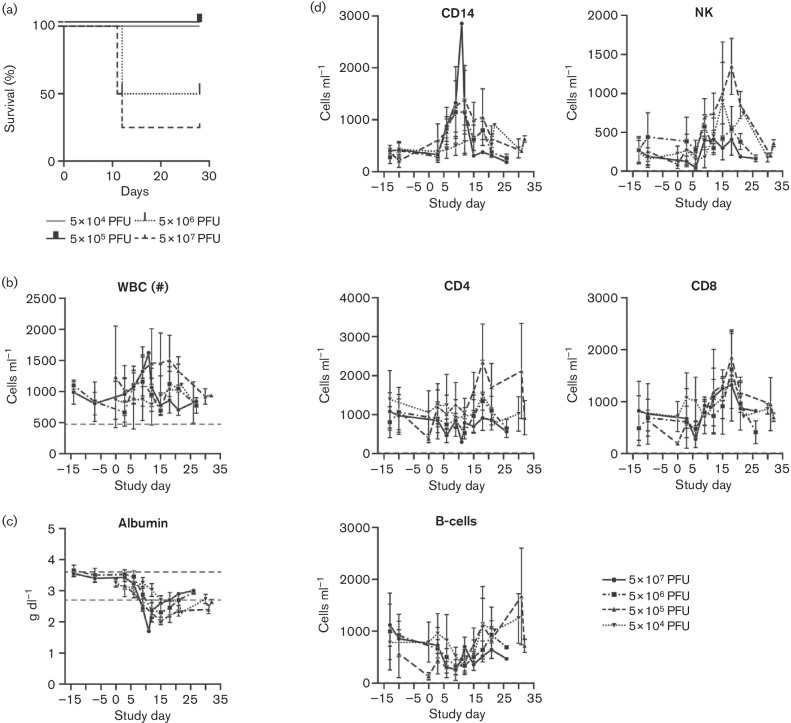
We previously demonstrated that small-particle (0.5-3.0 µm) aerosol infection of rhesus monkeys (Macaca mulatta) with cowpox virus (CPXV)-Brighton Red (BR) results in fulminant respiratory tract disease characterized by severe lung parenchymal pathology but only limited systemic virus dissemination and limited classic epidermal pox-like lesion development (Johnson et al., 2015). Based on these results, and to further develop CPXV as an improved model of human smallpox, we evaluated a novel large-particle aerosol (7.0-9.0 µm) exposure of rhesus monkeys to CPXV-BR and monitored for respiratory tract disease by serial computed tomography (CT). As expected, the upper respiratory tract and large airways were the major sites of virus-induced pathology following large-particle aerosol exposure. Large-particle aerosol CPXV exposure of rhesus macaques resulted in severe upper airway and large airway pathology with limited systemic dissemination.


Figures

Fig. 1.
Fig. 1.
(a) Kaplan–Meier analysis of the large-particle aerosol exposure of rhesus monkeys. Three out of four (75 %) NHPs receiving 5×10⁷ p.f.u. of CPXV succumbed by day 12 post-exposure, and 50 % of NHPs receiving 5×10⁶ p.f.u. of CPXV succumbed by day 12 post-exposure. Groups receiving 5×10⁵ and 5×10⁴ p.f.u. survived to study end. (b) WBC concentrations during the course of the experiment. Dashed lines indicate normal value ranges. Subjects did not consistently demonstrate leucocytosis. (c) Serum chemistry value of albumin indicates a decrease across all groups, suggesting a response to exposure. (d) The major PBMC populations increased in response to infection. Various PBMC cell populations were assayed periodically by TruCount analysis as described in the Methods.
Fig. 2.
Fig. 2.
Histopathology of CPXV large-particle aerosol-exposed NHPs. (a) Histological evaluation of the upper respiratory tract. Cross-sections of the trachea are shown and demonstrate the severe inflammatory response of the non-survivors when compared to the survivors. Placement of the endotracheal (ET) tube displaced significant intraluminal exudates from subjects 13, 15, 9 and 11 (an ET tube was not placed in subject 16 prior to necropsy). Bars, 1 mm. (b) Severe, consolidating fibrinosuppurative and necrotizing bronchopneumonia for subject 13. Inset: Ulcerated bronchus (note supporting hyaline cartilage at left) with smooth muscle. The bronchial lumen is filled with a necrotic fibrinosuppurative exudate. Bars (including inset), 100 µm. (c) Right caudal lung lobe. The bronchus and surrounding pulmonary parenchyma exhibit mild, locally extensive, peribronchial and peribronchiolar organizing pneumonia. Inset: Focal bronchiolization and squamous metaplasia of affected alveoli. Bars (including inset), 100 µm. (d) Non-survivor epiglottis demonstrating severe necrosuppurative and ulcerative epiglottitis observed in subject 15. The junction of intact epithelium exhibited hydropic degeneration and necrotic epithelium with myriad, intracytoplasmic, intraepithelial inclusion bodies (black arrow). Inset: subgross cross-section of the epiglottis. Bars, 10 µm and 1 mm (inset). (e) Epiglottis from surviving subject (subject 10) which appears normal. Inset: subgross cross-section of the epiglottis. Bars, 100 µm and 1 mm (inset).
Fig. 3.
Fig. 3.
(a) Haematoxylin and eosin (H&E)-stained sections of trachea show severe inflammation and necrosis involving the mucosal epithelial cells and the submucosal glands. (b) Immunohistochemistry for CPXV virus shows abundant virus antigen staining (brown) in both attached and free (necrotic) mucosal epithelial cells, and within the subjacent submucosal glands (inset). (c) H&E-stained sections of lung show severe inflammation and necrosis involving bronchioles and the surrounding alveolar interstitium. Alveolar spaces are filled with fibrin, inflammatory cells and karyorrhectic cellular debris (inset). (d) Immunohistochemistry for CPXV shows abundant virus antigen labelling (brown) within sloughed epithelial cells and within the cytoplasm of cells (macrophages) associated with damaged and necrotic alveolar walls. Magnification (a–d), main image ×4; inset ×40. Bars (a–d), 100 µm. (e) Histopathology of CPXV lung infection (i), characterized by severe and necrotizing brochiolitis and alveolitis. Alveolar spaces are filled with fibrin, cellular debris and macrophages. Bright-field immunohistochemistry (BF-IHC) (ii) shows CPXV-antigen-positive cells clustered within alveolar spaces; cells are were often large, round to polygonal with abundant virus-positive cytoplasm. Immunofluorescence images (iii–vi) show the same location with IBA1 (iii; pan-macrophage), CPXV (iv; virus antigen) and DAPI (v; nuclear) immunohistochemical markers, and a merged image (vi) demonstrating CPXV-antigen-positive macrophages (yellow) associated with regions of severe lung damage. (f) Histopathology of CPXV infection of the trachea (i), characterized by severe necrotizing tracheitis and massive cellular infiltration of the submucosal stroma. Bright-field immunohistochemistry (ii) shows CPXV antigen within mucosal epithelial cells; inflammatory cells within the subjacent stroma frequently display cytoplasmic positivity. Immunofluorescence images (iii–vi) show the same location with IBA1 (iii; pan-macrophage), CPXV (iv; virus antigen) and DAPI (v; nuclear) immunohistochemical markers, and a merged image (vi) demonstrating CPXV-antigen-positive macrophages (yellow) associated with regions of severe tracheal damage. Bars (e, f), 100 µm.
Fig. 4.
Fig. 4.
CT indicates lung pathology and resolution. The selected images are representative of non-survivors or survivors from the two groups in which NHPs met endpoint criteria. The data shown demonstrates the CT image at the observed peak lung pathology for that subject: day 11 for panel (a), day 12 for panel (b), day 12 for panel (c), and day 12 for panel (d). Corresponding CT images from previous or subsequent days were selected for comparison of disease progression and regression within the same anatomical location. Subjects that succumbed or met endpoint criteria demonstrated similar CT findings (a–c). Subjects that survived demonstrated pathology followed by resolution (d). (a) Subject 16, 5×10⁷ p.f.u. group, succumbed at day 11. (b) Subject 15, 5×10⁷ p.f.u. group, euthanized on day 12. (c) Subject 9, 5×10⁶ p.f.u. group, euthanized on day 12. (d) Subject 10, 5×10⁶ p.f.u. group, survivor.
Fig. 5.
Fig. 5.
Quantitative analysis of the CT data indicates progressive and resolving lung pathology that corresponds to dose. CT data was analysed as described in Methods. Individual subjects are shown by group. (a) 5×10⁷ p.f.u., (b) 5×10⁶ p.f.u., (c) 5×10⁵ p.f.u., (d) 5×10⁴ p.f.u. Asterisk indicates that the day 3 data points were dropped due to incorrect placement of the ET tube.
Fig. 6.
Fig. 6.
Comparison of NHP models of smallpox to human smallpox. The different NHP models are aligned against human smallpox. Incubation period, rash onset and staging, and lymphadenopathy and development of other clinical signs are noted for cross-model comparison. IB, Intrabronchial; IN, intranasal.

References

    1. Aebersold P.(2012). FDA experience with medical countermeasures under the Animal Rule. Adv Prev Med 2012507571. 10.1155/2012/507571 - DOI - PMC - PubMed
    1. Americo J. L., Moss B., Earl P. L.(2010). Identification of wild-derived inbred mouse strains highly susceptible to monkeypox virus infection for use as small animal models. J Virol 848172–8180. 10.1128/JVI.00621-10 - DOI - PMC - PubMed
    1. Bai H., Buller R. M., Chen N., Green M., Nuara A. A.(2005). Biosynthesis of the IFN-gamma binding protein of ectromelia virus, the causative agent of mousepox. Virology 33441–50. 10.1016/j.virol.2005.01.015 - DOI - PubMed
    1. Barnewall R. E., Fisher D. A., Robertson A. B., Vales P. A., Knostman K. A., Bigger J. E.(2012). Inhalational monkeypox virus infection in cynomolgus macaques. Front Cell Infect Microbiol 2117. 10.3389/fcimb.2012.00117 - DOI - PMC - PubMed
    1. Bohannon J. K., Lackemeyer M. G., Kuhn J. H., Wada J., Bollinger L., Jahrling P. B., Johnson R. F.(2015). Generation and characterization of large-particle aerosols using a center flow tangential aerosol generator with a non-human-primate, head-only aerosol chamber. Inhal Toxicol 27247–253. 10.3109/08958378.2015.1033570 - DOI - PMC - PubMed

Publication types

LinkOut - more resources