. 2016 Jul 5;7(27):41432-41444.
doi: 10.18632/oncotarget.9180.

Endoplasmic reticulum stress and IRE-1 signaling cause apoptosis in colon cancer cells in response to andrographolide treatment

Affiliations

Endoplasmic reticulum stress and IRE-1 signaling cause apoptosis in colon cancer cells in response to andrographolide treatment

Aditi Banerjee et al. Oncotarget. .

Abstract

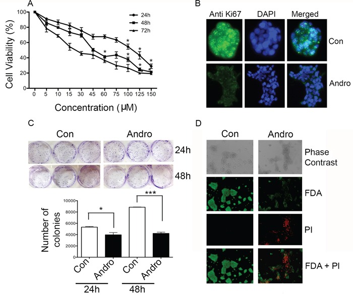
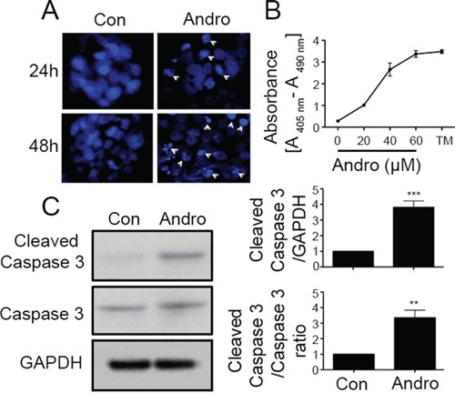
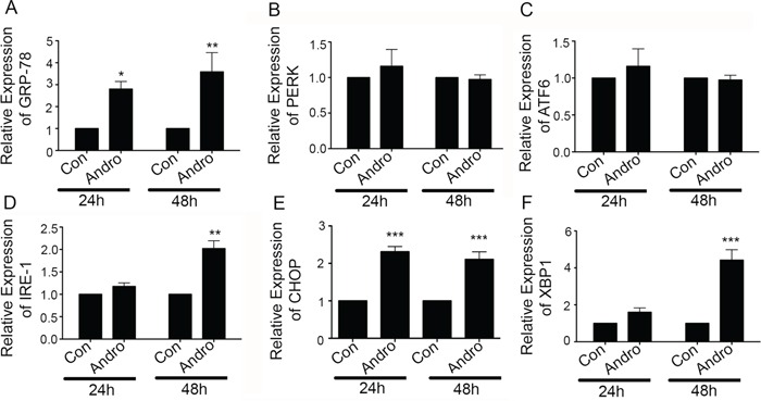
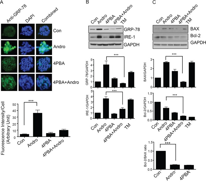
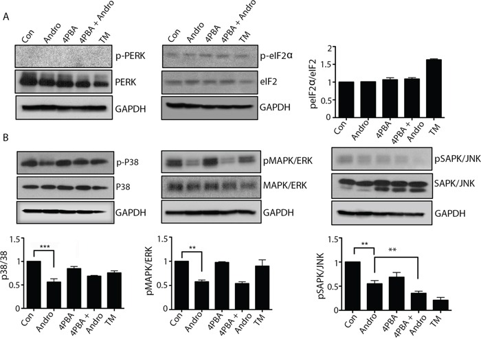
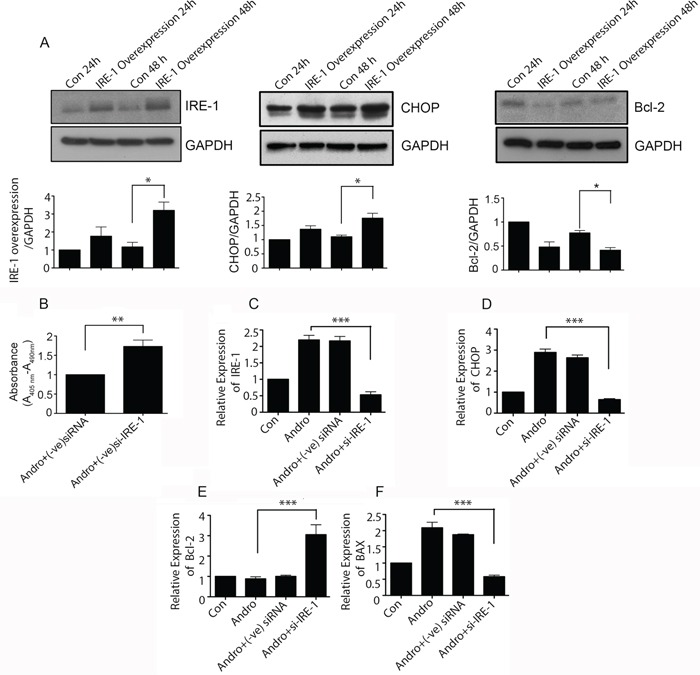
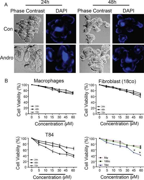
The plant metabolite andrographolide induces cell cycle arrest and apoptosis in cancer cells. The mechanism(s) by which andrographolide induces apoptosis however, have not been elucidated. The present study was performed to determine the molecular events that promote apoptosis in andrographolide treated cells using T84, HCT116 and COLO 205 colon cancer cell lines. Andrographolide was determined to limit colony formation and Ki67 expression, alter nuclear morphology, increase cytoplasmic histone-associated-DNA-fragments, and increase cleaved caspase-3 levels. Andrographolide also induced significantly higher expression of endoplasmic reticulum (ER) stress proteins GRP-78 and IRE-1 by 48 h but not PERK or ATF6. Apoptosis signaling molecules BAX, spliced XBP-1 and CHOP were also significantly increased. Moreover, chemical inhibition of ER stress or IRE-1 depletion with siRNA in andrographolide treated cells significantly limited expression of IRE-1 and CHOP as determined by immunofluorescence staining, real time PCR, or immunobloting. This was accompanied by a decreased BAX/Bcl-2 ratio. Andrographolide significantly promotes cancer cell death compared to normal cells. These data demonstrate that andrographolide associated ER stress contributes to apoptosis through the activation of a pro-apoptotic GRP-78/IRE-1/XBP-1/CHOP signaling pathway.

Keywords: andrographolide; apoptosis; chemotherapy; endoplasmic reticulum stress; unfolded protein response.


Conflict of interest statement

None of the authors participating in this study have any conflicts of interest in this work or publication.

Figures

Figure 1
Figure 1. Andrographolide suppresses cell proliferation and clonogenicity in T84 cells
A. Cells were treated with andrographolide for 24, 48 and 72 h and cell viability was quantified by MTT assay. B. T84 cells were stained with FITC labeled anti-Ki67 antibodies and DAPI and evaluated by fluorescence microscopy. C. T84 cells were diluted and treated with andrographolide at IC50. Growth was measured by direct counting of clonal clusters stained in multiwell plates with crystal violet at 24 and 48 h. Representative photomicrographs are shown. D. Fluorescence microscopy images showing the viability of T84 cells cultured in vitro with or without andrographolide (from top to bottom: phase contrast image, FDA staining, PI staining, composite of FDA and PI staining). (scale bar: 200 μm). Experiments were performed either two times (B and C) or three times (A and D). (*P < 0.05, **P < 0.01.)
Figure 2
Figure 2. Andrographolide induces cell apoptosis in colon cancer T84 cells
A. Cells were treated with andrographolide IC50 dose for either 24 (upper panels) or 48 h (lower panels) and stained with DAPI. Apoptotic cells were identified by condensation and fragmentation (arrows) of nuclei using inverted fluorescence microscope. B. Detection of nucleosomes in cytoplasmic fractions at increasing doses of andrographolide and TM. 104 cells were treated with or without andrographolide (20, 40, or 60 μM) and TM for 48 h at 37°C. 20 μl of cell lysates were analyzed in the ELISA. C. Cleaved caspase-3 and caspase-3 protein expression was evaluated by immunoblotting of T84 cell lysates after 48 h of andrographolide IC50 treatment. Staining was normalized using GAPDH expression and the ratio of cleaved caspase and caspase-3 and amount of relative staining was determined by densitometry. (**P < 0.01, *** P < 0.001)
Figure 3
Figure 3. Andrographolide induces ER stress-related IRE-1 and associated proteins
T84 cells were treated with Andrographolide at IC50 (45 μM) for 24 and 48 h and the transcriptional level of expression for ER Stress and apoptosis associated genes was determined by qRT-PCR for A. GRP-78, B. PERK, C. ATF6, D. IRE1, E. CHOP, and F. XBP-1. Bar graphs show quantitative results normalized to GAPDH mRNA levels. Results are from three independent experiments. Statistical significance was determined using one way-ANOVA followed by Bonferroni test. (*P < 0.05, **P < 0.01, ***P < 0.001)
Figure 4
Figure 4. Andrographolide induced apoptosis signaling is dependent on ER Stress
A. T84 cells were grown on coverslips and treated with Andrographolide IC50 in the presence or absence of 4-PBA and GRP-78 expression was evaluated by immunofluorescent staining. Nuclei were stained using DAPI and cells were examined by fluorescence microscopy. Fluorescence intensity was determined and compared with untreated T84 cells. B. T84 cells treated with Andrographolide IC50 for 48 h were lysed and protein expression was determined by immunoblotting for GRP-78, IRE1, and GAPDH. Densitometry analysis was performed and normalized with GAPDH expression to demonstrate significant reductions in expression of GRP-78, IRE1, in the present of 4-PBA. C. Cell lysates were analyzed for pro-apoptotic BAX and anti-apoptotic Bcl-2 expression by immunoblot analysis and quantified by densitometry. The lower graph shows the ratio of Bcl-2/BAX. The results shown are from three independent experiments. (***P < 0.001)
Figure 5
Figure 5. Andrographolide induced ER stress does not activate PERK or ATF-6 pathways, and downregulates MAPK pathways
T84 cells were treated with Andrographolide at IC50 (45 μM) for 24 and 48 h and evaluated by western blot for A. phospho-PERK, phospho-eIF2α and GAPDH or B. phospho-p38, phospho-ERK1/2, and phospho-SAPK/JNK and quantified by densitometry. The results shown are from three independent experiments. (**P < 0.001)
Figure 6
Figure 6. Andrographolide induced apoptosis signaling is dependent on IRE-1
A. T84 cells were transfected with plasmid for overexpression of IRE-1 and then treated with Andrographolide. Cell lysates were analyzed by western blot and quantified by densitometry for expression of IRE-1, CHOP, and Bcl-2. Expression is normalized against GAPDH expression. T84 cells were transfected with siRNA for IRE-1 or control siRNA and treated with Andrographolide for 48 h and B. DNA fragmentation was compared using a colorimetric assay. (C) Cells were also evaluated for mRNA expression by qRT-PCR for C. IRE-1, D. CHOP, E. Bcl-2, and F. BAX. (***P < 0.001)
Figure 7
Figure 7. Andrographolide associated reductions in viability of are less pronounced in noncancer cells
A. Three dimensional primary cultures of mouse intestinal epithelial cells organoids were expanded and treated with Andrographolide IC50 for 24 and 48 h and then stained with DAPI to evaluate nuclear morphology. B. Primary cultures of mouse bone marrow macrophages and human fibroblasts were treated with Andrographolide at the indicated dose range and compared to Andrographolide treated T84 cells for cell viability using the MTT assay. The data are expressed as the percentage of viable cells relative to untreated control cells. Data shown are from three independent experiments. (P < 0.05)

Similar articles

Cited by

References

    1. Lao VV, Grady WM. Epigenetics and colorectal cancer. Nat Rev Gastroenterol Hepatol. 2011;8:686–700. - PMC - PubMed
    1. Anand P, Kunnumakkara AB, Newman RA, Aggarwal BB. Bioavailability of curcumin: problems and promises. Mol Pharmaceutics. 2007;4:807–18. - PubMed
    1. Mohanty C, Acharya S, Mohanty AK, Dilnawaz F, Sahoo SK. Curcumin-encapsulated MePEG/PCL diblock copolymeric micelles: a novel controlled delivery vehicle for cancer therapy. Nanomedicine. 2010;5:433–49. - PubMed
    1. Gali-Muhtasib HU, Younes IH, Karchesy JJ, el-Sabban ME. Plant tannins inhibit the induction of aberrant crypt foci and colonic tumors by 1,2-dimethylhydrazine in mice. Nutrition and Cancer. 2001;39:108–16. - PubMed
    1. Chiou WF, Lin JJ, Chen CF. Andrographolide suppresses the expression of inducible nitric oxide synthase in macrophage and restores the vasoconstriction in rat aorta treated with lipopolysaccharide. Br J Pharmacol. 1998;125:327–34. - PMC - PubMed

MeSH terms