. 2016 Aug 19;44(14):6924-34.
doi: 10.1093/nar/gkw401. Epub 2016 May 10.

Rational engineering of the Neurospora VS ribozyme to allow substrate recognition via different kissing-loop interactions

Affiliations

Rational engineering of the Neurospora VS ribozyme to allow substrate recognition via different kissing-loop interactions

Julie Lacroix-Labonté et al. Nucleic Acids Res. .

Abstract

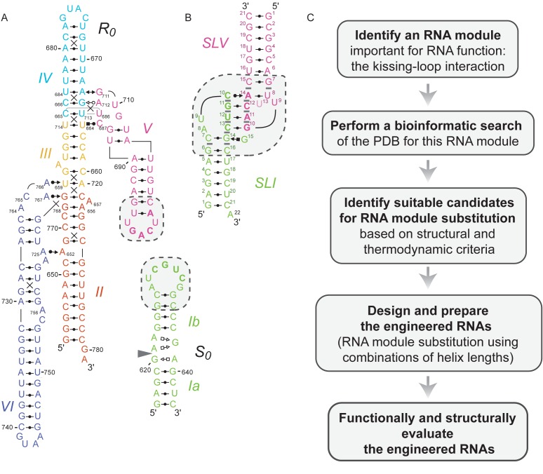
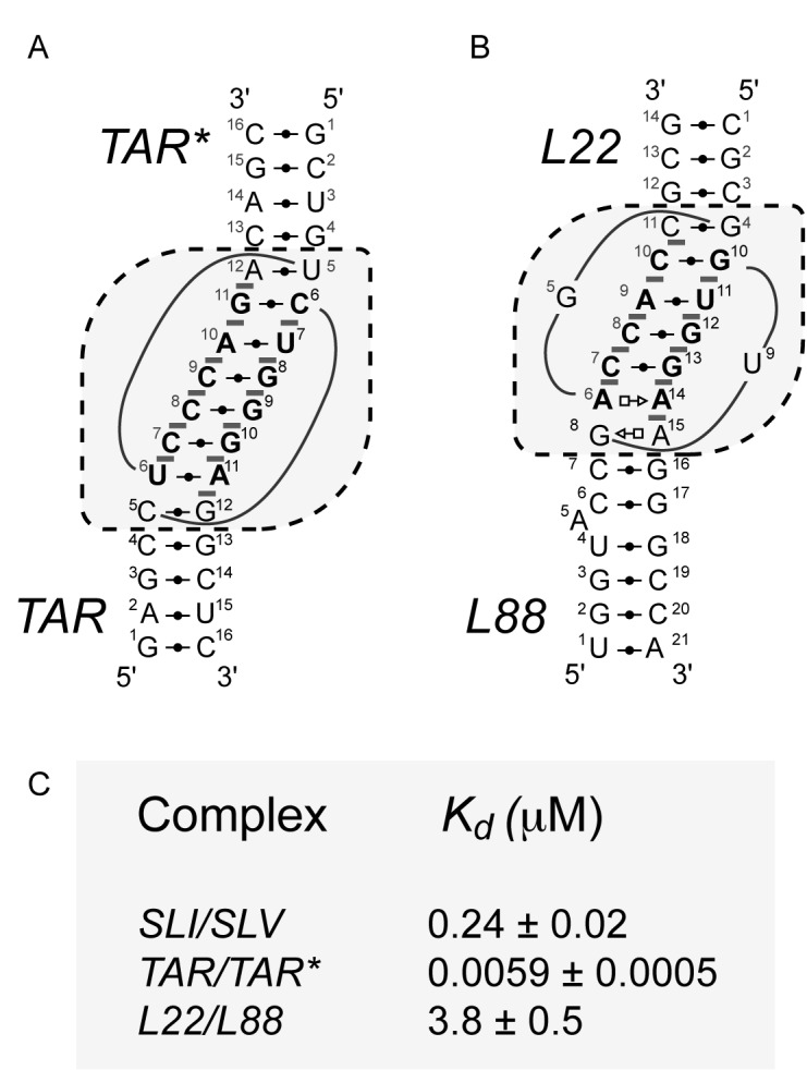
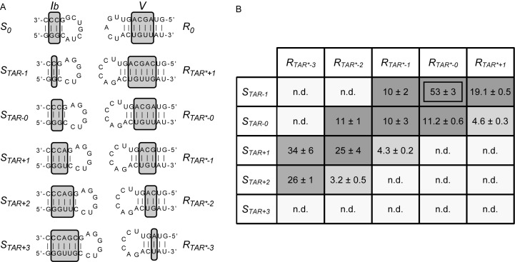
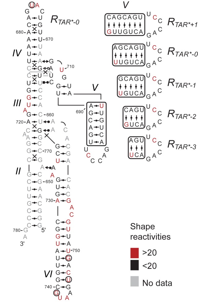
The Neurospora VS ribozyme is a catalytic RNA that has the unique ability to specifically recognize and cleave a stem-loop substrate through formation of a highly stable kissing-loop interaction (KLI). In order to explore the engineering potential of the VS ribozyme to cleave alternate substrates, we substituted the wild-type KLI by other known KLIs using an innovative engineering method that combines rational and combinatorial approaches. A bioinformatic search of the protein data bank was initially performed to identify KLIs that are structurally similar to the one found in the VS ribozyme. Next, substrate/ribozyme (S/R) pairs that incorporate these alternative KLIs were kinetically and structurally characterized. Interestingly, several of the resulting S/R pairs allowed substrate cleavage with substantial catalytic efficiency, although with reduced activity compared to the reference S/R pair. Overall, this study describes an innovative approach for RNA engineering and establishes that the KLI of the trans VS ribozyme can be adapted to cleave other folded RNA substrates.


Figures

Figure 1.
Figure 1.
Rational engineering of KLIs for substrate recognition by the Neurospora VS ribozyme. (A) Structural schematic of a trans VS ribozyme system formed by the S0 substrate and the R0 ribozyme (32). The schematic is derived from NMR and X-ray structures (14,43). The cleavage site is located between G620 and A621 as indicated by the gray arrowhead. (B) Structural characteristics of the VS ribozyme I/V KLI derived from the NMR structure of the SLI/SLV complex [PDB ID: 2MI0; (13)]. (C) Flowchart of the engineering strategy used in this study. In (A and B), the dashed boxes enclose residues that contribute to the KLI and the closing base pairs of the two stems, whereas base-pairing interactions are represented using the Leontis-Westhof notation (12). In addition, base-pairing residues at the KLI are in bold. In (B), gray bars represent base stacking at the KLI.
Figure 2.
Figure 2.
Schematics and thermodynamic stability of selected KLIs. Structural characteristics of the selected KLIs: (A) the HIV-1 TAR/TAR* KLI [PDB ID: 1KIS; (35)] and (B) the large ribosomal subunit L22/L88 KLI from Deinococcus radiodurans [PDB ID: 4IOA; (36)]. (C) Dissociation constants (Kd) of the selected KLIs determined in a previous study for the SLI/SLV and TAR/TAR* complexes (18) and here for the L22/L88 complex (Supplementary Figure S2). These Kd values were all determined at 25°C in 10 mM Tris pH 7.0 and 20 mM MgCl2. In (A and B), the dashed boxes enclose residues of the KLI and the closing base pairs of the two stems and base-pairing interactions are represented using the Leontis-Westhof notation (12). In addition, base-pairing residues at the KLI are in bold and gray bars highlight base stacking at the KLI.
Figure 3.
Figure 3.
Sequences and kinetic data of the STAR/RTAR*variants. (A) Sequences of the STAR/RTAR* variants. Only the regions of the S0 substrate and R0 ribozyme (stem-loops Ib and V shown above; Figure 1A) that were substituted in the STAR and RTAR* variants are shown. The number of base pairs varies in stems Ib and V, as indicated by the gray boxes on the putative secondary structures. (B) Grayscale heat map of the kcat/KM values (10−2 min−1μM−1) for the STAR/RTAR* variants. Dark gray shading corresponds to kcat/KM values within 5-fold of the most active STAR/RTAR* pair (STAR-1/RTAR*-0); medium gray shading corresponds to lower kcat/KM values (5–17-fold lower than the most active STAR/RTAR* pair), and light gray shading reflects the absence of detectable cleavage activity (n.d.: kobs ≤ 0.002 min−1) after 24 min at [R] ≤ 15 μM.
Figure 4.
Figure 4.
SHAPE analysis of the RTAR* ribozyme variants. The normalized NMIA reactivity of each nucleotide within RTAR*-0 is color-coded on its structural schematic as per the SHAPE reactivities key. For the other RTAR*variants, the normalized NMIA reactivity is within the same category as for RTAR*-0, except for a few nucleotides in the variable SLV region (right panel) and outside this region (black circles indicate inverted reactivity compared to RTAR*-0; U675 for RTAR-1 and G741, U746 and G749 for RTAR-3).
Figure 5.
Figure 5.
Three-dimensional models of substrate/ribozyme complexes. (A) Superposition of three-dimensional models of the variant S/R pairs on the X-ray structure of the VS ribozyme. Individual representations from this superposition are zoomed in and described in details for the subsequent panels. (B) X-ray structure of the VS ribozyme [PDB ID: 4R4P; (14)]. In this representation, the substrate of one protomer is shown in dark gray sticks with the phosphorus at the scissile phosphate as a sphere and the trans VS ribozyme (helical domains II–VI) of the other protomer is shown as a white surface, except for residue A756, which is shown as red sticks. (C) Model of the S0/R0 pair derived from the NMR structure of a VS ribozyme SLI/SLV complex [lime green; PDB ID: 2MI0; (13)]. (D) Model of the SL88-0/RL22-0 pair derived from the X-ray structure of the ribosomal L22/L88 KLI [purple; PDB ID: 4IOA; (36)]. (E) Model of the STAR-0/RTAR*-0 pair derived from the NMR structure of the TAR/TAR* complex [mustard yellow; PDB ID: 1KIS; (35)]. In (A–E), the trans VS ribozyme is shown as a white surface with A756 in red sticks and the individual substrates are shown as sticks with the phosphorus at the scissile phosphate as a sphere.

Similar articles

Cited by

References

    1. Lilley D.M. Catalysis by the nucleolytic ribozymes. Biochem. Soc. Trans. 2011;39:641–646. - PubMed
    1. Jimenez R.M., Polanco J.A., Luptak A. Chemistry and biology of self-cleaving ribozymes. Trends Biochem. Sci. 2015;40:648–661. - PMC - PubMed
    1. Weinberg Z., Kim P.B., Chen T.H., Li S., Harris K.A., Lunse C.E., Breaker R.R. New classes of self-cleaving ribozymes revealed by comparative genomics analysis. Nat. Chem. Biol. 2015;11:606–610. - PMC - PubMed
    1. Harris K.A., Lunse C.E., Li S., Brewer K.I., Breaker R.R. Biochemical analysis of pistol self-cleaving ribozymes. RNA. 2015;21:1852–1858. - PMC - PubMed
    1. Li S., Lunse C.E., Harris K.A., Breaker R.R. Biochemical analysis of hatchet self-cleaving ribozymes. RNA. 2015;21:1845–1851. - PMC - PubMed

Publication types

Grants and funding