. 2016 May 10;110(9):2016-25.
doi: 10.1016/j.bpj.2016.04.005.

Lipid-Bilayer Dynamics Probed by a Carbon Dot-Phospholipid Conjugate

Affiliations

Lipid-Bilayer Dynamics Probed by a Carbon Dot-Phospholipid Conjugate

Sukhendu Nandi et al. Biophys J. .

Abstract

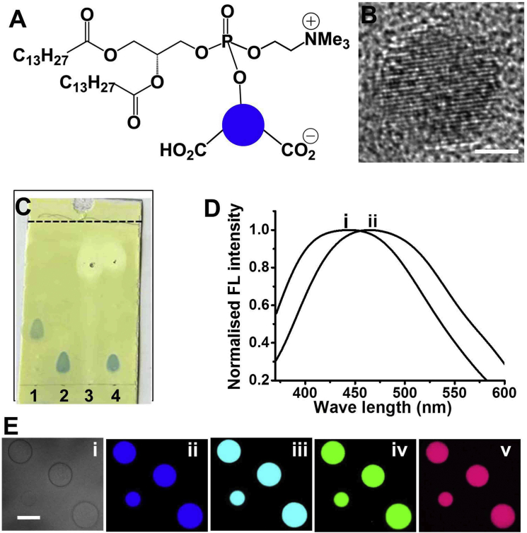
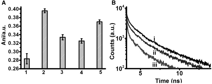
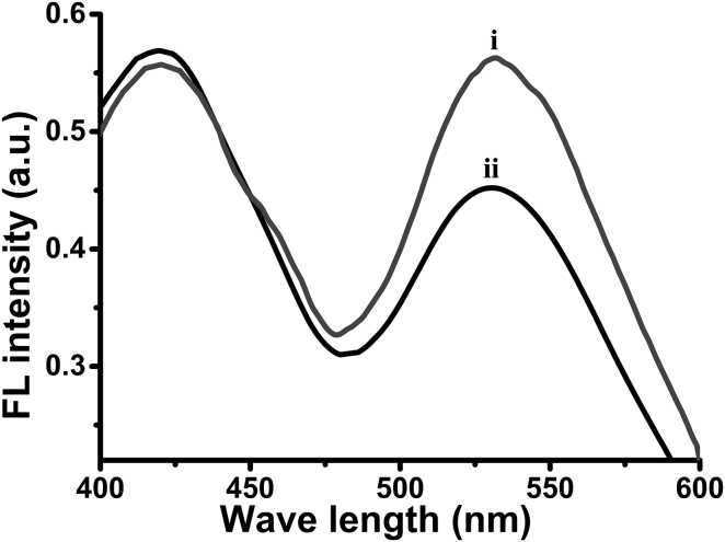
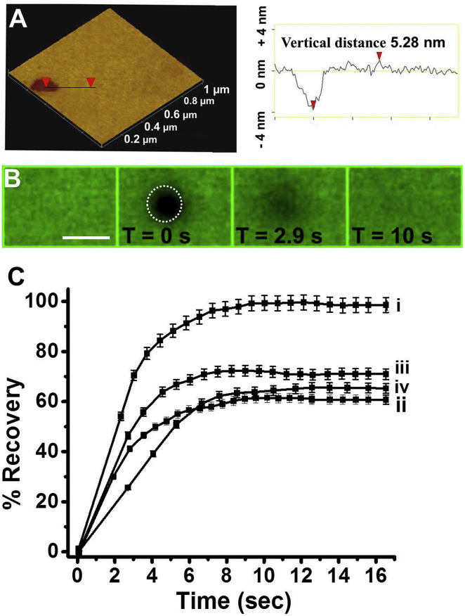
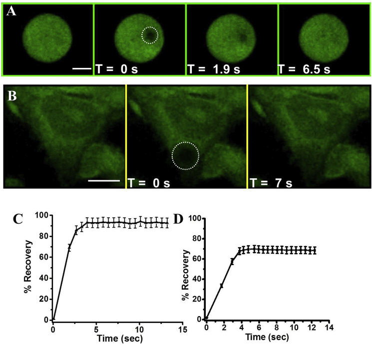
Elucidating the dynamic properties of membranes is important for understanding fundamental cellular processes and for shedding light on the interactions of proteins, drugs, and viruses with the cell surface. Dynamic studies of lipid bilayers have been constrained, however, by the relatively small number of pertinent molecular probes and the limited physicochemical properties of the probes. We show that a lipid conjugate comprised of a fluorescent carbon dot (C-dot) covalently attached to a phospholipid constitutes a versatile and effective vehicle for studying bilayer dynamics. The C-dot-modified phospholipids readily incorporated within biomimetic membranes, including solid-supported bilayers and small and giant vesicles, and inserted into actual cellular membranes. We employed the C-dot-phospholipid probe to elucidate the effects of polymyxin-B (a cytolytic peptide), valproic acid (a lipophilic drug), and amyloid-β (a peptide associated with Alzheimer's disease) upon bilayer fluidity and lipid dynamics through the application of various biophysical techniques.


Figures

Figure 1
Figure 1
Synthesis and characterization of the C-dot-DMPC conjugate. (A) Chemical structure of the C-dot-DMPC molecule; the blue circle corresponds to the C-dot. (B) HRTEM image of a typical C-dot used in the synthesis. Scale bar, 1 nm. (C) Image of a TLC plate after elution, spraying with visualization reagent for detection of phosphate groups. The different columns correspond to 1) the C-dot-DMPC reaction product, 2) DMPC only, 3) C-dots only, and 4) a physical mixture of DMPC and C-dots that did not react. (D) Fluorescence emission spectra of the C-dots (excitation at 350 nm) before (i) and after (ii) coupling to DMPC in phosphate buffer (10 mM, pH 7.4). (E) Confocal microscopy images of GVs composed of DOPC (1 mM) and C-dot-DMPC (2 mg/mL). Bright-field (i) and confocal fluorescence microscopy images were recorded upon excitation at 405 nm, emission filter EM 445/60 (ii); excitation at 440 nm, emission filter EM 477/45 (iii); excitation at 488 nm, emission filter EM 525/50 (iv); and excitation at 514 nm, emission filter EM 525/50 (v). Scale bar, 10 μm. To see this figure in color, go online.
Figure 2
Figure 2
Bilayer dynamics in C-dot-DMPC/DOPC SUVs. (A) Fluorescence anisotropy (excitation 375 nm, emission 463 nm) of C-dot-DMPC embedded in DOPC SUVs. 1) Control C-dot-DMPC/DOPC SUVs before addition of membrane-active compounds. Anisotropy was recorded after addition of 2) PMB (0.1 mM), 3) valproic acid (0.1 mM), or 4) Aβ1-40 (30 μM). 5) C-dot-DMPC embedded in SUVs composed of DOPC and cholesterol (7:3 mol ratio of DOPC/cholesterol). (B) Fluorescence lifetime decays of C-dot-DMPC embedded in the SUVs. (i) Control vesicles before addition of membrane-active compounds. (ii) C-dot-DMPC embedded in SUVs composed of DOPC and cholesterol (7:3 mol ratio of DOPC/cholesterol). (iii) After addition of PMB (0.1 mM) to the C-dot-DMPC/DOPC SUVs.
Figure 3
Figure 3
FRET experiment. Emission spectra (excitation at 350 nm) of SUVs composed of DOPC, NBD-PE (100:1 mol ratio), and C-dot-DMPC before (i) and after (ii) addition of PMB.
Figure 4
Figure 4
FRAP experiments in C-dot-DMPC/DOPC SSBs. (A) AFM image of an SSB (left) and the corresponding height profile (right). The size of the AFM image is 1 μm × 1 μm. (B) Confocal fluorescence microscopy images (excitation at 405 nm, emission filter EM 445/60) recorded at different times after photobleaching by a 405 nm laser (time immediately after laser irradiation is defined as T = 0 s). The bleached region is indicated by the circle. Scale bar, 5 μm. (C) Recovery curves recorded upon incubation of the SSBs with different molecules: control SSB (i), PMB (ii), valproic acid (iii), and Aβ1-40 (iv). Each experiment was repeated five times. To see this figure in color, go online.
Figure 5
Figure 5
FRAP using C-dot-DMPC embedded in GVs and cell membranes. (A and B) Confocal fluorescence microscopy images (excitation at 405 nm, emission filter EM 445/60) recorded at different times after photobleaching by a 405 nm laser (time immediately after laser irradiation defined as T = 0 s). (A) C-dot-DMPC/DOPC GVs. (B) HeLa cells incubated with C-dot-DMPC. Scale bars, 5 μm. (C) Recovery curve corresponding to the FRAP experiment shown in (A). (D) Recovery curve corresponding to the FRAP experiment shown in (B). Each experiment was repeated five times. To see this figure in color, go online.

Similar articles

Cited by

References

    1. Pucadyil T.J., Mukherjee S., Chattopadhyay A. Organization and dynamics of NBD-labeled lipids in membranes analyzed by fluorescence recovery after photobleaching. J. Phys. Chem. B. 2007;111:1975–1983. - PubMed
    1. Guo L., Har J.Y., Wohland T. Molecular diffusion measurement in lipid bilayers over wide concentration ranges: a comparative study. ChemPhysChem. 2008;9:721–728. - PubMed
    1. Tero R. Substrate effects on the formation process, structure and physicochemical properties of supported lipid bilayers. Materials (Basel) 2012;5:2658.
    1. Singer S.J., Nicolson G.L. The fluid mosaic model of the structure of cell membranes. Science. 1972;175:720–731. - PubMed
    1. Ma Y., Jiang R., Sun X.-L. Chemoselectively surface funtionalizable tethered bilayer lipid membrane for versatile membrane mimetic systems fabrication. J. Mater. Chem. 2012;22:6148–6155.

Publication types

MeSH terms

LinkOut - more resources