

Electron Paramagnetic Resonance of a Single NV Nanodiamond Attached to an Individual Biomolecule
- PMID: 27166812
- PMCID: PMC4940449
- DOI: 10.1016/j.bpj.2016.03.022
Electron Paramagnetic Resonance of a Single NV Nanodiamond Attached to an Individual Biomolecule
Abstract
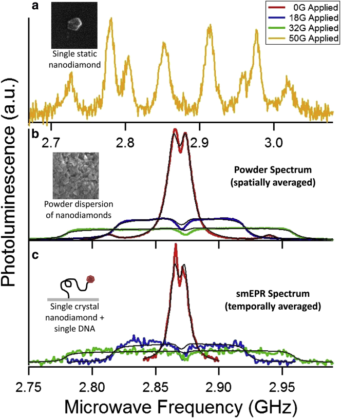
Electron paramagnetic resonance (EPR), an established and powerful methodology for studying atomic-scale biomolecular structure and dynamics, typically requires in excess of 10(12) labeled biomolecules. Single-molecule measurements provide improved insights into heterogeneous behaviors that can be masked in ensemble measurements and are often essential for illuminating the molecular mechanisms behind the function of a biomolecule. Here, we report EPR measurements of a single labeled biomolecule. We selectively label an individual double-stranded DNA molecule with a single nanodiamond containing nitrogen-vacancy centers, and optically detect the paramagnetic resonance of nitrogen-vacancy spins in the nanodiamond probe. Analysis of the spectrum reveals that the nanodiamond probe has complete rotational freedom and that the characteristic timescale for reorientation of the nanodiamond probe is slow compared with the transverse spin relaxation time. This demonstration of EPR spectroscopy of a single nanodiamond-labeled DNA provides the foundation for the development of single-molecule magnetic resonance studies of complex biomolecular systems.
Copyright © 2016 Biophysical Society. Published by Elsevier Inc. All rights reserved.
Figures


Similar articles
-
High-Frequency Electron Paramagnetic Resonance Spectroscopy of Nitroxide-Functionalized Nanodiamonds in Aqueous Solution.Cell Biochem Biophys. 2017 Jun;75(2):151-157. doi: 10.1007/s12013-016-0739-4. Epub 2016 Jun 21. Cell Biochem Biophys. 2017. PMID: 27324044
-
Investigating Functional DNA Grafted on Nanodiamond Surface Using Site-Directed Spin Labeling and Electron Paramagnetic Resonance Spectroscopy.J Phys Chem B. 2016 May 5;120(17):4003-8. doi: 10.1021/acs.jpcb.6b00790. Epub 2016 Apr 20. J Phys Chem B. 2016. PMID: 27058261
-
Nanodiamond molecular imaging with enhanced contrast and expanded field of view.J Biomed Opt. 2014 Jan;19(1):011015. doi: 10.1117/1.JBO.19.1.011015. J Biomed Opt. 2014. PMID: 24057337
-
Nanoscale quantum sensing with Nitrogen-Vacancy centers in nanodiamonds - A magnetic resonance perspective.Prog Nucl Magn Reson Spectrosc. 2023 Apr-Jun;134-135:20-38. doi: 10.1016/j.pnmrs.2022.12.001. Epub 2022 Dec 13. Prog Nucl Magn Reson Spectrosc. 2023. PMID: 37321756 Review.
-
High-field EPR on membrane proteins - crossing the gap to NMR.Prog Nucl Magn Reson Spectrosc. 2013 Nov;75:1-49. doi: 10.1016/j.pnmrs.2013.07.002. Epub 2013 Jul 29. Prog Nucl Magn Reson Spectrosc. 2013. PMID: 24160760 Review.
Cited by
-
Graphitic and oxidised high pressure high temperature (HPHT) nanodiamonds induce differential biological responses in breast cancer cell lines.Nanoscale. 2018 Jul 5;10(25):12169-12179. doi: 10.1039/c8nr02177e. Nanoscale. 2018. PMID: 29917033 Free PMC article.
-
Self-Aligned Multilayered Nitrogen Vacancy Diamond Nanoparticles for High Spatial Resolution Magnetometry of Microelectronic Currents.Nano Lett. 2025 Jun 11;25(23):9204-9213. doi: 10.1021/acs.nanolett.5c00656. Epub 2025 May 19. Nano Lett. 2025. PMID: 40387807 Free PMC article.
-
Single and double quantum transitions in spin-mixed states under photo-excitation.Sci Rep. 2024 Sep 28;14(1):22421. doi: 10.1038/s41598-024-73118-z. Sci Rep. 2024. PMID: 39341935 Free PMC article.
-
High-throughput nitrogen-vacancy center imaging for nanodiamond photophysical characterization and pH nanosensing.Nanoscale. 2020 Nov 5;12(42):21821-21831. doi: 10.1039/d0nr05931e. Nanoscale. 2020. PMID: 33103692 Free PMC article.
-
Optical Magnetometry Based on Nanodiamonds with Nitrogen-Vacancy Color Centers.Materials (Basel). 2019 Sep 11;12(18):2951. doi: 10.3390/ma12182951. Materials (Basel). 2019. PMID: 31514463 Free PMC article.
References
-
- Cornish P.V., Ha T. A survey of single-molecule techniques in chemical biology. ACS Chem. Biol. 2007;2:53–61. - PubMed
-
- Allemand J.-F., Bensimon D., Croquette V. Stretching DNA and RNA to probe their interactions with proteins. Curr. Opin. Struct. Biol. 2003;13:266–274. - PubMed
-
- Haustein E., Schwille P. Single-molecule spectroscopic methods. Curr. Opin. Struct. Biol. 2004;14:531–540. - PubMed
-
- Santos N.C., Castanho M.A.R.B. An overview of the biophysical applications of atomic force microscopy. Biophys. Chem. 2004;107:133–149. - PubMed
Publication types
MeSH terms
Substances
Grants and funding
LinkOut - more resources
Full Text Sources
Other Literature Sources
