Meta-Analysis
. 2016 May 11;11(5):e0155277.
doi: 10.1371/journal.pone.0155277. eCollection 2016.

Muscle Activity Onset Prior to Landing in Patients after Anterior Cruciate Ligament Injury: A Systematic Review and Meta-Analysis

Affiliations
Meta-Analysis

Muscle Activity Onset Prior to Landing in Patients after Anterior Cruciate Ligament Injury: A Systematic Review and Meta-Analysis

Daniel Theisen et al. PLoS One. .

Abstract

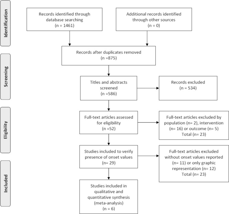
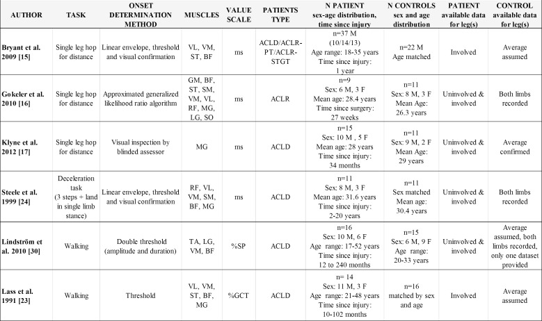
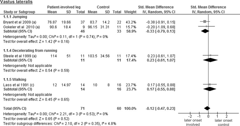
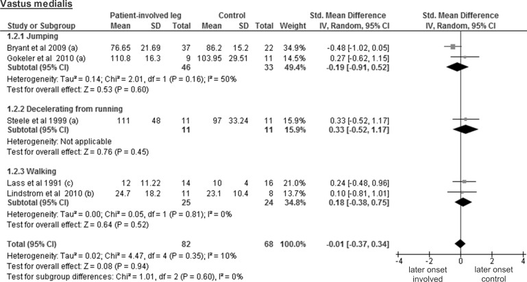
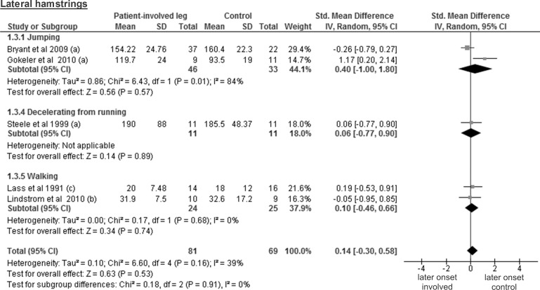
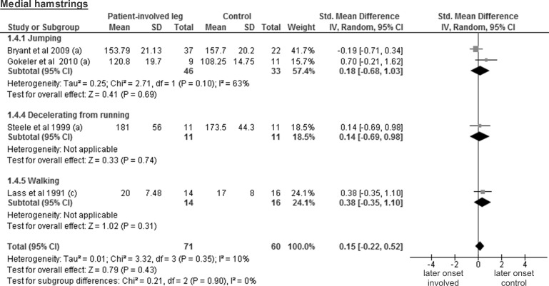
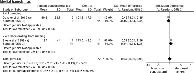
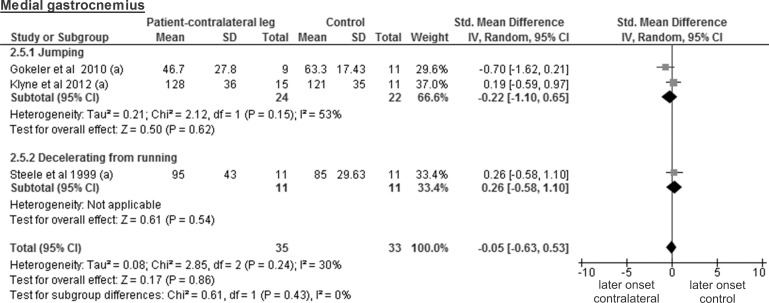
Muscle activation during landing is paramount to stabilise lower limb joints and avoid abnormal movement patterns. Delayed muscle activity onset measured by electromyography (EMG) has been suggested to be associated with anterior cruciate ligament (ACL) injury. Therefore, the aim of this systematic review and meta-analysis was to test the hypothesis if ACL-injured patients display different results for muscle onset timing during standard deceleration tasks compared to healthy control participants. PubMed, Embase, Scopus and ScienceDirect databases were systematically searched over the period from January 1980 to February 2015, yielding a total of 1461 citations. Six studies meeting inclusion criteria underwent quality assessment, data extraction and re-computing procedures for the meta-analysis. The quality was rated "moderate" for 2 studies and "poor" for 4. Patients included and procedures used were highly heterogeneous. The tasks investigated were single leg hopping, decelerating from running or walking, tested on a total of 102 ACL-injured participants and 86 controls. EMG analyses of the muscles vastus lateralis, vastus medialis, lateral and medial hamstrings revealed trivial and non-significant standardised mean differences (SMD<0.20; p>0.05) between patients and control participants. Furthermore, no differences were found between the contralateral leg of patients and controls for muscle activity onset of the medial and lateral gastrocnemius (SMD<0.20; p>0.05). Based on 3 studies, the involved legs of ACL-injured patients showed overall earlier muscle activity onset compared to control participants for the medial gastrocnemius (SMD = 0.5; p = 0.05). Similar results were found for the lateral gastrocnemius (SMD = 2.1; p<0.001), with a greater effect size but based only on a single study. We conclude that there are no differences between leg muscles of ACL-injured patients and healthy controls regarding the muscle activity onset during landing. However, current evidence is scarce and weak, which highlights the need for further research in this area.


Conflict of interest statement

Competing Interests: The authors have declared that no competing interests exist.

Figures

Fig 1
Fig 1. Search results throughout the review process.
Fig 2
Fig 2. Overview of the main studies characteristics.
ACLD, ACL-deficient; ACLR, ACL-reconstructed; PT (patellar tendon graft); STGT (semitendinosus-gracilis tendon graft); ms, milliseconds; %SP, percentage of stance phase; %GCT, percentage of gait cycle time; VL, vastus lateralis; VM, vastus medialis; RF, rectus femoris; ST, semitendinosus; SM, semimembranosus; BF, biceps femoris; GM, gluteus maximus; MG, medial gastrocnemius; LG, lateral gastrocnemius; SO, soleus; TA, tibialis anterior. Note that in the study of Lindstrom et al, the final number of included participants is lower due to technical issues related to the EMG recordings.
Fig 3
Fig 3. Forest plot illustrating muscle activity onset times of both groups and group differences for vastus lateralis.
SMD between ACL patients’ involved leg (Patient-involved leg) and control participants (Control). (a), values in milliseconds; (c), values in percentage of gait cycle time.
Fig 4
Fig 4. Forest plot illustrating muscle activity onset times of both groups and group differences for vastus medialis.
SMD between ACL patients’ involved leg (Patient-involved leg) and control participants (Control). (a), values in milliseconds; (b), values in percentage of stance phase; (c), values in percentage of gait cycle time.
Fig 5
Fig 5. Forest plot illustrating muscle activity onset times of both groups and group differences for lateral hamstrings.
SMD between ACL patients’ involved leg (Patient-involved leg) and control participants (Control). (a), values in milliseconds; (b), values in percentage of stance phase; (c), values in percentage of gait cycle time.
Fig 6
Fig 6. Forest plot illustrating muscle activity onset times of both groups and group differences for medial hamstrings.
SMD between ACL patients’ involved leg (Patient-involved leg) and control participants (Control). (a), values in milliseconds; (c), values in percentage of gait cycle time.
Fig 7
Fig 7. Forest plot illustrating muscle activity onset times of both groups and group differences for medial gastrocnemius.
SMD between ACL patients’ involved leg (Patient-involved leg) and control participants (Control). (a), values in milliseconds.
Fig 8
Fig 8. Forest plot illustrating muscle activity onset times of both groups and group differences for vastus lateralis.
SMD between ACL patients’ healthy contralateral leg (Patient-contralateral leg) and control participants (Control). (a), values in milliseconds.
Fig 9
Fig 9. Forest plot illustrating muscle activity onset times of both groups and group differences for vastus medialis.
SMD between ACL patients’ healthy contralateral leg (Patient-contralateral leg) and control participants (Control). (a), values in milliseconds; (b) values in percentage of stance phase.
Fig 10
Fig 10. Forest plot illustrating muscle activity onset times of both groups and group differences for lateral hamstrings.
SMD between ACL patients’ healthy contralateral leg (Patient-contralateral leg) and control participants (Control). (a), values in milliseconds; (b) values in percentage of stance phase.
Fig 11
Fig 11. Forest plot illustrating muscle activity onset times of both groups and group differences for medial hamstrings.
SMD between ACL-injured patients’ healthy contralateral leg (Patient-contralateral leg) and control participants (Control). (a), values in milliseconds.
Fig 12
Fig 12. Forest plot illustrating muscle activity onset times of both groups and group differences for medial gastrocnemius.
SMD between ACL patients’ healthy contralateral legs (Patient-contralateral leg) and control participants (Control). (a), values in milliseconds.

References

    1. Hewett TE, Myer GD, Ford KR, Heidt RS Jr., Colosimo AJ, McLean SG, et al. Biomechanical measures of neuromuscular control and valgus loading of the knee predict anterior cruciate ligament injury risk in female athletes: a prospective study. Am J Sports Med. 2005;33(4):492–501. 10.1177/0363546504269591 . - DOI - PubMed
    1. Cochrane JL, Lloyd DG, Buttfield A, Seward H, McGivern J. Characteristics of anterior cruciate ligament injuries in Australian football. Journal of science and medicine in sport / Sports Medicine Australia. 2007;10(2):96–104. 10.1016/j.jsams.2006.05.015 . - DOI - PubMed
    1. Boden BP, Torg JS, Knowles SB, Hewett TE. Video analysis of anterior cruciate ligament injury: abnormalities in hip and ankle kinematics. Am J Sports Med. 2009;37(2):252–9. 10.1177/0363546508328107 . - DOI - PubMed
    1. Olsen OE, Myklebust G, Engebretsen L, Bahr R. Injury mechanisms for anterior cruciate ligament injuries in team handball: a systematic video analysis. Am J Sports Med. 2004;32(4):1002–12. . - PubMed
    1. Fox AS, Bonacci J, McLean SG, Spittle M, Saunders N. What is normal? Female lower limb kinematic profiles during athletic tasks used to examine anterior cruciate ligament injury risk: a systematic review. Sports medicine (Auckland, NZ). 2014;44(6):815–32. Epub 2014/04/01. 10.1007/s40279-014-0168-8 . - DOI - PubMed

MeSH terms