. 2016 Jun 14;7(24):36800-36813.
doi: 10.18632/oncotarget.9191.

C-terminus of MUC16 activates Wnt signaling pathway through its interaction with β-catenin to promote tumorigenesis and metastasis

Affiliations

C-terminus of MUC16 activates Wnt signaling pathway through its interaction with β-catenin to promote tumorigenesis and metastasis

Qi Liu et al. Oncotarget. .

Abstract

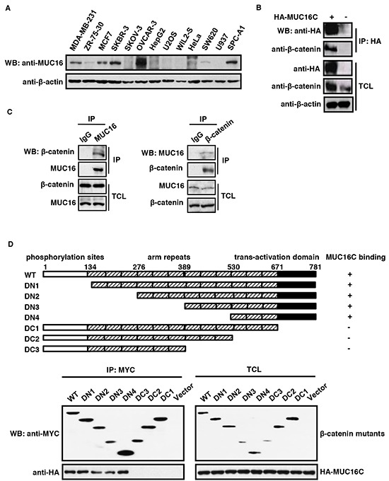
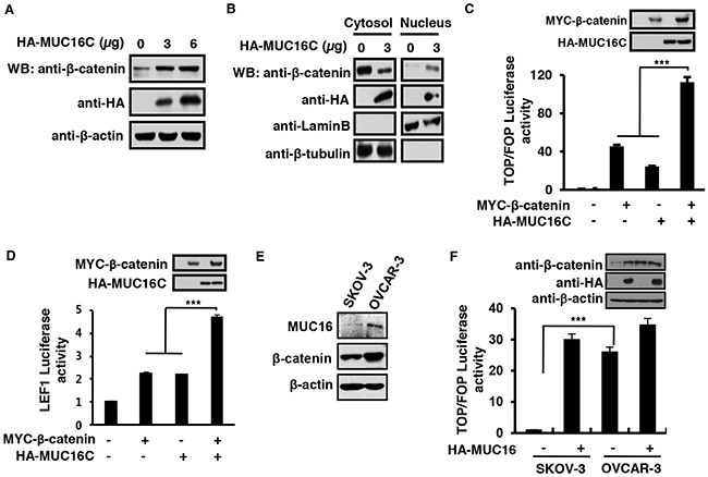
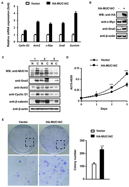
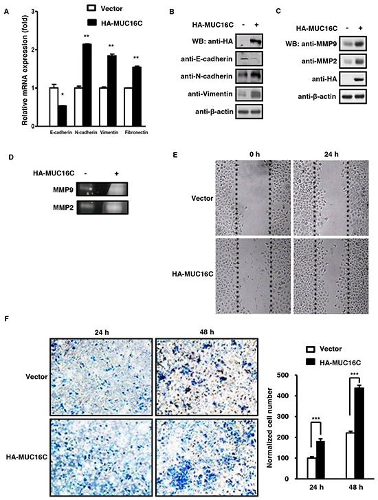
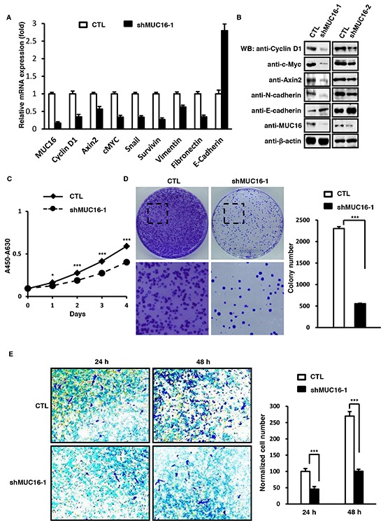
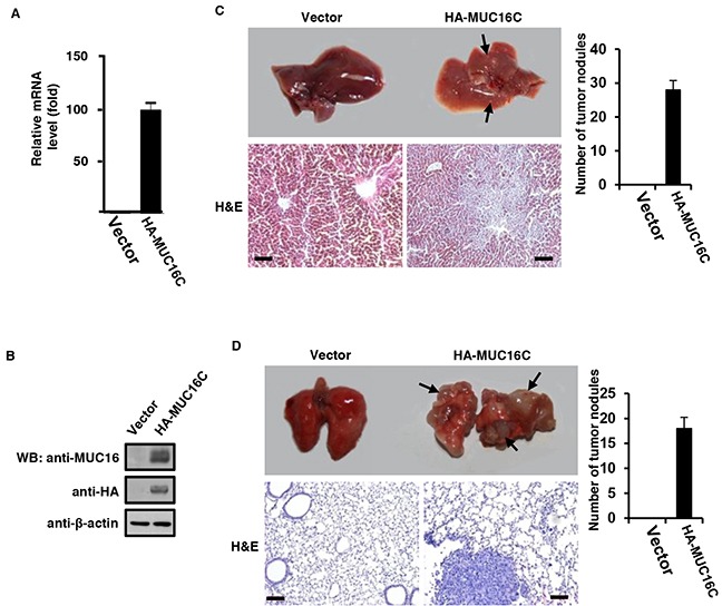
MUC16/CA125 has been identified as a prominent cancer biomarker, especially for epithelial ovarian cancers, in clinical test for over three decades. Due to its huge mass, limited knowledge of MUC16 was acquired previously. By utilizing a well characterized self-made MUC16 monoclonal antibody, we identified the endogenous interaction between a C-terminal fragment of MUC16 (MUC16C) and β-catenin for the first time, and further elucidated that trans-activation domain of β-catenin is required for this interaction. Such interaction could activate the Wnt/β-catenin signaling pathway by facilitating cytosol-nucleus transportation of β-catenin, consequently induce cell proliferation and the migration, eventually lead to tumorigenesis and metastasis in nude mice. Consistently, knockdown of MUC16 significantly weakened the capabilities of cells for proliferation and migration. Based on our discovery, we suggest that MUC16 appears as an attractive target for the development of effective anticancer drugs.

Keywords: C-terminus; MUC16; Wnt signaling; tumorigenesis; β-catenin.


Conflict of interest statement

The authors declare that they have no conflicts of interest.

Figures

Figure 1
Figure 1. MUC16/MUC16C interacts with β-catenin
A. Expression profile of MUC16 in different cell lines. The cell lysates were analyzed by Western blot with the self-made mouse anti-MUC16 (40 μg input) or mouse anti-β-actin (8 μg input) antibody respectively. B. Ectopically expressed MUC16C interacts with endogenous β-catenin. HeLa cells were transfected with pcDNA3.3-HA-MUC16C or the empty vector as a control. At 24 h post-transfection, cells were lysed and subjected to immunoprecipitation with mouse anti-HA antibody, followed by Western blot with mouse anti-HA and rabbit anti-β-catenin antibodies separately. C. MUC16 and β-catenin interact with each other in vivo. Lysates of SKBR-3 cells were subjected to immunoprecipitation with the control IgG, rabbit anti-β-catenin and mouse anti-MUC16 antibodies respectively. The precipitates were then detected with indicated antibodies. D. C-terminus of β-catenin is essential for its interaction with MUC16. For the domain mapping experiment, structures of deletion mutants of β-catenin are shown on the top of the panel. Functional domains of β-catenin are indicated above the schema; the remaining fragments of each deletion mutant are shown in the diagram. HEK293T cells were co-transfected with pcDNA3.3-HA-MUC16C and different MYC-β-catenin mutants or the empty vector as a control. At 24 h post-transfection, cells were lysed and subjected to immunoprecipitation with rabbit anti-MYC antibody, followed by Western blot with mouse anti-HA or anti-MYC antibody.
Figure 2
Figure 2. MUC16C promotes β-catenin-dependent transcriptional activity through enhanced cytosol-nucleus transportation
A. MUC16C increases the protein level of β-catenin. SKOV-3 cells were transfected with different amount of pcDNA3.3-HA-MUC16C vectors (0, 3 and 6 μg). At 24 h post-transfection, total cell lysates were harvested and subjected to Western blot with indicated antibodies. B. MUC16C enhances the cytosol-nucleus transportation of β-catenin. SKOV-3 cells were transfected with different amount of pcDNA3.3-HA-MUC16C vectors (0 and 3 μg). At 72 h post-transfection, cells were harvested and subjected to nuclear/cytosol fractionation, followed by Western blot with rabbit anti-β-catenin, mouse anti-HA, goat anti-Lamin B and rabbit anti-β-tubulin antibodies respectively. MUC16C activates β-catenin-dependent TOP/FOP luciferase reporter (C) and LEF1 reporter (D). HEK293T cells were transfected with the expression vectors of MYC-β-catenin and HA-MUC16C alone or in combination, together with pRL-TK and TOP/FOP luciferase reporter C. or pGL3-fos-7LEF-luciferase/pCG-LEF1 D. At 24 h post-transfection, luciferase activities were determined, and the results are presented as mean±SD of three independent experiments. The protein levels of ectopically expressed β-catenin and MUC16C were analyzed by Western blot. E. In ovary cancer cell lines SKOV-3 and OVCAR-3, the protein levels of MUC16 are closely correlated with that of β-catenin. Protein levels were determined by using Western blot. F. Wnt signaling is significantly activated in OVCAR-3 cells rather than in SKOV-3 cells. SKOV-3 and OVCAR-3 cells transfected with or without HA-MUC16C, together with TOP/FOP luciferase reporter were determined for relative luciferase activities. ***P<0.0001, student's T-test.
Figure 3
Figure 3. MUC16C up-regulates expression of Wnt target genes and promotes cell proliferation
A. MUC16C increases mRNA levels of classic downstream genes of the Wnt signaling pathway. SKOV-3 cells were infected with lentivirus containing pBOBI-HA-MUC16C or pBOBI vector as a control. At 36 h post-infection, cells were harvested and the mRNA levels of Cyclin D1, Axin2, c-Myc, Snail and Survivin were determined by quantitative RT-PCR. B. MUC16C up-regulates protein levels of Wnt target genes c-Myc and Snail. SKOV-3 cells were processed as in (A). At 36 h post-infection, cells were lysed and subjected to Western blot with indicated antibodies. C. The expression of Wnt target genes is relative to MUC16 in three human breast cancer samples. The homogenates of these three pathological samples (N=normal, C=carcinoma) were analyzed by Western blot with indicated antibodies. D. MUC16C promotes cell proliferation as assessed by MTT assay. SKOV-3 cells were processed as in (A). Proliferation of cells was measured by MTT staining during a three-day culture period. Results were shown as mean±SEM of three independent experiments. Significance was calculated by the student's T-test (*P<0.01, ***P<0.0001). E. MUC16C promotes cell proliferation as assessed by colony formation assay, in which 2 × 10² cells of SKOV-3 stably expressing HA-MUC16C or the empty vector as a control were seeded on 100 mm culture dishes. After 2 weeks, colonies were fixed and stained with crystal violet. Colonies numbers were counted and showed as mean±SD of three independent experiments. Significance was calculated by the student's T-test (***P<0.0001).
Figure 4
Figure 4. MUC16C promotes cell migration and invasion
A. MUC16C influences mRNA levels of novel EMT markers like E-cadherin, N-cadherin, Vimentin and Fibronectin. SKOV-3 cells with or without over-expressed MUC16C were harvested at 36 h post-infection and the mRNA levels of above EMT markers were determined by RT-PCR. B. MUC16C influences protein levels of EMT markers. SKOV-3 cells described in (A) were lysed at 36 h post-infection, followed by Western blot with indicated antibodies. MUC16C promotes expression of MMP family members, MMP2 and MMP9, as assessed by Western blot in C. or gelatin zymography assay in D.. SKOV-3 cells for assays in (D) were cultured further in the serum-free medium for another 48 h, and then the culture medium was collected for detecting activated MMP9 and MMP2. E. MUC16C promotes migration of SKOV-3 cells. The SKOV-3 cells described above in (A) were subjected to wound healing assay. Photographs were taken at different time points (0, 24 h). F. MUC16C promotes invasion of SKOV-3 cells. The SKOV-3 cells described above in (A) were subjected to transwell invasion assays. Data are reported as normalized number of cells that invaded through the transwell membrane relative to that of the control (100%), and represent the mean mean±SD of three independent experiments. Significance was calculated by the student's T-test (***P<0.0001).
Figure 5
Figure 5. MUC16 knockdown reduces proliferation and metastasis of SKBR-3 cells
A. MUC16 knockdown influences mRNA levels of classic Wnt target genes and EMT markers. SKBR-3 cells were infected with lentivirus containing pLV-LacZ (CTL) or pLV-MUC16 (shMUC16-1). At 36 h post-infection, cells were harvested and the mRNA levels of MUC16, Cyclin D1, Axin2, c-Myc, Snail, Survivin, Vimentin, E-cadherin and Fibronectin were determined by RT-PCR. B. MUC16 knockdown in SKBR-3 cells decreases protein levels of Wnt target genes. SKBR-3 cells were transfected with two independent shRNA against MUC16 (shMUC16-1 and shMUC16-2) individually. At 36 h post-infection, cells were lysed and subjected to Western blot with indicated antibodies. C. MUC16 knockdown in SKBR-3 cells inhibits cell proliferation as assessed by CCK8 test. Proliferations of cells described above were measured by CCK8 staining during a four-day culture period. The results were shown as mean±SEM of three independent experiments. Significance was calculated by the student's T-test (*P<0.01, ***P<0.0001). D. MUC16 knockdown in SKBR-3 cells inhibits cell proliferation as showed by colony formation assay. 2 × 10² of SKBR-3 cells were seeded on 100 mm culture dishes. After 2 weeks, colonies were fixed and stained with crystal violet. Numbers of colonies were counted and showed as mean±SD of three independent experiments. Significance was calculated by the student's T-test (***P<0.0001). E. MUC16 knockdown inhibits invasion of SKBR-3 cells. The SKBR-3 cells described above in (A) were subjected to transwell invasion assays. Data are reported as normalized cell number that invaded through the transwell membrane relative to the empty vector control (100%), and represented as mean±SD of three independent experiments. Significance was calculated by the student's T-test (***P<0.0001).
Figure 6
Figure 6. Over-expressed MUC16C enhances metastasis capability of SKOV-3 cells in vivo
1 × 10⁶ of SKOV-3 cells with or without over-expressed MUC16C were injected intravenously into nude mice (n=5) once a week for consecutive 3 weeks. After another 5 weeks, mice were sacrificed to analyze their main organs and tissues. A. Livers from nude mice described above were homogenated followed by Western blot with antibodies indicated. B. Livers from nude mice described above were homogenated and then the total RNA was extracted for fluorescence quantitative RT-PCR analysis of the RNA level of MUC16/MUC16C. Representative photographs (upper) of livers C. and lungs D. from nude mice described above and the corresponding histological analyses (lower) are shown. Sections were stained with H&E. The black arrows indicate the metastasis stoves. Scale bars 100 μm. Metastatic tumor nodules on the surfaces of livers and lungs were counted and statistical results were presented as histograms in the right (n=5).

Similar articles

Cited by

References

    1. Kui Wong N, Easton RL, Panico M, Sutton-Smith M, Morrison JC, Lattanzio FA, Morris HR, Clark GF, Dell A, Patankar MS. Characterization of the oligosaccharides associated with the human ovarian tumor marker CA125. The Journal of biological chemistry. 2003;278:28619–28634. - PubMed
    1. Hollingsworth MA, Swanson BJ. Mucins in cancer: protection and control of the cell surface. Nature reviews Cancer. 2004;4:45–60. - PubMed
    1. Corfield AP. Mucins: a biologically relevant glycan barrier in mucosal protection. Biochimica et biophysica acta. 2015;1850:236–252. - PubMed
    1. Bramwell ME, Bhavanandan VP, Wiseman G, Harris H. Structure and function of the Ca antigen. British journal of cancer. 1983;48:177–183. - PMC - PubMed
    1. Gendler SJ, Burchell JM, Duhig T, Lamport D, White R, Parker M, Taylor-Papadimitriou J. Cloning of partial cDNA encoding differentiation and tumor-associated mucin glycoproteins expressed by human mammary epithelium. Proceedings of the National Academy of Sciences of the United States of America. 1987;84:6060–6064. - PMC - PubMed