. 2016 Oct 11;7(41):66525-66539.
doi: 10.18632/oncotarget.9089.

Double mutant P53 (N340Q/L344R) promotes hepatocarcinogenesis through upregulation of Pim1 mediated by PKM2 and LncRNA CUDR

Affiliations

Double mutant P53 (N340Q/L344R) promotes hepatocarcinogenesis through upregulation of Pim1 mediated by PKM2 and LncRNA CUDR

Mengying Wu et al. Oncotarget. .

Abstract

P53 is frequently mutated in human tumors as a novel gain-of-function to promote tumor development. Although dimeric (M340Q/L344R) influences on tetramerisation on site-specific post-translational modifications of p53, it is not clear how dimeric (M340Q/L344R) plays a role during hepatocarcinogenesis. Herein, we reveal that P53 (N340Q/L344R) promotes hepatocarcinogenesis through upregulation of PKM2. Mechanistically, P53 (N340Q/L344R) forms complex with CUDR and the complex binds to the promoter regions of PKM2 which enhances the expression, phosphorylation of PKM2 and its polymer formation. Thereby, the polymer PKM2 (tetramer) binds to the eleventh threonine on histone H3 that increases the phosphorylation of the eleventh threonine on histone H3 (pH3T11). Furthermore, pH3T11 blocks HDAC3 binding to H3K9Ac that prevents H3K9Ac from deacetylation and stabilizes the H3K9Ac modification. On the other hand, it also decreased tri-methylation of histone H3 on the ninth lysine (H3K9me3) and increases one methylation of histone H3 on the ninth lysine (H3K9me1). Moreover, the combination of H3K9me1 and HP1 α forms more H3K9me3-HP1α complex which binds to the promoter region of Pim1, enhancing the expression of Pim1 that enhances the expression of TERT, oncogenic lncRNA HOTAIR and reduces the TERRA expression. Ultimately, P53 (N340Q/L344R) accerlerates the growth of liver cancer cells Hep3B by activating telomerase and prolonging telomere through the cascade of P53 (N340Q/L344R)-CUDR-PKM2-pH3T11- (H3K9me1-HP1α)-Pim1- (TERT-HOTAIR-TERRA). Understanding the novel functions of P53 (N340Q/L344R) will help in the development of new liver cancer therapeutic approaches that may be useful in a broad range of cancer types.

Keywords: LncRNA CUDR; PKM2; hepatoma; mutant P53 (N340Q/L344R).


Conflict of interest statement

CONFLICTS OF INTEREST

The authors disclose no conflicts.

Figures

Figure 1
Figure 1. Double mutant P53 (N340Q/L344R) accelerlates liver cancer cell Hep3B growth in vitro
A. a. The Western blotting analysis of P53 (N340Q/L344R) in stable mutant P53 (N340Q/L344R) overexpressed Hep3B cell lines infected with pLVX-Tet-On-P53 (N340Q/L344R) virus via Tet-On Advanced Inducible Expression System[tetracycline (Tc) :05-2μg/ml]. β-actin as internal control. b. The Nuclear Run on analysis of P53 (N340Q/L344R) in stable mutant P53 (N340Q/L344R) overexpressed Hep3B cell lines. β-actin as internal control. B. Cells growth assay using CCK8. Each value was presented as mean±standard error of the mean (SEM). C. S phase cells assay using BrdU. Each value was presented as mean±standard error of the mean (SEM). D. Cells soft agar colony formation assay. Each value was presented as mean±standard error of the mean (SEM).
Figure 2
Figure 2. Double mutant P53 (N340Q/L344R) promotes liver cancer cell Hep3B growth in vivo
A. The mice were stratified and the tumors were recovered. The photography of xenograft tumors in the five groups (indicated in left). B. The wet weight of each tumor was determined for each mouse. Each value was presented as mean±standard error of the mean (SEM). C. The Xenograft appearance time (days). Each value was presented as mean±standard error of the mean (SEM). D. A portion of each tumor was fixed in 4% paraformaldehyde and embedded in paraffin for anti-PCNA immunostainning. (original magnification×100). E. PCNA positive cells analysis. Each value was presented as mean±standard error of the mean (SEM).
Figure 3
Figure 3. mutant P53 (N340Q/L344R) enhances PKM2 expression dependent on SPONGE CUDR
A. RNA Immunoprecipitation (RIP) with anti-P53 (N340Q/L344R) followed by RT-PCR with CUDR mRNA primers in P53 (N340Q/L344R) overexpressed Hep3B cell lines infected with pLVX-Tet-On-P53 (N340Q/L344R) virus via Tet-On Advanced Inducible Expression System[tetracycline (Tc) :05-2μg/ml]. IgG RIP as negative control. CUDR mRNA as INPUT. B. Chromatin Immunoprecipitation (CHIP) with anti- anti-P53 (N340Q/L344R) followed by PCR with PKM2 promoter primers in in P53 (N340Q/L344R) overexpressed Hep3B cell lines. IgG CHIP as negative control. PMK2promoter DNA as INPUT. C. Chromatin Immunoprecipitation (CHIP) with anti- anti-P53 (N340Q/L344R) followed by PCR with PKM2 promoter primers in in P53 (N340Q/L344R) overexpressed and CUDR knocked-down Hep3B cell lines. IgG CHIP as negative control. PMK2 promoter DNA as INPUT. D. PKM2 promoter luciferase activity assay in mutant P53 (N340Q/L344R) overexpressed Hep3B cell lines. Each value was presented as mean±standard error of the mean (SEM). E. PKM2 expression analysis by RT-PCR in mutant P53 (N340Q/L344R) overexpressed Hep3B cell lines. β-actin as internal control. F. Western blotting with anti-PKM2 and anti-pPKM2 in mutant P53 (N340Q/L344R) overexpressed Hep3B cell lines. β-actin as internal control.
Figure 4
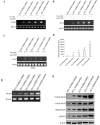
Figure 4. mutant P53 (N340Q/L344R) promotes the interplay between H3K9me1 and HP1α
A. anti-PKM2 Co-Immunoprecipitation (IP) followed by Western blotting with Histone H3 in P53 (N340Q/L344R) overexpressed Hep3B cell lines infected with pLVX-Tet-On-P53 (N340Q/L344R) virus via Tet-On Advanced Inducible Expression System[tetracycline (Tc) :05-2.0μg/ml]. IgG IP as negative control. INPUT refers to Western blotting with anti-PKM2. B. Western blotting with anti-P53 (N340Q/L344R), anti-pH3K11, anti-HDAC3, anti-H3K9Ac, anti-H3K9me3, anti-H3K9me1, anti-HP1α.β-actin as internal control. C. anti-H3K9me1 Co-Immunoprecipitation (IP) followed by Western blotting with anti-HP1α in P53 (N340Q/L344R) overexpressed Hep3B cell lines infected with pLVX-Tet-On-P53 (N340Q/L344R) virus via Tet-On Advanced Inducible Expression System[tetracycline (Tc):05-2μg/ml]. IgG IP as negative control. INPUT refers to Western blotting with anti-H3K9me1.
Figure 5
Figure 5. Mutant P53 (N340Q/L344R) enhances Pim1 expression triggered by the increased interplay between H3K9me1 and HP1α
A. Chromatin Immunoprecipitation (CHIP) with anti-H3K9me1, anti-HP1α followed by PCR with Pim1 promoter primers anti-Histone in P53 (N340Q/L344R) overexpressed Hep3B cell lines infected with pLVX-Tet-On-P53 (N340Q/L344R) virus via Tet-On Advanced Inducible Expression System[tetracycline (Tc) :05-2μg/ml]. IgG CHIP as negative control. Pim1 promoter as INPUT. B. Super-EMSA (gel-shift) with biotin- pim1 promoter probe and anti-H3k9me1 antibody. The intensity of the band was examined by Western blotting with anti-Bioton. C. Pim1 promoter luciferase activity assay in mutant P53 (N340Q/L344R) overexpressed HepG2 cell lines. Each value was presented as mean±standard error of the mean (SEM). D. pim1 expression analysis by RT-PCR (upper) and Western blotting (lower) with anti-Pim1 in mutant P53 (N340Q/L344R) overexpressed HepG2 cell lines. β-actin as internal control.
Figure 6
Figure 6. mutant P53 (N340Q/L344R) enhances HOTAIR expression and stimulates telomerase activity through pim1
A. Nuclear run on assay for HOTAIR and TERRA in P53 (N340Q/L344R) overexpressed Hep3B cell lines infected with pLVX-Tet-On-P53 (N340Q/L344R) virus via Tet-On Advanced Inducible Expression System[tetracycline (Tc) :05-2μg/ml]. β-actin as internal control. B. The telomerase activity was measured by using Quantitative Telomerase Detection. Each value was presented as mean±standard error of the mean (SEM). C. The telomere length was measured by using Quantitative PCR. Each value was presented as mean±standard error of the mean (SEM).
Figure 7
Figure 7. The rescued experiment of carcinogenesis effect of the mutant P53 (N340Q/L344R)
PKM2 knockdown abrogated the Mutant P53 (N340Q/L344R) oncogenic function in Hep3B cell lines infected with control plasmid, pLVX-Tet-On-P53 (N340Q/L344R) [Tc: 2μg/ml], pLVX-Tet-On-P53 (N340Q/L344R) plus pGFP-V-RS-PKM2. A. The western blotting analysis with anti-P53 (fulllength) and anti-PKM2.β -actin as internal control. B. Cells growth assay using CCK8. Each value was presented as mean±standard error of the mean (SEM). C. Cells soft agar colony formation assay. Each value was presented as mean±standard error of the mean (SEM). D. In vivo test in Hep3B cell lines infected with control plasmid, pLVX-Tet-On-P53 (N340Q/L344R) [Tc: 2μg/ml], pLVX-Tet-On-P53 (N340Q/L344R) plus pGFP-V-RS-PKM2. The mice were stratified and the tumors were recovered. The photography of xerograft tumor in the three groups (indicated in left). E. The wet weight of each tumor was determined for each mouse. Each value was presented as mean±standard error of the mean (SEM). F. The Xenograft appearance time. Each value was presented as mean±standard error of the mean (SEM).
Figure 8
Figure 8. The schematic diagram illustrates a model that P53 (N340Q/L344R) promotes hepatocarcinogenesis through upregulation of PKM2
Both P53 (N340Q/L344R) and PKM2 are upregulated in human hepatocellular carcinoma tissues, and present the positive correlation. And the P53 (N340Q/L344R) promotes the growth of hepatocellular carcinoma cells in vitro and in vivo. Mechanistically, P53 (N340Q/L344R) forms complex with CUDR and the complex binds to the promoter regions of PKM2 which enhances the expression, phosphorylation of PKM2 and its polymer formation. Thereby, the polymer PKM2 (tetramer) binds to the eleventh threonine on histone H3 that increases the phosphorylation of the eleventh lysine on histone H3 (pH3T11). Furthermore, pH3T11 blocks HDAC3 binding to H3K9Ac that prevents H3K9Ac from deacetylation and stabilizes the H3K9Ac modification. On the other hand, it also decreased tri-methylation of the ninth lysine ninth on histone3 (H3K9me3) and increases one methylation of the ninth lysine ninth on histone H3 (H3K9me1). Moreover, the combination of H3K9me1 and HP1 α forms more H3K9me3-HP1α complex. This increased complex binds to the promoter region of Pim1, enhancing the expression of Pim1 that increases the expression of TERT, oncogenic lncRNA HOTAIR and reduced expression of TERRA expression. Ultimately, P53 (N340Q/L344R) accerlerates the growth of hepatocellular carcinoma cells by activated telomerase and prolonged telomere through the cascade of P53 (N340Q/L344R)-CUDR-PKM2-pH3T11- (H3K9me1-HP1α)-Pim1- (TERT-HOTAIR-TERRA).

References

    1. Nguyen TA, Menendez D, Resnick MA, Anderson CW. Mutant TP53 posttranslational modifications: challenges and opportunities. Hum Mutat. 2014;35:738–55. - PMC - PubMed
    1. Greenblatt MS, Feitelson MA, Zhu M, Bennett WP, Welsh JA, Jones R, Borkowski A, Harris CC. Integrity of p53 in hepatitis B x antigen-positive and -negative hepatocellular carcinomas. Cancer Res. 1997;57:426–32. - PubMed
    1. Feitelson MA, Zhu M, Duan LX, London WT. patitis B x antigen and p53 are associated in vitro and in liver tissues from patients with primary hepatocellular carcinoma. Oncogene. 1993;8:1109–17. - PubMed
    1. Muller PA, Vousden KH. Mutant p53 in cancer: new functions and therapeutic opportunities. Cancer Cell. 2014;25:304–17. - PMC - PubMed
    1. Ó hAinmhire E, Quartuccio SM, Cheng W, Ahmed RA, King SM, Burdette JE. Mutation or loss of p53 differentially modifies TGFβ action in ovarian cancer. PLoS One. 2014;9:e89553. - PMC - PubMed

MeSH terms