

Review
doi: 10.1213/ANE.0000000000001368.
Mapping General Anesthetic Sites in Heteromeric γ-Aminobutyric Acid Type A Receptors Reveals a Potential For Targeting Receptor Subtypes
Affiliations
- PMID: 27167687
- PMCID: PMC5073028
- DOI: 10.1213/ANE.0000000000001368
Item in Clipboard
Review
Mapping General Anesthetic Sites in Heteromeric γ-Aminobutyric Acid Type A Receptors Reveals a Potential For Targeting Receptor Subtypes
Anesth Analg.
2016 Nov.
Abstract
IV general anesthetics, including propofol, etomidate, alphaxalone, and barbiturates, produce important actions by enhancing γ-aminobutyric acid type A (GABAA) receptor activation. In this article, we review scientific studies that have located and mapped IV anesthetic sites using photoaffinity labeling and substituted cysteine modification protection. These anesthetics bind in transmembrane pockets between subunits of typical synaptic GABAA receptors, and drugs that display stereoselectivity also show remarkably selective interactions with distinct interfacial sites. These results suggest strategies for developing new drugs that selectively modulate distinct GABAA receptor subtypes.
Figures
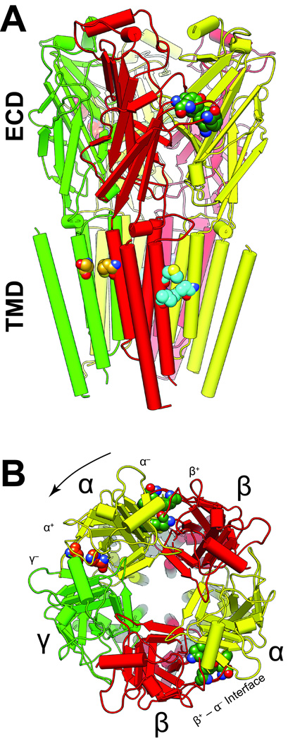
The structure shown was obtained by homology modeling based on the GluCl chloride channel (3RHW.pdb) (7,26). The five subunits are arranged around a central ion–conducting pore and colored as follows: α1, yellow; β3, red, and γ2 green. The α-helices are represented as cylinders, β-sheets as flat planks and loops as strings. A. A side view of the α1β3γ2L GABAAR homology model shows the extracellular domain (ECD) and the transmembrane domain (TMD). The intracellular domain is not modeled because no structural information is available. B. The extracellular domain viewed from the synapse, showing residues involved in agonist and BDZ binding. By convention, subunit order is counted in an anticlockwise direction (arrow). Important amino acids associated with various sites are distinguished by the color of their carbon atoms: agonist site, dark green (α1 Phe-65; β3 Tyr-157 & −205, Phe-200); benzodiazepine site (BDZ), orange (α1 His-102 & −210); azietomidate site, cyan (β3 Met-286 & Val-290, α1 Met-236); main R–mTFD-MPAB site, goldenrod (γ2 Ser-301, β3 Met-227). Red and blue atoms are oxygen and nitrogen respectively.
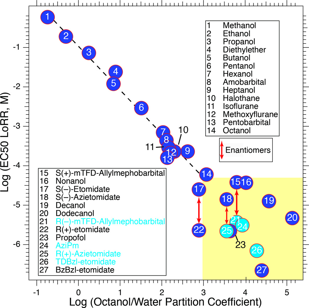
The correlation between EC50 for loss of righting reflexes (LoRR) in tadpoles and octanol/water partition coefficient holds well for anesthetics with EC50s above ~50 µM (Pearson correlation coefficient = −0.987; P < 0.0001). More potent agents display weak dependence on partition coefficient (Pearson correlation coefficient = −0.209; P = 0.49; yellow box). Some of the most successful photolabels lie in this group and are more potent than predicted by hydrophobicity (points with light blue fill). Data sources are given in (46).
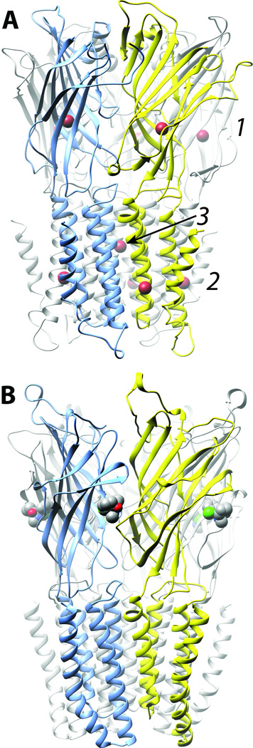
A. Bromoform (brown), a low potency drug, occupies eleven sites in three different classes on ELIC (33): (1) 5 homologous intrasubunit sites in the extracellular domain (ECD); (2) 5 homologous sites in the lipid-protein interface of the transmembrane domain, and (3) a single site in the channel lumen at the interface of all five subunits. B. Ketamine (carbons grey, nitrogen blue, chlorine green) occupies a single class of 5 intersubunit sites on GLIC (34), which potently and stereoselectively inhibits. Channel subunits are shown as ribbons and are colored arbitrarily. The anesthetics are shown space filled and colored conventionally.
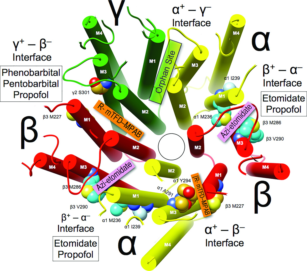
The homology model and coloring are as in Figure 1, and the view of the transmembrane domain () is from the synaptic side with the extracellular domain removed. Subunit interfaces are labeled (note that “+” corresponds to M3 helices and “−” corresponds to M1 helices). Photolabels that selectively bind in each interface are identified in colored boxes, and white boxes identify the general anesthetics that compete with photolabels for occupancy of that interface.
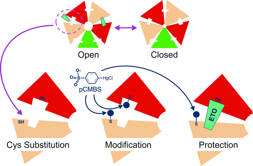
Top: Transmembrane cross-sections of both open and closed receptors are diagrammed. Anesthetic site affinity or accessibility is enhanced by activating GABAA receptors with GABA. Bottom: A close-up view of one interfacial pocket is shown with engineered sulfhydryls, one of which is in the anesthetic site. Modification of both sulfhydryls by p-chloromercuribenzene sulfonate (pCMBS), and protection of one sulfhydryl by bound anesthetic are also depicted. Experiments are performed with one cysteine substitution at a time.
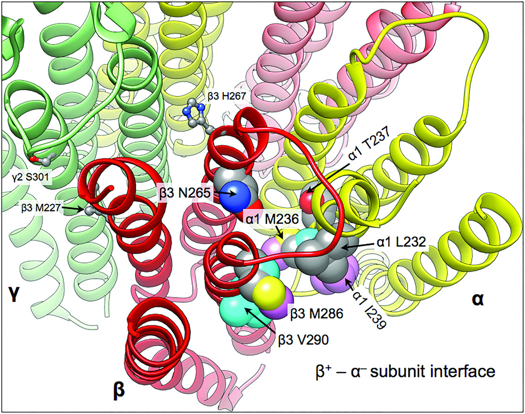
The homology model is based on the GluCl chloride channel (3RHW.pdb) (7,26). The five subunits are colored as follows: α1, yellow; β3, red, and γ2 green. The transmembrane helical backbones are depicted as ribbons. Residues in this site identified with either photolabeling or SCAMP are shown as space-filling atomic models and labeled. Contact residues are coded by coloring their carbons. Residues photolabeled by etomidate and propofol derivatives show cyan and pink carbons, respectively. Grey carbons indicate binding site residues identified by SCAMP with either etomidate or propofol. Red and blue atoms are oxygen and nitrogen respectively. Also shown in ball and stick mode are three residues in the γ+ – β− inter-subunit region that are discussed in the text: γ2S301, β3M227, and β3H267.
References
-
- Rudolph U, Antkowiak B. Molecular and neuronal substrates for general anaesthetics. Nat Rev Neurosci. 2004;5:709–720. - PubMed
-
- Sieghart W. Allosteric modulation of GABAA receptors via multiple drug-binding sites. Adv Pharmacol. 2015;72:53–96. - PubMed
-
- Miller PS, Smart TG. Binding, activation and modulation of Cys-loop receptors. Trends Pharmacol Sci. 2010;31:161–174. - PubMed
Publication types
MeSH terms
Substances
Grants and funding
LinkOut - more resources
Full Text Sources
Other Literature Sources
