. 2016 Jul;157(7):2883-93.
doi: 10.1210/en.2015-1914. Epub 2016 May 11.

Progesterone-Dependent Induction of Phospholipase C-Related Catalytically Inactive Protein 1 (PRIP-1) in Decidualizing Human Endometrial Stromal Cells

Affiliations

Progesterone-Dependent Induction of Phospholipase C-Related Catalytically Inactive Protein 1 (PRIP-1) in Decidualizing Human Endometrial Stromal Cells

Joanne Muter et al. Endocrinology. 2016 Jul.

Abstract

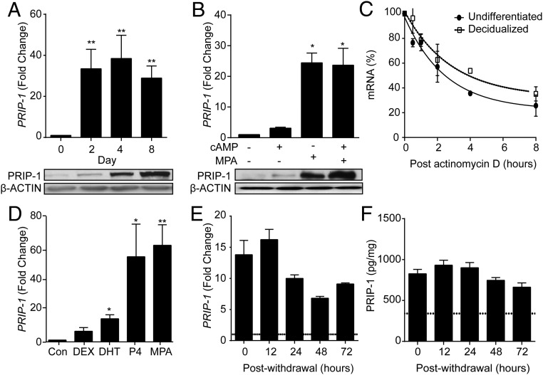
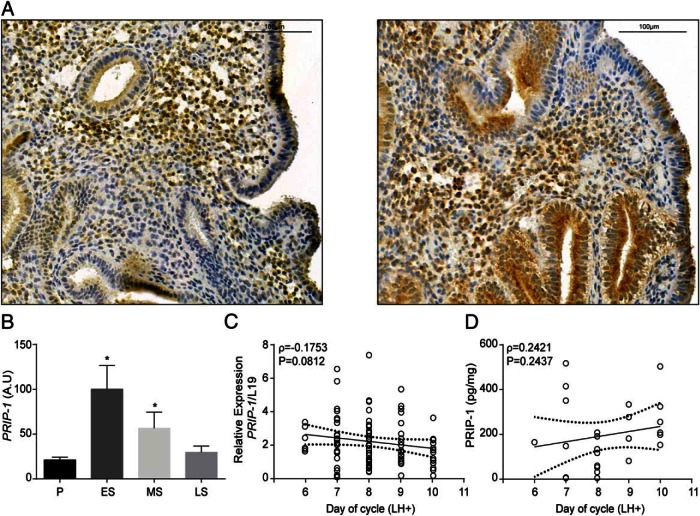
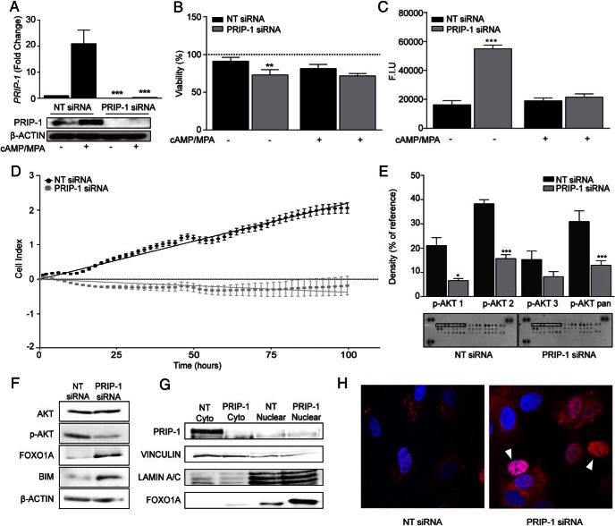
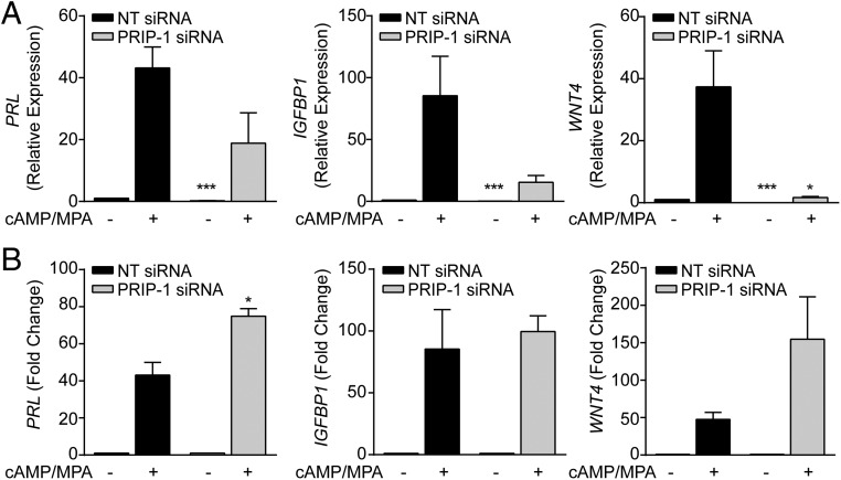
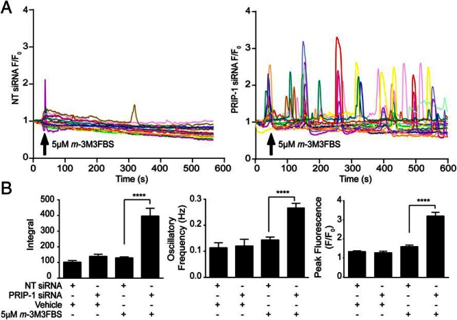
Decidualization denotes the transformation of endometrial stromal cells into specialized decidual cells. In pregnancy, decidual cells form a protective matrix around the implanting embryo, enabling coordinated trophoblast invasion and formation of a functional placenta. Continuous progesterone (P4) signaling renders decidual cells resistant to various environmental stressors, whereas withdrawal inevitably triggers tissue breakdown and menstruation or miscarriage. Here, we show that PLCL1, coding phospholipase C (PLC)-related catalytically inactive protein 1 (PRIP-1), is highly induced in response to P4 signaling in decidualizing human endometrial stromal cells (HESCs). Knockdown experiments in undifferentiated HESCs revealed that PRIP-1 maintains basal phosphoinositide 3-kinase/Protein kinase B activity, which in turn prevents illicit nuclear translocation of the transcription factor forkhead box protein O1 and induction of the apoptotic activator BIM. By contrast, loss of this scaffold protein did not compromise survival of decidual cells. PRIP-1 knockdown did also not interfere with the responsiveness of HESCs to deciduogenic cues, although the overall expression of differentiation markers, such as PRL, IGFBP1, and WNT4, was blunted. Finally, we show that PRIP-1 in decidual cells uncouples PLC activation from intracellular Ca(2+) release by attenuating inositol 1,4,5-trisphosphate signaling. In summary, PRIP-1 is a multifaceted P4-inducible scaffold protein that gates the activity of major signal transduction pathways in the endometrium. It prevents apoptosis of proliferating stromal cells and contributes to the relative autonomy of decidual cells by silencing PLC signaling downstream of Gq protein-coupled receptors.


Figures

Figure 1.
Figure 1.. P4 regulates PRIP-1 expression in HESCs.
A, PRIP-1 transcript and protein levels were measured in undifferentiated HESCs and cells decidualized with 8-br-cAMP and MPA for 2, 4, or 8 days. The upper panel shows fold induction of PRIP-1 mRNA (mean ± SEM) in 3 independent primary cultures relative to expression in undifferentiated cells. Total protein lysates from parallel cultures were subjected to Western blotting (lower panel). β-Actin served as a loading control. B, Primary HESC cultures were treated with either 8-br-cAMP, MPA, or a combination for 96 hours. The upper panel shows the relative increase in PRIP-1 mRNA levels (mean ± SEM) compared with vehicle-treated undifferentiated HESC cultures established from 3 biopsies. The lower panel shows the corresponding protein levels, determined by Western blotting. β-Actin served as a loading control. C, Primary cultures (n = 3) remained undifferentiated or were decidualized for 96 hours before treatment with 2μM actinomycin D. RNA was extracted at the indicated time points and subjected to qRT-PCR analysis. D, Primary HESCs isolated from 3 different biopsies were treated with DEX, DHT, P4, MPA, or vehicle (control). Total RNA was harvested 96 hours later and subjected to qRT-PCR analysis. The data show induction of PRIP-1 transcripts (mean ± SEM) relative to control cells. E, PRIP-1 transcripts were measured after withdrawal of MPA for the indicated time points in 3 separate cultures first decidualized with 8-br-cAMP and MPA for 96 hours. PRIP-1 mRNA levels were normalized to the level of expression in undifferentiated cells (dotted line). F, Total protein lysates were harvested from parallel cultures PRIP-1 protein levels determined by ELISA; *, P < .05; **, P < .01.
Figure 2.
Figure 2.. PRIP-1 expression in midluteal endometrium.
A, Immunohistochemistry of midluteal endometrial biopsies demonstrates heterogeneous expression of PRIP-1 in both stromal and epithelial cells. B, Comparison of endometrial PRIP-1 transcripts, expressed in arbitrary units (A.U.), in proliferative, early-, mid-, and late-secretory endometrium. The data were derived from in silico analysis of publicly available microarray data (GEO Profiles; ID, GDS2052); *, P < .05. C, PRIP-1 transcript levels were measured by qRT-PCR in 73 endometrial biopsies obtained between 6 and 10 days after the LH surge (LH+). D, PRIP-1 protein levels were measured, in triplicate, in 25 whole endometrial samples by ELISA. Dotted lines represent 95% confidence intervals.
Figure 3.
Figure 3.. PRIP-1 is a survival factor in undifferentiated HESCs.
A, Three independent primary cultures were transfected with either NT or PRIP-1 siRNA. After 48 hours, some cultures were decidualized for 96 hours, whereas others remained untreated. PRIP-1 mRNA and protein levels were measured in parallel cultures by qRT-PCR (upper panel) and Western blotting (lower panel), respectively. Transcript levels were normalized to expression in undifferentiated cells transfected with NT siRNA; ***, P < .001. B, Cell viability as measured by trypan blue exclusion assay in 3 independent primary cultures first transfected with either NT or PRIP-1 siRNA. After transfection, the cultures remained either untreated or were decidualized for 96 hours. Data normalized to untransfected control (dotted line); **, P < .01. C, In parallel experiments, caspase 3/7 activity was measured and expressed in fluorescent intensity units (F.I.U.). The data represent mean (±SEM) activity in 3 independent cultures; **, P < .01 and ***, P < .001. D, Real-time monitoring of the growth of undifferentiated HESCs over 100 hours after transfection with NT or PRIP-1 siRNA. E, Protein lysates from undifferentiated HESC transfected with either NT or PRIP-1 siRNA were subjected to proteome profiler MAPK array membranes and analyzed by densitometry; *, P < .05 and ***, P < .01. F, Western blot analysis of total protein lysates from undifferentiated HESCs 48 hours after transfection with either NT or PRIP-1 siRNA. G, Nuclear accumulation of FOXO1 was confirmed by Western blot analysis of cytoplasmic and nuclear cell fractions. VINCULIN and LAMIN A/C confirmed enrichment of the cytoplasmic and nuclear proteins, respectively. H, Confocal microscopy showing FOXO1 immunoreactivity in primary HESCs transfected with either NT or PRIP-1 siRNA. Arrowheads indicate cells characterized by marked nuclear FOXO1 accumulation.
Figure 4.
Figure 4.. PRIP-1 blunts the overall expression of differentiation markers in decidualizing HESCs.
A, Three independent primary HESCs were first transfected with NT or PRIP-1 siRNA. The cultures then remained untreated or were decidualized with 8-br-cAMP and MPA. The data shows relative expression (mean ± SEM) of decidual marker genes. B, The same data expressed as fold induction in transcript level relative to the basal levels in undifferentiated cells transfected with NT or PRIP-1 siRNA; *, P < .05 and ***, P < .01.
Figure 5.
Figure 5.. PRIP-1 blocks PLC-dependent Ca2+ signaling in decidual cells.
A, HESCs cultured in glass bottomed Petri dishes were transfected with NT (left panel) or PRIP-1 (right panel) siRNA and decidualized for 4 days. Cells were then loaded with 5μM Fluo-4-AM and challenged with 5μM m-3M3FBS or vehicle (data not shown) at the indicated time point. Cytosolic fluorescence recorded by confocal microscopy over 10 minutes was used as an index of [Ca2+]i. Traces showing fluorescence within individual cells, transfected either with NT (left panel) or PRIP-1 siRNA (right panel) are expressed as a fold increase over fluorescence at time 0 (F/F0). Data were obtained from 4 independent cultures. B, Traces were analyzed to assess the peak changes in fluorescence, the integral, and oscillation frequency (Hz). Data show mean ± SEM; ****, P < .0001.

References

    1. Gellersen B, Brosens JJ. Cyclic decidualization of the human endometrium in reproductive health and failure. Endocr Rev. 2014;35:851–905. - PubMed
    1. Bernardini L, Moretti-Rojas I, Brush M, Rojas FJ, Balmaceda JP. Changes in expression of adenyl cyclase activity in human endometrium during hormone replacement therapy and ovarian stimulation. Mol Hum Reprod. 1999;5:955–960. - PubMed
    1. Jones MC, Fusi L, Higham JH, et al. Regulation of the SUMO pathway sensitizes differentiating human endometrial stromal cells to progesterone. Proc Natl Acad Sci USA. 2006;103:16272–16277. - PMC - PubMed
    1. Brosens JJ, Hayashi N, White JO. Progesterone receptor regulates decidual prolactin expression in differentiating human endometrial stromal cells. Endocrinology. 1999;140:4809–4820. - PubMed
    1. Evans J, Salamonsen LA. Inflammation, leukocytes and menstruation. Rev Endocr Metab Disord. 2012;13:277–288. - PubMed

Publication types

Substances